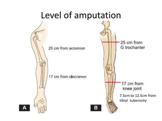
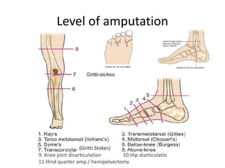
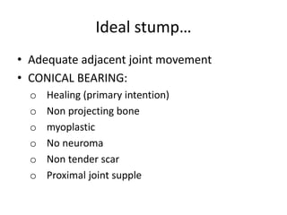
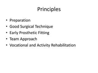
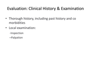
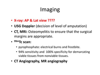
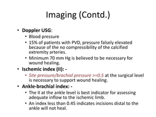
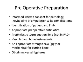
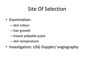
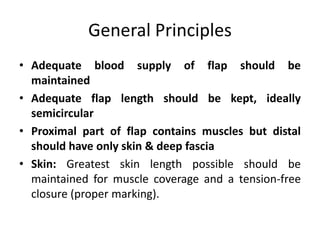
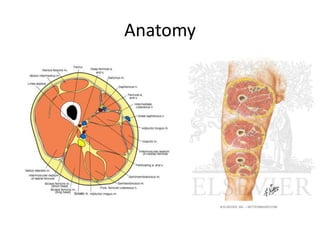
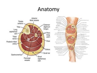
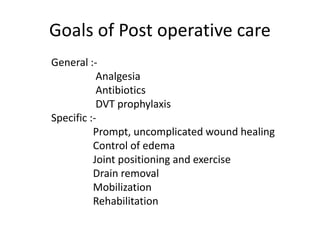
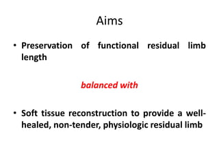
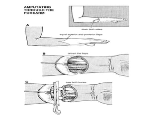
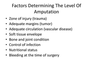
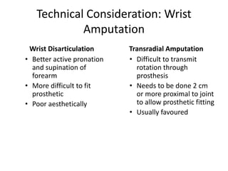
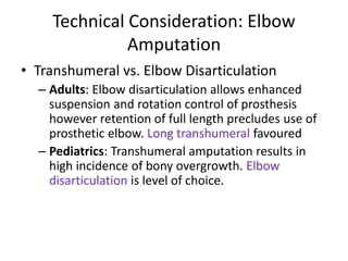
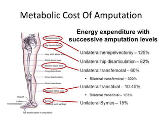
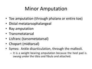
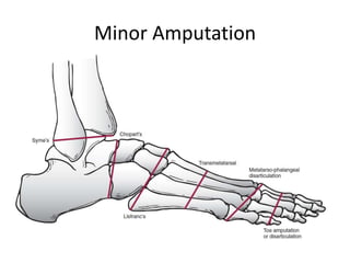
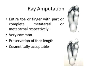
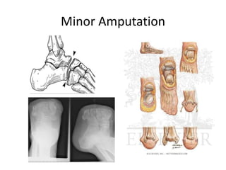
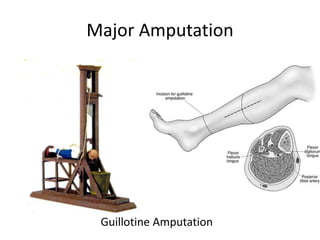
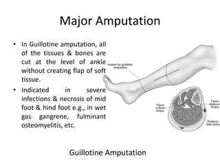
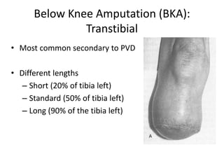
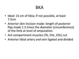
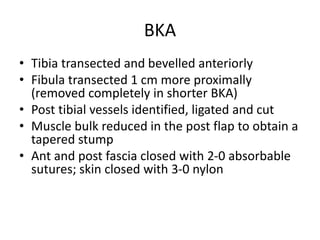
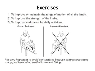
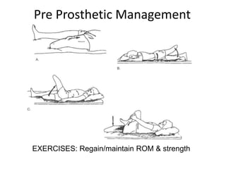
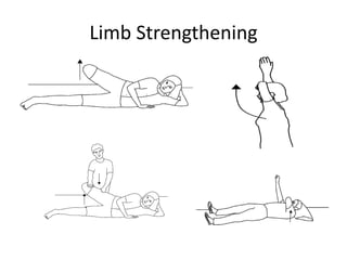
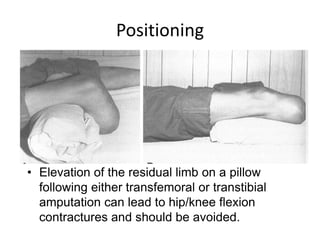
AMPUTATION AND
REHABILITATION
Dr. Rajendra M
Dept. of Surgical
Disciplines
Outline
• Introduction And General Principles
• Pre Operative Preparations
• Upper Limb Amputation
• Lower Limb Amputation
• Rehabilitation
Definition
• Amputation:“Surgical removal of limb or part
of the limb through a bone or multiple
bones”
Versus
• Disarticulation:“Surgical removal of whole
limb or part of the limb through a joint”
History
• Most ancient of surgical procedure.
• Stimulated by the aftermath of war.
• Crude procedure - limb was rapidly
severed from unanesthetized
patient.
• The open stump was then crushed
or dipped in boiling oil to obtain
hemostasis.
• Hippocrates was the first to use
ligature.
• Ambroise Pare (a French military
surgeon) introduced artery forceps.
He also designed prosthesis.
Etiology
WHO manual of amputation and rehabilitation,2004
Etiology
• Peripheral Vascular Disease
• Trauma
• Malignant Tumors
• Burns
• Neurologic Conditions
• Infections
• Congenital Deformities
Dead
Deadly
Dam Nuisance
Put in order of frequency
Indications
• DEAD LIMB :
Gangrene
• DEADLY LIMB
Wet gangrene
Spreading cellulitis
Arteriovenous fistula
Other (e.g. malignancy)
• ‘DEAD LOSS’ LIMB
Severe rest pain with unreconstructable
critical leg ischaemia
Paralysis
Other (e.g. contracture, trauma)
Types of stump
I. • Non end bearing/side bearing.
• End bearing/cone bearing.
II. • Weight bearing.
• Nonweight bearing.
Types of flaps
• Long posterior flap in below-knee amputation.
• Equal flaps in above-knee amputation.
Level of amputation
WHO manual of amputation and rehabilitation,2004
Level of amputation
7.5cm to 12.5cm from
tibial tuberosity
Level of amputation
9. Knee joint disarticulation 10.Hip diarticulatio
11.Hind quarter amp./ hemipelvectomy
(Gritti Stokes)
Ideal stump
• It should be of optimum length.
• Should be rounded, smooth, with gentle
contour.
• Should be firm and heal adequately (thin scar).
• The opposing groups of muscle should be
sutured together over the end of bone.
• The muscle should be sutured in a such way
that they will be converted into fibrous tissue
and serve as in effective cushion.
Ideal stump….
• Vascularity of flap should be normal.
• There should be no projecting of spur bone.
• The stump should not be under tension.
• The position of scar should be avoided of
pressure and should be transverse to avoid
pulling up two bones in AP scar.
• In case of UL amputation, the scar can be
terminal, but in LL, a posterior scar is desirable to
avoid pressure of weight of artificial limb.
Ideal stump…
• Adequate adjacent joint movement
• CONICAL BEARING:
o Healing (primary intention)
o Non projecting bone
o myoplastic
o No neuroma
o Non tender scar
o Proximal joint supple
Principles
• Preparation
• Good Surgical Technique
• Early Prosthetic Fitting
• Team Approach
• Vocational and Activity Rehabilitation
Evaluation: Clinical History & Examination
• Thorough history, including past history and co
morbidities
• Local examination:
-Inspection
–Palpation
Evaluation: Lab Studies
• Hb, Hct (control of anemia)
• S. Creatinine, S. Albumin
• K+, Ca2+ (elevated levels cause cardiac arrhythmias
and seizures).
• WBC count, CRP, ESR (control of infection using
antibiotics).
• CRP is the first laboratory value which respond to
treatment.
• Platelets, Coagulation profile
• U. Myoglobinuria
Imaging
• X-ray: AP & Lat view ????
• USG Doppler (decision of level of amputation)
• CT, MRI: Osteomyelitis to ensure that the surgical
margins are appropriate.
• 99mTc scan:
• pyrophosphate: electrical burns and frostbite.
• 94% sensitivity and 100% specificity for demarcating
viable tissues from nonviable tissues.
• CT Angiography, MR angiography
Imaging (Contd.)
• Doppler USG:
• Blood pressure
• 15% of patients with PVD, pressure falsely elevated
because of the no compressibility of the calcified
extremity arteries.
• Minimum 70 mm Hg is believed to be necessary for
wound healing.
• Ischemic index (II): -
• Site pressure/brachial pressure >=0.5 at the surgical level
is necessary to support wound healing.
• Ankle-brachial index: -
• The II at the ankle level is best indicator for assessing
adequate inflow to the ischemic limb.
• An index less than 0.45 indicates incisions distal to the
ankle will not heal.
Pre Operative Preparation
• Informed written consent for pathology,
inevitability of amputation & its complications
• Identification of patient and limb
• Appropriate preoperative antibiotics
• Prophylactic tourniquet on limb (not in PAD)
• Vascular and bone instruments
• An appropriate strength saw (gigly or
mechanical)for cutting bone
• Obtaining vessel ligatures
Site Of Selection
• Examination:
– skin colour
– hair growth
– lowest palpable pulse
– skin temperature
• Investigation: USG Doppler/ angiography
General Principles
• Adequate blood supply of flap should be
maintained
• Adequate flap length should be kept, ideally
semicircular
• Proximal part of flap contains muscles but distal
should have only skin & deep fascia
• Skin: Greatest skin length possible should be
maintained for muscle coverage and a tension-free
closure (proper marking).
General Principles (Contd.)
• Muscle:
• Muscle should be placed over the cut end of bones
via myodesis
• OR a long posterior flap should be sutured anteriorly
• OR a well-balanced myoplasty should be done (i.e.,
antagonistic muscle and fascia sutured together)
General Principles (Contd.)
• Nerves:
– Pulled distally, cut sharply (knife) & allowed to
retract
– Ligation of large nerves can be performed when
an associated vessel is present
• Arteries and veins:
– Dissected and separately ligated
– Prevents the development of arteriovenous
fistulas and aneurysms
General Principles (Contd.)
• Bone:
– Bony prominences around disarticulations are removed
with a saw
– In distal ends, cut with bevelling anteriorly and filed
smooth
– Maintain the maximal extremity length possible
• For non-ischemic limbs, below-knee amputations are best
performed 12-18 cm below the joint line
• For ischemic limbs, a higher level of 10-12.5 cm below the
joint line is used because making limbs longer than this can
interfere with prosthetic use and design
General Principles (Contd.)
• In crush injury/ entrapment injury/ sepsis-
Guillotine amputation should be done.
Later definitive closure is planned.
• Knowledge of anatomy of respective level of
limb is a must.
Anatomy
Anatomy
Goals of Post operative care
General :-
Analgesia
Antibiotics
DVT prophylaxis
Specific :-
Prompt, uncomplicated wound healing
Control of edema
Joint positioning and exercise
Drain removal
Mobilization
Rehabilitation
Upper Limb
• Upper extremity non weight bearing
• Less durable skin acceptable
• Decreased sensation better tolerated
• Joint deformity better tolerated
• Late amputations rare
• Transplants now being performed
Indications in Upper limb
Most Common Cause: Trauma
• Cause of 90 % upper extremity amputations
• Male: Female = 4:1
• Most amputations are at level of digits
• Major limb amputations less common
• Revascularization possible for incomplete
amputation
• Replantation possible for complete
amputation
Contraindications for Replantation
1. Severely crushed or mangled parts
2. Multiple levels
3. Other serious injuries or diseases
4. Atherosclerotic vessels
5. Mentally unstable
6. > 6 hours ischemic time
7. Severe contamination
Decision Making
• Limb injury score (LIS): >6
• Mangled Extremity Severity Score (MESS) (most
useful) : >7
• Injury severity score (ISS) > 50
• To avoid metabolic overload and secondary organ
failure in attempts to salvage a severely injured limb
• NISSSA: >11
• HFS-98: >11
• PSI: >8
Scores Used In Amputation Decision
Making
• Mangled Extremity Severity Score (MESS)1,3
• Limb Salvage Index (LSI)5
• Predictive Salvage Index (PSI)2
• Nerve Injury, Ischemia, Soft-Tissue Injury,
Skeletal Injury, Shock, and Age of Patient Score
(NISSSA)4
• Hannover Fracture Scale-98 (HFS-98)6,
• Limb Injury Score
Mangled Extremity Severity Score
(MESS)
A. Skeletal/Soft tissue injury
1. Low energy (stab wound, simple
fracture, low-energy gunshot wound)
2. Medium energy (open or multiple
fractures, dislocation)
3. High energy (high-speed motor vehicle
collision or rifle gunshot wound)
4. Very high energy (above plus gross
contamination)
B. Limb ischemia*
1. Pulse reduced or absent but perfusion
normal
2. Pulse less; paresthesia, diminished
capillary refill
3. Cool, paralyzed, insensate, numb
C. Shock
0. Systolic blood pressure always > 90 mm
Hg
1. Systolic blood pressure transiently < 90
mm Hg
2. Systolic blood pressure persistently < 90
mm Hg
D. Age (years)
0. <30
1. 30 -50
2. >50
*Score doubled for ischemia time > 6 hours
Aims
• Preservation of functional residual limb
length
balanced with
• Soft tissue reconstruction to provide a well-
healed, non-tender, physiologic residual limb
Factors Determining The Level Of
Amputation
• Zone of injury (trauma)
• Adequate margins (tumor)
• Adequate circulation (vascular disease)
• Soft tissue envelope
• Bone and joint condition
• Control of infection
• Nutritional status
• Bleeding at the time of surgery
Technical Consideration: Wrist
Amputation
Wrist Disarticulation
• Better active pronation
and supination of
forearm
• More difficult to fit
prosthetic
• Poor aesthetically
Transradial Amputation
• Difficult to transmit
rotation through
prosthesis
• Needs to be done 2 cm
or more proximal to joint
to allow prosthetic fitting
• Usually favoured
Technical Consideration: Elbow
Amputation
• Transhumeral vs. Elbow Disarticulation
– Adults: Elbow disarticulation allows enhanced
suspension and rotation control of prosthesis
however retention of full length precludes use of
prosthetic elbow. Long transhumeral favoured
– Pediatrics: Transhumeral amputation results in
high incidence of bony overgrowth. Elbow
disarticulation is level of choice.
Technical Consideration
• Preservation of Elbow function is a priority
• Consider replantation/salvage of parts to maintain
elbow function
• 4-5 cm of proximal ulna necessary for elbow
function
• For very proximal amputations, it may be
necessary to attach bicep tendon to ulna
Krukenberg Procedure
• More than 80 years ago,
Krukenberg described a
technique -a forearm stump into
a pincer motorized by the
pronator teres muscle.
• Used to be done for bilateral
upper-extremity amputations, in
those who are also blind.
• Not recommended as a primary
procedure at the time of an
amputation,
• To consider this surgical option,
the ulna and radius must extend
distal to the majority of the
pronator teres (the motor for
pinching) and an elbow flexion
contracture of less than 70°.
Forequarter Amputation
• It entails the surgical
removal of entire upper
extremity & shoulder
girdle including the
scapula & portion of the
clavicle.
• MC performed in high
grade bone sarcomas of
the proximal humerus &
scapula
• Mortality: 10%
Musculoskeletal Cancer Surgery, 2001
Pathophysiology
• As the level of the amputation moves proximally
• Greater the energy expenditure that is required
• Walking speed of the individual decreases
• Oxygen consumption increases
• Transtibial amputations: Energy cost for walking
similar to non amputee
• Transfemoral amputations: Energy required is 50-
65% greater than that required for those who
have not undergone amputations .
Metabolic Cost Of Amputation
Minor Amputation
• Toe amputation (through phalanx or entire toe)
• Distal metatarsophalangeal
• Ray amputation
• Transmetatarsal
• Lisfranc (tarsometatarsal)
• Chopart (midtarsal)
• Symes: Ankle disarticulation, through the malleoli.
– It is a weight bearing amputation because the heel pad is
swung under the tibia and fibula and attached.
Minor Amputation
Ray Amputation
• Entire toe or finger with part or
complete metatarsal or
metacarpal respectively
• Very common
• Preservation of foot length
• Cosmetically acceptable
Minor Amputation
Major Amputation
Guillotine Amputation
Major Amputation
Guillotine Amputation
• In Guillotine amputation, all
of the tissues & bones are
cut at the level of ankle
without creating flap of soft
tissue.
• Indicated in severe
infections & necrosis of mid
foot & hind foot e.g., in wet
gas gangrene, fulminant
osteomyelitis, etc.
Below Knee Amputation (BKA):
Transtibial
• Most common secondary to PVD
• Different lengths
– Short (20% of tibia left)
– Standard (50% of tibia left)
– Long (90% of the tibia left)
BKA
• Ideal: 15 cm of tibia; if not possible, at least
7.5cm
• Anterior skin incision made: length of posterior
flap made 1.5 times the diameter (circumference)
of the limb at level of amputation.
• Ant compartment muscles (TA, EHL, EDL) cut
• Anterior tibial artery and vein ligated and divided
BKA
• Tibia transected and bevelled anteriorly
• Fibula transected 1 cm more proximally
(removed completely in shorter BKA)
• Post tibial vessels identified, ligated and cut
• Muscle bulk reduced in the post flap to obtain a
tapered stump
• Ant and post fascia closed with 2-0 absorbable
sutures; skin closed with 3-0 nylon
Incision-Burgess
BKA(Contd.)
BKA (Contd.)
BKA-Skew Flap
Above Knee Amputation
• Relatively uncommon
• < 1½ inches of tibia is viable
• Intact femur results in good weight bearing surface
• Lengths
– Long (>60% of femur left)
– Standard (60%-35% of femur left)
– Short (<35% of femur left)
Above Knee Amputation
• Skin and subcutaneous tissue cut
• Ant femoral muscles (sartorius, quadriceps)
cut first, f/b medial femoral muscles (gracilis,
pectineus, adductor)
• Superficial femoral art and vein ligated and cut
• Post femoral muscles (biceps femoris,
semimembranosus, semitendinosus) cut
AKA contd…
• Sciatic nerve (b/w
adductor magnus and
biceps femoris) ligated
and cut
• Periostium stripped and
femur transected
• Ant and post fascia close
with 2-0 absorbable; skin
closed with 3-0 nylon;
drain±
AKA
Complications
• General:
–hemorrhage
–hematoma
–infection
Complications
• Specific:
– Flap breakdown
– Flexion contracture
– Residual pain
– Stump pain
– Phantom pain
– Stump ulceration
– Ring sequestrum
formation
– Painful scar
– Joint contracture
• Others:
– Scar hypertrophy
– Thickening
– Hyperkeratosis
– Papilloma
– Eczema
– Lympoedema
– Boils
– Bursae
– Spur, osteophytes
formation, jactitation,
aneurysm, stump # etc.
Pain management
• Post op Pain: Analgesics + Limb elevation
• Watch for infection
• Pain after healing: treat cause + mechanical
stimulation
• Prosthetic pain: best fit prosthesis
• Phantom Pain: Pharmacotherapy, surgical
procedures, adjuvant therapy
Psychological adjustment
• Provide information
• Assess social support
• Address both the amputee and the family
• Peer Counselling and support groups
• Return to work and previous life roles.
Skin Care
Skin hygiene and lubrication Skin Inspection
Skin Mobilization Skin desensitization
Physiotherapy
1. Residual Limb Shrinkage and Shaping
2. Limb Desensitization
3. Maintain joint range of motion
4. Strengthen residual limb
5. Maximize Self reliance
6. Patient education: Future goals and prosthetic
options
Exercises
Pre Prosthetic Management
EXERCISES: Regain/maintain ROM & strength
Limb Strengthening
Positioning
• Elevation of the residual limb on a pillow
following either transfemoral or transtibial
amputation can lead to hip/knee flexion
contractures and should be avoided.
Mobility
• Reaching for an object promotes weight shifting on/off the
prosthesis. Mirror reduces tendency to look at the floor.
Self Care
Ideal Prosthesis
• Comfortable
• Functional
• Cosmetic
• Exoprosthesis: external replacement for a lost
part of the limb
Types available
• Syme’s elephant boot, Canadian Syme’s
prosthesis
• BKA: PTB (patellar tendon bearing) prosthesis,
SACH (solid ankle cushion heel)
• AKA: suction type
• Hemipelvectomy: TTP (tilting table prosthesis)
• Bionics
• CAD-CAM (computer assisted designing and
computer assisted manufacturing)
• Exo- vs. endoskeleton
Types available
Round/elephant boot canadian type syme’s boots PTB prosthesis
Temporary Prosthesis
• Cosmetically unfinished
prosthesis that has been
fitted and aligned
• Used when amputee’s
ability to wear a prosthesis
is in doubt
• Can help shape limb better
rather than dressing
Part of Prosthesis
Strap or belt holding
prosthesis to stump
Soft Foam or silicone
Contact with the skin
Connects socket with foot
Contact with ground
Classification
• Passive
– Cosmetic
• Body Powered
– Harnesses and cables
• Myoelectric
– Surface EMG
– Activation delay
• Neuroprosthetics
– Investigational
Jaipur Foot
Bhagwan Mahavir Viklang Sahyata Samiti Dr. P. K . Sethi
•The articulation at the 'ankle' allows not
only Inversion-Eversion movements but
also dorsiflexion (essential for squatting,
standing up from prone position etc.)
•A shorter keel helps achieve this. Also,
the materials used at the foot end are
waterproof and moderately mimic a real
foot.
Conclusion
• Fewer procedure in surgery evoke more fear in
patient than a major amputation.
• Careful selection of level based on circulation and
functional issues, attention to detail in the operating
room, and careful perioperative care are required to
obtain good results.
• Surgeon who performs a major limb amputation
owes the patient debt of rehabilitation
• Improved prosthetic design does not compensate for
a poorly performed surgical procedure
Amputation is not the end of life. It is the first day of
new beginning!!
Quiz
Which of the following
deformities is most
common after
amputation shown in
the figure?
a) Pes cavus
b) Pes planus
c) Hind foot valgus
d) Equinovarus
e) Calcaneovalgus

Amputation and Rehabilitation

  • 1.
    AMPUTATION AND REHABILITATION Dr. RajendraM Dept. of Surgical Disciplines
  • 2.
    Outline • Introduction AndGeneral Principles • Pre Operative Preparations • Upper Limb Amputation • Lower Limb Amputation • Rehabilitation
  • 3.
    Definition • Amputation:“Surgical removalof limb or part of the limb through a bone or multiple bones” Versus • Disarticulation:“Surgical removal of whole limb or part of the limb through a joint”
  • 4.
    History • Most ancientof surgical procedure. • Stimulated by the aftermath of war. • Crude procedure - limb was rapidly severed from unanesthetized patient. • The open stump was then crushed or dipped in boiling oil to obtain hemostasis. • Hippocrates was the first to use ligature. • Ambroise Pare (a French military surgeon) introduced artery forceps. He also designed prosthesis.
  • 5.
    Etiology WHO manual ofamputation and rehabilitation,2004
  • 6.
    Etiology • Peripheral VascularDisease • Trauma • Malignant Tumors • Burns • Neurologic Conditions • Infections • Congenital Deformities Dead Deadly Dam Nuisance Put in order of frequency
  • 7.
    Indications • DEAD LIMB: Gangrene • DEADLY LIMB Wet gangrene Spreading cellulitis Arteriovenous fistula Other (e.g. malignancy) • ‘DEAD LOSS’ LIMB Severe rest pain with unreconstructable critical leg ischaemia Paralysis Other (e.g. contracture, trauma)
  • 8.
    Types of stump I.• Non end bearing/side bearing. • End bearing/cone bearing. II. • Weight bearing. • Nonweight bearing. Types of flaps • Long posterior flap in below-knee amputation. • Equal flaps in above-knee amputation.
  • 9.
    Level of amputation WHOmanual of amputation and rehabilitation,2004
  • 10.
    Level of amputation 7.5cmto 12.5cm from tibial tuberosity
  • 11.
    Level of amputation 9.Knee joint disarticulation 10.Hip diarticulatio 11.Hind quarter amp./ hemipelvectomy (Gritti Stokes)
  • 12.
    Ideal stump • Itshould be of optimum length. • Should be rounded, smooth, with gentle contour. • Should be firm and heal adequately (thin scar). • The opposing groups of muscle should be sutured together over the end of bone. • The muscle should be sutured in a such way that they will be converted into fibrous tissue and serve as in effective cushion.
  • 13.
    Ideal stump…. • Vascularityof flap should be normal. • There should be no projecting of spur bone. • The stump should not be under tension. • The position of scar should be avoided of pressure and should be transverse to avoid pulling up two bones in AP scar. • In case of UL amputation, the scar can be terminal, but in LL, a posterior scar is desirable to avoid pressure of weight of artificial limb.
  • 14.
    Ideal stump… • Adequateadjacent joint movement • CONICAL BEARING: o Healing (primary intention) o Non projecting bone o myoplastic o No neuroma o Non tender scar o Proximal joint supple
  • 15.
    Principles • Preparation • GoodSurgical Technique • Early Prosthetic Fitting • Team Approach • Vocational and Activity Rehabilitation
  • 17.
    Evaluation: Clinical History& Examination • Thorough history, including past history and co morbidities • Local examination: -Inspection –Palpation
  • 18.
    Evaluation: Lab Studies •Hb, Hct (control of anemia) • S. Creatinine, S. Albumin • K+, Ca2+ (elevated levels cause cardiac arrhythmias and seizures). • WBC count, CRP, ESR (control of infection using antibiotics). • CRP is the first laboratory value which respond to treatment. • Platelets, Coagulation profile • U. Myoglobinuria
  • 19.
    Imaging • X-ray: AP& Lat view ???? • USG Doppler (decision of level of amputation) • CT, MRI: Osteomyelitis to ensure that the surgical margins are appropriate. • 99mTc scan: • pyrophosphate: electrical burns and frostbite. • 94% sensitivity and 100% specificity for demarcating viable tissues from nonviable tissues. • CT Angiography, MR angiography
  • 20.
    Imaging (Contd.) • DopplerUSG: • Blood pressure • 15% of patients with PVD, pressure falsely elevated because of the no compressibility of the calcified extremity arteries. • Minimum 70 mm Hg is believed to be necessary for wound healing. • Ischemic index (II): - • Site pressure/brachial pressure >=0.5 at the surgical level is necessary to support wound healing. • Ankle-brachial index: - • The II at the ankle level is best indicator for assessing adequate inflow to the ischemic limb. • An index less than 0.45 indicates incisions distal to the ankle will not heal.
  • 21.
    Pre Operative Preparation •Informed written consent for pathology, inevitability of amputation & its complications • Identification of patient and limb • Appropriate preoperative antibiotics • Prophylactic tourniquet on limb (not in PAD) • Vascular and bone instruments • An appropriate strength saw (gigly or mechanical)for cutting bone • Obtaining vessel ligatures
  • 22.
    Site Of Selection •Examination: – skin colour – hair growth – lowest palpable pulse – skin temperature • Investigation: USG Doppler/ angiography
  • 23.
    General Principles • Adequateblood supply of flap should be maintained • Adequate flap length should be kept, ideally semicircular • Proximal part of flap contains muscles but distal should have only skin & deep fascia • Skin: Greatest skin length possible should be maintained for muscle coverage and a tension-free closure (proper marking).
  • 24.
    General Principles (Contd.) •Muscle: • Muscle should be placed over the cut end of bones via myodesis • OR a long posterior flap should be sutured anteriorly • OR a well-balanced myoplasty should be done (i.e., antagonistic muscle and fascia sutured together)
  • 25.
    General Principles (Contd.) •Nerves: – Pulled distally, cut sharply (knife) & allowed to retract – Ligation of large nerves can be performed when an associated vessel is present • Arteries and veins: – Dissected and separately ligated – Prevents the development of arteriovenous fistulas and aneurysms
  • 26.
    General Principles (Contd.) •Bone: – Bony prominences around disarticulations are removed with a saw – In distal ends, cut with bevelling anteriorly and filed smooth – Maintain the maximal extremity length possible • For non-ischemic limbs, below-knee amputations are best performed 12-18 cm below the joint line • For ischemic limbs, a higher level of 10-12.5 cm below the joint line is used because making limbs longer than this can interfere with prosthetic use and design
  • 27.
    General Principles (Contd.) •In crush injury/ entrapment injury/ sepsis- Guillotine amputation should be done. Later definitive closure is planned. • Knowledge of anatomy of respective level of limb is a must.
  • 28.
  • 29.
  • 30.
    Goals of Postoperative care General :- Analgesia Antibiotics DVT prophylaxis Specific :- Prompt, uncomplicated wound healing Control of edema Joint positioning and exercise Drain removal Mobilization Rehabilitation
  • 32.
    Upper Limb • Upperextremity non weight bearing • Less durable skin acceptable • Decreased sensation better tolerated • Joint deformity better tolerated • Late amputations rare • Transplants now being performed
  • 33.
  • 34.
    Most Common Cause:Trauma • Cause of 90 % upper extremity amputations • Male: Female = 4:1 • Most amputations are at level of digits • Major limb amputations less common • Revascularization possible for incomplete amputation • Replantation possible for complete amputation
  • 35.
    Contraindications for Replantation 1.Severely crushed or mangled parts 2. Multiple levels 3. Other serious injuries or diseases 4. Atherosclerotic vessels 5. Mentally unstable 6. > 6 hours ischemic time 7. Severe contamination
  • 36.
    Decision Making • Limbinjury score (LIS): >6 • Mangled Extremity Severity Score (MESS) (most useful) : >7 • Injury severity score (ISS) > 50 • To avoid metabolic overload and secondary organ failure in attempts to salvage a severely injured limb • NISSSA: >11 • HFS-98: >11 • PSI: >8
  • 37.
    Scores Used InAmputation Decision Making • Mangled Extremity Severity Score (MESS)1,3 • Limb Salvage Index (LSI)5 • Predictive Salvage Index (PSI)2 • Nerve Injury, Ischemia, Soft-Tissue Injury, Skeletal Injury, Shock, and Age of Patient Score (NISSSA)4 • Hannover Fracture Scale-98 (HFS-98)6, • Limb Injury Score
  • 38.
    Mangled Extremity SeverityScore (MESS) A. Skeletal/Soft tissue injury 1. Low energy (stab wound, simple fracture, low-energy gunshot wound) 2. Medium energy (open or multiple fractures, dislocation) 3. High energy (high-speed motor vehicle collision or rifle gunshot wound) 4. Very high energy (above plus gross contamination) B. Limb ischemia* 1. Pulse reduced or absent but perfusion normal 2. Pulse less; paresthesia, diminished capillary refill 3. Cool, paralyzed, insensate, numb C. Shock 0. Systolic blood pressure always > 90 mm Hg 1. Systolic blood pressure transiently < 90 mm Hg 2. Systolic blood pressure persistently < 90 mm Hg D. Age (years) 0. <30 1. 30 -50 2. >50 *Score doubled for ischemia time > 6 hours
  • 39.
    Aims • Preservation offunctional residual limb length balanced with • Soft tissue reconstruction to provide a well- healed, non-tender, physiologic residual limb
  • 41.
    Factors Determining TheLevel Of Amputation • Zone of injury (trauma) • Adequate margins (tumor) • Adequate circulation (vascular disease) • Soft tissue envelope • Bone and joint condition • Control of infection • Nutritional status • Bleeding at the time of surgery
  • 42.
    Technical Consideration: Wrist Amputation WristDisarticulation • Better active pronation and supination of forearm • More difficult to fit prosthetic • Poor aesthetically Transradial Amputation • Difficult to transmit rotation through prosthesis • Needs to be done 2 cm or more proximal to joint to allow prosthetic fitting • Usually favoured
  • 43.
    Technical Consideration: Elbow Amputation •Transhumeral vs. Elbow Disarticulation – Adults: Elbow disarticulation allows enhanced suspension and rotation control of prosthesis however retention of full length precludes use of prosthetic elbow. Long transhumeral favoured – Pediatrics: Transhumeral amputation results in high incidence of bony overgrowth. Elbow disarticulation is level of choice.
  • 44.
    Technical Consideration • Preservationof Elbow function is a priority • Consider replantation/salvage of parts to maintain elbow function • 4-5 cm of proximal ulna necessary for elbow function • For very proximal amputations, it may be necessary to attach bicep tendon to ulna
  • 45.
    Krukenberg Procedure • Morethan 80 years ago, Krukenberg described a technique -a forearm stump into a pincer motorized by the pronator teres muscle. • Used to be done for bilateral upper-extremity amputations, in those who are also blind. • Not recommended as a primary procedure at the time of an amputation, • To consider this surgical option, the ulna and radius must extend distal to the majority of the pronator teres (the motor for pinching) and an elbow flexion contracture of less than 70°.
  • 46.
    Forequarter Amputation • Itentails the surgical removal of entire upper extremity & shoulder girdle including the scapula & portion of the clavicle. • MC performed in high grade bone sarcomas of the proximal humerus & scapula • Mortality: 10% Musculoskeletal Cancer Surgery, 2001
  • 48.
    Pathophysiology • As thelevel of the amputation moves proximally • Greater the energy expenditure that is required • Walking speed of the individual decreases • Oxygen consumption increases • Transtibial amputations: Energy cost for walking similar to non amputee • Transfemoral amputations: Energy required is 50- 65% greater than that required for those who have not undergone amputations .
  • 49.
  • 50.
    Minor Amputation • Toeamputation (through phalanx or entire toe) • Distal metatarsophalangeal • Ray amputation • Transmetatarsal • Lisfranc (tarsometatarsal) • Chopart (midtarsal) • Symes: Ankle disarticulation, through the malleoli. – It is a weight bearing amputation because the heel pad is swung under the tibia and fibula and attached.
  • 51.
  • 52.
    Ray Amputation • Entiretoe or finger with part or complete metatarsal or metacarpal respectively • Very common • Preservation of foot length • Cosmetically acceptable
  • 53.
  • 54.
  • 55.
    Major Amputation Guillotine Amputation •In Guillotine amputation, all of the tissues & bones are cut at the level of ankle without creating flap of soft tissue. • Indicated in severe infections & necrosis of mid foot & hind foot e.g., in wet gas gangrene, fulminant osteomyelitis, etc.
  • 56.
    Below Knee Amputation(BKA): Transtibial • Most common secondary to PVD • Different lengths – Short (20% of tibia left) – Standard (50% of tibia left) – Long (90% of the tibia left)
  • 57.
    BKA • Ideal: 15cm of tibia; if not possible, at least 7.5cm • Anterior skin incision made: length of posterior flap made 1.5 times the diameter (circumference) of the limb at level of amputation. • Ant compartment muscles (TA, EHL, EDL) cut • Anterior tibial artery and vein ligated and divided
  • 58.
    BKA • Tibia transectedand bevelled anteriorly • Fibula transected 1 cm more proximally (removed completely in shorter BKA) • Post tibial vessels identified, ligated and cut • Muscle bulk reduced in the post flap to obtain a tapered stump • Ant and post fascia closed with 2-0 absorbable sutures; skin closed with 3-0 nylon
  • 59.
  • 60.
  • 61.
  • 62.
  • 63.
    Above Knee Amputation •Relatively uncommon • < 1½ inches of tibia is viable • Intact femur results in good weight bearing surface • Lengths – Long (>60% of femur left) – Standard (60%-35% of femur left) – Short (<35% of femur left)
  • 64.
    Above Knee Amputation •Skin and subcutaneous tissue cut • Ant femoral muscles (sartorius, quadriceps) cut first, f/b medial femoral muscles (gracilis, pectineus, adductor) • Superficial femoral art and vein ligated and cut • Post femoral muscles (biceps femoris, semimembranosus, semitendinosus) cut
  • 65.
    AKA contd… • Sciaticnerve (b/w adductor magnus and biceps femoris) ligated and cut • Periostium stripped and femur transected • Ant and post fascia close with 2-0 absorbable; skin closed with 3-0 nylon; drain±
  • 66.
  • 67.
  • 68.
    Complications • Specific: – Flapbreakdown – Flexion contracture – Residual pain – Stump pain – Phantom pain – Stump ulceration – Ring sequestrum formation – Painful scar – Joint contracture • Others: – Scar hypertrophy – Thickening – Hyperkeratosis – Papilloma – Eczema – Lympoedema – Boils – Bursae – Spur, osteophytes formation, jactitation, aneurysm, stump # etc.
  • 70.
    Pain management • Postop Pain: Analgesics + Limb elevation • Watch for infection • Pain after healing: treat cause + mechanical stimulation • Prosthetic pain: best fit prosthesis • Phantom Pain: Pharmacotherapy, surgical procedures, adjuvant therapy
  • 71.
    Psychological adjustment • Provideinformation • Assess social support • Address both the amputee and the family • Peer Counselling and support groups • Return to work and previous life roles.
  • 72.
    Skin Care Skin hygieneand lubrication Skin Inspection Skin Mobilization Skin desensitization
  • 73.
    Physiotherapy 1. Residual LimbShrinkage and Shaping 2. Limb Desensitization 3. Maintain joint range of motion 4. Strengthen residual limb 5. Maximize Self reliance 6. Patient education: Future goals and prosthetic options
  • 74.
  • 75.
    Pre Prosthetic Management EXERCISES:Regain/maintain ROM & strength
  • 76.
  • 77.
    Positioning • Elevation ofthe residual limb on a pillow following either transfemoral or transtibial amputation can lead to hip/knee flexion contractures and should be avoided.
  • 78.
    Mobility • Reaching foran object promotes weight shifting on/off the prosthesis. Mirror reduces tendency to look at the floor.
  • 79.
  • 80.
    Ideal Prosthesis • Comfortable •Functional • Cosmetic • Exoprosthesis: external replacement for a lost part of the limb
  • 81.
    Types available • Syme’selephant boot, Canadian Syme’s prosthesis • BKA: PTB (patellar tendon bearing) prosthesis, SACH (solid ankle cushion heel) • AKA: suction type • Hemipelvectomy: TTP (tilting table prosthesis) • Bionics • CAD-CAM (computer assisted designing and computer assisted manufacturing) • Exo- vs. endoskeleton
  • 82.
    Types available Round/elephant bootcanadian type syme’s boots PTB prosthesis
  • 83.
    Temporary Prosthesis • Cosmeticallyunfinished prosthesis that has been fitted and aligned • Used when amputee’s ability to wear a prosthesis is in doubt • Can help shape limb better rather than dressing
  • 84.
    Part of Prosthesis Strapor belt holding prosthesis to stump Soft Foam or silicone Contact with the skin Connects socket with foot Contact with ground
  • 85.
    Classification • Passive – Cosmetic •Body Powered – Harnesses and cables • Myoelectric – Surface EMG – Activation delay • Neuroprosthetics – Investigational
  • 86.
    Jaipur Foot Bhagwan MahavirViklang Sahyata Samiti Dr. P. K . Sethi •The articulation at the 'ankle' allows not only Inversion-Eversion movements but also dorsiflexion (essential for squatting, standing up from prone position etc.) •A shorter keel helps achieve this. Also, the materials used at the foot end are waterproof and moderately mimic a real foot.
  • 87.
    Conclusion • Fewer procedurein surgery evoke more fear in patient than a major amputation. • Careful selection of level based on circulation and functional issues, attention to detail in the operating room, and careful perioperative care are required to obtain good results. • Surgeon who performs a major limb amputation owes the patient debt of rehabilitation • Improved prosthetic design does not compensate for a poorly performed surgical procedure
  • 88.
    Amputation is notthe end of life. It is the first day of new beginning!!
  • 89.
    Quiz Which of thefollowing deformities is most common after amputation shown in the figure? a) Pes cavus b) Pes planus c) Hind foot valgus d) Equinovarus e) Calcaneovalgus

Editor's Notes

  • #37 Skeletal / soft-tissue injury       Low energy (stab; simple fracture; pistol gunshot wound): 1       Medium energy (open or multiple fractures, dislocation): 2       High energy (high speed MVA or rifle GSW): 3       Very high energy (high speed trauma + gross contamination): 4  Limb ischemia       Pulse reduced or absent but perfusion normal: 1*       Pulseless; paresthesias, diminished capillary refill: 2       Cool, paralyzed, insensate, numb: 3*  Shock       Systolic BP always > 90 mm Hg: 0       Hypotensive transiently: 1       Persistent hypotension: 2  Age (years)       < 30: 0       30-50: 1       > 50: 2 
  • #62 Ata ,deep peroneal nerve Pta and tibial nerve