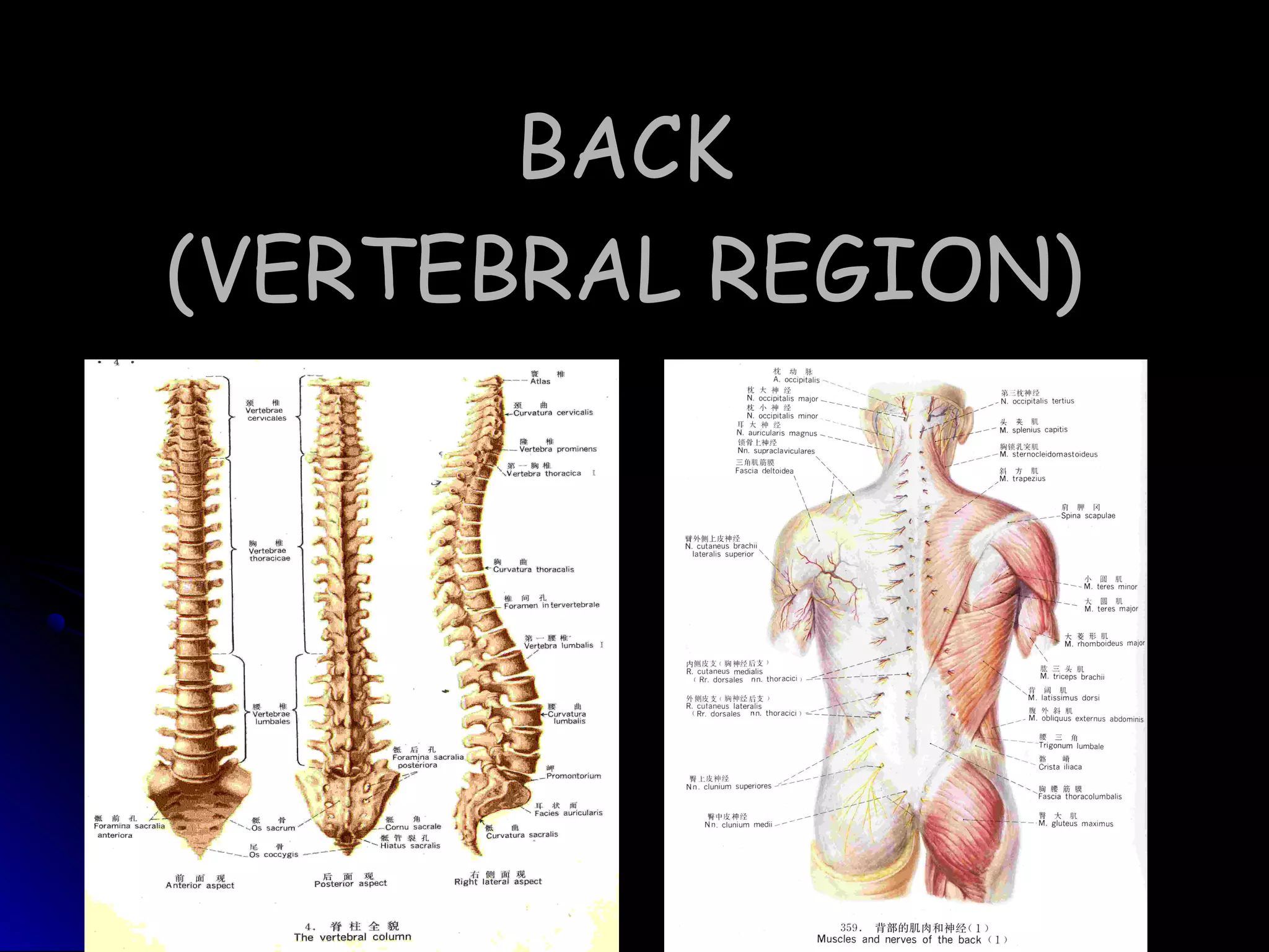
BACKBACK
(VERTEBRAL REGION)(VERTEBRAL REGION)
BoundariesBoundaries
superiorsuperior :: externalexternal
occipital protuberance andoccipital protuberance and
superior nuchal linesuperior nuchal line
inferior:inferior: line from theline from the
coccyx apex to posteriorcoccyx apex to posterior
iliac spineiliac spine
laterallateral ::
ant. border of trapezius,ant. border of trapezius,
pos. border of deltoid,pos. border of deltoid,
pos. wall of axillary fossa,pos. wall of axillary fossa,
pos. axillary linepos. axillary line
Divisions:Divisions:
The napeThe nape
The back of thoraxThe back of thorax
The lumberThe lumber
The sacrococcygealThe sacrococcygeal
acromionacromion
SurfaceSurface
AnatomyAnatomy
SurfaceSurface
AnatomyAnatomy
1.Skin incision1.Skin incision
External occipitalExternal occipital
protuberance lineprotuberance line
Acromion lineAcromion line
Inf. Angle ofInf. Angle of
scapula linescapula line
Iliac crest lineIliac crest line
Dissection ProceduresDissection Procedures
Med. vertical lineMed. vertical line
2.Reflect the skin flaps laterally2.Reflect the skin flaps laterally
Method of DissectionMethod of Dissection
Make out the line ofMake out the line of
incision by the scalpelincision by the scalpel
Hold the blade at rightHold the blade at right
angle to the skin, and driveangle to the skin, and drive
its point through the skinits point through the skin
till the superficial fascia istill the superficial fascia is
reached. Then incline thereached. Then incline the
blade to an angle of 45blade to an angle of 4500
toto
the surfacethe surface
Only reflect the skin flaps, but not superficial fasciaOnly reflect the skin flaps, but not superficial fascia
LAYERSLAYERS
 Superficial layersSuperficial layers
 Deep fasciaDeep fascia
 MusclesMuscles
Superficial layersSuperficial layers
 SkinSkin
 Superficial fasciaSuperficial fascia
SkinSkin
ThickThick and highly protectiveand highly protective
Full of hairFull of hair folliclesfollicles,, sweat glands & sebaceous glandssweat glands & sebaceous glands
Superficial fasciaSuperficial fascia
 Dense, especially the Nuchal regionDense, especially the Nuchal region
 Full of FatFull of Fat
 Cutaneous nerve & Superficial bloodCutaneous nerve & Superficial blood
vesselsvessels
Cutaneous nerveCutaneous nerve
Spinal nerveSpinal nerve
Pos. ramusPos. ramus
Ant. ramusAnt. ramus
ganglion
Greater occipital nerveGreater occipital nerve
3rd occipital nerve3rd occipital nerve
Spinal nerve (cutaneousSpinal nerve (cutaneous
branches of dorsal rami)branches of dorsal rami)
Superior cluneal nerveSuperior cluneal nerve
(cutaneous branches of(cutaneous branches of
dorsal rami L1-3)dorsal rami L1-3)
Middle cluneal nerveMiddle cluneal nerve
(cutaneous branches of(cutaneous branches of
dorsal rami S1-3)dorsal rami S1-3)
Inferior cluneal nerveInferior cluneal nerve
((posterior femoralposterior femoral cutaneouscutaneous
nerve(sacral plexus)nerve(sacral plexus)
Dorsal Rami of Spinal NerveDorsal Rami of Spinal Nerve
Superficial Blood VesselsSuperficial Blood Vessels
NuchalNuchal
Occipital arteryOccipital artery
Superficial cervical arterySuperficial cervical artery
dorsal scapular arterydorsal scapular artery
Thoracic dorsalThoracic dorsal
Posterior intercostals arteryPosterior intercostals artery
dorsal scapular arterydorsal scapular artery
thoracodorsal arterythoracodorsal artery
LumberLumber Lumbar arteryLumbar artery
SacrococcygealSacrococcygeal
Superior Cluneal arterySuperior Cluneal artery
Inferior cluneal arteryInferior cluneal artery
Deep FasciaDeep Fascia
 Nuchal fasciaNuchal fascia: a deep investing membrane: a deep investing membrane
which covers the deep muscles of the backwhich covers the deep muscles of the back
of the neck.of the neck.
 Thoracolumbar fasciaThoracolumbar fascia: a deep investing: a deep investing
membrane which covers the deep musclesmembrane which covers the deep muscles
of the back of the trunk.of the back of the trunk.
ThoracolumbarThoracolumbar
FasciaFascia
a deep investinga deep investing
membrane whichmembrane which
covers the deepcovers the deep
muscles of themuscles of the
back of the trunkback of the trunk
 3 layers in lumbar3 layers in lumbar
regionregion
Q.L
Erector spinae
MUSCLESMUSCLES
intrinsic backintrinsic back
musclesmuscles
movement andmovement and
stabilization ofstabilization of
the vertebralthe vertebral
columncolumn
respiration andrespiration and
movements of themovements of the
upper extremityupper extremity
extrinsic back musclesextrinsic back muscles
IntermediateIntermediate
groupgroup
DeepDeep
groupgroup
SuperficialSuperficial
groupgroup
TrapeziusTrapezius
RhomboideusRhomboideus
Latissimus dorsiLatissimus dorsi
Semispinalis capitisSemispinalis capitis
Splenius capitisSplenius capitis
levator scapulaelevator scapulae
SerratusSerratus
posteriorposterior
superiorsuperior
erector spinaeerector spinae
SerratusSerratus
posteriorposterior
inferiorinferior
ObliquusObliquus
externusexternus
abdominisabdominis
Serratus posterior superiorSerratus posterior superior
Serratus posterior inferiorSerratus posterior inferior
Intermediate GroupIntermediate Group
DEEP GROUPDEEP GROUP
Spinotransversales MusclesSpinotransversales Muscles
Splenius CapitisSplenius Capitis
Splenius CervicisSplenius Cervicis
ErectorErector Spine MusclesSpine Muscles
IliocostalisIliocostalis
LongissimusLongissimus
SpinalisSpinalis
DEEP GROUPDEEP GROUP
Transversospinales musclesTransversospinales muscles
SemispinalisSemispinalis
RotatoresRotatores
MultifidusMultifidus
Segmental back musclesSegmental back muscles
Levatores costarumLevatores costarum
InterspinalesInterspinales
IntertransversariiIntertransversarii
P59P59
DEEP GROUPDEEP GROUP
Suboccipital MusclesSuboccipital Muscles
Rectus Capitis Posterior MinorRectus Capitis Posterior Minor
Rectus Capitis Posterior MajorRectus Capitis Posterior Major
Superior ObliqueSuperior Oblique
Inferior ObliqueInferior Oblique
Triangle Of AuscultationTriangle Of Auscultation
BoundariesBoundaries
TrapeziusTrapezius
Latissimus dorsiLatissimus dorsi
Medial border of scapulaMedial border of scapula
Characteristic:Characteristic:
Free of overlying muscleFree of overlying muscle
Clinical note:Clinical note:
It is particularly suited forIt is particularly suited for
auscultationauscultation
 Rectus capitisRectus capitis
posterior majorposterior major
 Superior RectusSuperior Rectus
ObliqueOblique
 Inferior RectusInferior Rectus
ObliqueOblique
 Vertebral Artery
 Suboccipital N (Dorsal
Ramus Of C )Ⅰ
Semispinalis capitisSemispinalis capitis
Splenius capitisSplenius capitis
Suboccipital TriangleSuboccipital Triangle
Vertebral ArteryVertebral Artery
Superior Lumbar TriangleSuperior Lumbar Triangle
(Triangle Of Grynfeltt-Lesshaft )(Triangle Of Grynfeltt-Lesshaft )
 twelfth ribtwelfth rib
 SerratusSerratus
posterior inferiorposterior inferior
 Erector spinaeErector spinae
 internal obliqueinternal oblique
Superior Lumbar TriangleSuperior Lumbar Triangle
 Covered by latissimusCovered by latissimus
dorsidorsi
 The depth is transversesThe depth is transverses
abdominisabdominis
 Three nerves:Three nerves:
subcostal n.subcostal n.
iliohypogastric n.iliohypogastric n.
ilioinguinal n.ilioinguinal n.
Clinical note:Clinical note:
kidneykidney surgical via loin : obliquesurgical via loin : oblique
incision between the 12th ribincision between the 12th rib
and iliac crestand iliac crest
 ExternalExternal
AbdominalAbdominal
ObliqueOblique
 Latissimus DorsiLatissimus Dorsi
 Iliac CrestIliac Crest
Inferior Lumbar TriangleInferior Lumbar Triangle
(Petit's Lumbar Triangle)(Petit's Lumbar Triangle)
Deep ArteriesDeep Arteries
Nuchal RegionNuchal Region
Occipital ArteryOccipital Artery
Superficial Cervical ArterySuperficial Cervical Artery
Dorsal Scapular ArteryDorsal Scapular Artery
Vertebral ArteryVertebral Artery
Thoracic RegionThoracic Region
Posterior Intercostals ArteryPosterior Intercostals Artery
Dorsal Scapular ArteryDorsal Scapular Artery
Thoracodorsal ArteryThoracodorsal Artery
Lumber RegionLumber Region
Lumbar ArteryLumbar Artery
Subcostal ArterySubcostal Artery
SacrococcygealSacrococcygeal
RegionRegion
Superior Cluneal ArterySuperior Cluneal Artery
Inferior Cluneal ArteryInferior Cluneal Artery
Vertebral ArteryVertebral Artery
NuchalNuchal
Vertebral veinVertebral vein
Internal jugular veinInternal jugular vein
Subclavian veinSubclavian vein
ThoracicThoracic
Posterior Intercostals vein – azygos veinPosterior Intercostals vein – azygos vein
Subclavian veinSubclavian vein
Axillary veinAxillary vein
LumberLumber Lumbar vein – inferior vena cavaLumbar vein – inferior vena cava
SacrocoSacroco
ccygealccygeal
Internal iliac veinInternal iliac vein
DEEP VEINSDEEP VEINS
Vertebral VeinsVertebral Veins
Deep NerversDeep Nervers
 Dorsal rami of spinal nerveDorsal rami of spinal nerve
 Accessory nerveAccessory nerve
 Thoracodorsal nerveThoracodorsal nerve
 Dorsal scapular nerveDorsal scapular nerve
Dorsal Rami of Spinal NerveDorsal Rami of Spinal Nerve
Accessory NerveAccessory Nerve
Thoracodorsal
Nerve
Dorsal Scapular NerveDorsal Scapular Nerve
VERTEBRAL CANALVERTEBRAL CANAL
CONSTRUCTIONCONSTRUCTION
 Anterior Wall:Anterior Wall:
 Vertebral bodides,Vertebral bodides,
 Posterior Border OfPosterior Border Of
Intervertebral DiscsIntervertebral Discs
 Posterior LongitudinalPosterior Longitudinal
LigamentLigament
 Posterior Wall:Posterior Wall:
 LaminaLamina
 Ligament Flava;Ligament Flava;
 Bilateral Wall:Bilateral Wall:
 PediclePedicle
 Intervertebral ForaminaIntervertebral Foramina
Posterior LongitudinalPosterior Longitudinal
LigamentLigament
Intervertebral DiscIntervertebral Disc
Vertebral bodyVertebral body
Anterior Wall:Anterior Wall:
Ligament FlavaLigament Flava
LaminaLamina
Posterior Wall:Posterior Wall:
CONTENTSCONTENTS
Spinal CordSpinal Cord
MeningesMeninges
Spinal Nerve RootSpinal Nerve Root
Cauda EquinaCauda Equina
Blood VesselsBlood Vessels
NervesNerves
Lymphatic VesselsLymphatic Vessels
Connective TissuesConnective Tissues
SPINAL CORDSPINAL CORD
 31 spinal cord segments31 spinal cord segments
8 cervical segments8 cervical segments
12 thoracic segments12 thoracic segments
5 lumbar segments5 lumbar segments
5 sacral segments5 sacral segments
1 coccygeal segment1 coccygeal segment
 Two enlargementsTwo enlargements
Cervical enlargement (C5-T1)Cervical enlargement (C5-T1)
Lumbosacral enlargement (L2-Lumbosacral enlargement (L2-
S3)S3)
MENINGESMENINGES
Spinal dura materSpinal dura mater
Spinal arachnoid materSpinal arachnoid mater
Spinal pia materSpinal pia mater
MENINGESMENINGES
DURA MATERDURA MATER
 The dura mater forms aThe dura mater forms a
tube whose upper end istube whose upper end is
attached to the edge ofattached to the edge of
the foramen magnum,the foramen magnum,
The dural tube narrowsThe dural tube narrows
at the lower border ofat the lower border of
the second sacralthe second sacral
vertebra. It invests thevertebra. It invests the
thin spinal filumthin spinal filum
terminale, descends toterminale, descends to
the back of the coccyx,the back of the coccyx,
and blends with theand blends with the
periosteum.periosteum.
ARACHNOID MATERARACHNOID MATER
 closelyclosely
applied to theapplied to the
deep aspectdeep aspect
of the duraof the dura
mater.mater.
 A delicateA delicate
avascularavascular
membranemembrane
PIA MATERPIA MATER
The spinal pia mater closely invests theThe spinal pia mater closely invests the
surface of the spinal cord and passes intosurface of the spinal cord and passes into
the anterior median fissure.the anterior median fissure.
C1
T1
T12
L5
S1
Conus medullaris
terminale filum
Cauda
equina
coccyxcoccyx
DenticulateDenticulate
LigamentLigament
 Average 21 pairsAverage 21 pairs
 Attach pia materAttach pia mater
to the arachnoidto the arachnoid
and dura matersand dura maters
 Provide stabilityProvide stability
for the spinal cordfor the spinal cord
?
?
?
?
?
?
SPACESSPACES
Epidural spaceEpidural space
Subdural spaceSubdural space
Subarachnoid spaceSubarachnoid space
Epidural spaceEpidural space
Arachnoid materArachnoid mater
Dura materDura mater
subarachnoid spacssubarachnoid spacs
Pia materPia mater
Epidural spaceEpidural space
 The epidural space lies between the spinal duraThe epidural space lies between the spinal dura
mater and the tissues which line the vertebralmater and the tissues which line the vertebral
canal.canal.
 It is closed above by fusion of the spinal duraIt is closed above by fusion of the spinal dura
with the edge of the foramen magnum, andwith the edge of the foramen magnum, and
below by the posterior sacrococcygeal ligamentbelow by the posterior sacrococcygeal ligament
which closes the sacral hiatus.which closes the sacral hiatus.
 It contains loosely packed connective tissue, fat,It contains loosely packed connective tissue, fat,
venous plexus, small arterial branches,venous plexus, small arterial branches,
lymphatic vesselslymphatic vessels
 Clinical anaesthetics injected in the space canClinical anaesthetics injected in the space can
block the conduction of spinal nerves.block the conduction of spinal nerves.
Subdural spaceSubdural space
 The subdural space is a potential space in theThe subdural space is a potential space in the
normal spine because the arachnoid and duranormal spine because the arachnoid and dura
are closely apposed. It does not connect with theare closely apposed. It does not connect with the
subarachnoid space, but continues for a shortsubarachnoid space, but continues for a short
distance along the cranial and spinal nerves.distance along the cranial and spinal nerves.
Accidental subdural catheterization may occurAccidental subdural catheterization may occur
during extradural injections. Injection of fluid intoduring extradural injections. Injection of fluid into
the subdural space may either damage the cordthe subdural space may either damage the cord
by direct toxic effects or by compression of theby direct toxic effects or by compression of the
vasculature.vasculature.
Subarachnoid spaceSubarachnoid space
 The subarachnoid space lies between theThe subarachnoid space lies between the
arachnoid and the pia mater. It containsarachnoid and the pia mater. It contains
cerebrospinal fluid (CSF)cerebrospinal fluid (CSF)
 The terminal cistern is more expansiveThe terminal cistern is more expansive
spaces from Lspaces from L11 to Sto S2 .2 .and it is the best siteand it is the best site
for puncture to obtain CSF or spinalfor puncture to obtain CSF or spinal
anaesthesiaanaesthesia
Epidural AnaesthesiaEpidural Anaesthesia
L4
Epidural Venous PlexusEpidural Venous Plexus
Lumbar CisternLumbar Cistern
LUMBAR PUNCTURE
SPINAL NERVE ROOTSSPINAL NERVE ROOTS
Cisterna MagnaCisterna Magna
(Cerebellomedullary(Cerebellomedullary
Cistern)Cistern)
RelationshipRelationship
with intervertebralwith intervertebral
foramen & discforamen & disc
OPERATING DECOMPRESSIONOPERATING DECOMPRESSION
Segmental
Artery
VEINSVEINS
Spinal CordSpinal Cord VertebraVertebra
C1-4C1-4 C1-4C1-4
C5-8, T1-4C5-8, T1-4 Cn-1Cn-1
T5-8T5-8 Tn-2Tn-2
T9-12T9-12 Tn-3Tn-3
L1-5L1-5 T10-11T10-11
S1-5, CoccygealS1-5, Coccygeal T12, L1T12, L1
Segments of Spinal CordSegments of Spinal Cord
C1-C4C1-C4 head and neck.head and neck.
C3-C5C3-C5 diaphragm (chest and breathing)diaphragm (chest and breathing)
C5-T1C5-T1 shoulders, arms and handsshoulders, arms and hands
T2-T12T2-T12
chest and abdomenchest and abdomen
(excluding internal organs)(excluding internal organs)
L1-L4L1-L4 abdomen (excluding internal organs),abdomen (excluding internal organs),
buttocks, genitals, and upper legsbuttocks, genitals, and upper legs
L4-S1L4-S1 legslegs
S2-S4S2-S4 genitals and muscles of the perineumgenitals and muscles of the perineum
DISTRIBUTIONDISTRIBUTION
2 back

2 back

  • 1.
  • 2.
    BoundariesBoundaries superiorsuperior :: externalexternal occipitalprotuberance andoccipital protuberance and superior nuchal linesuperior nuchal line inferior:inferior: line from theline from the coccyx apex to posteriorcoccyx apex to posterior iliac spineiliac spine laterallateral :: ant. border of trapezius,ant. border of trapezius, pos. border of deltoid,pos. border of deltoid, pos. wall of axillary fossa,pos. wall of axillary fossa, pos. axillary linepos. axillary line
  • 3.
    Divisions:Divisions: The napeThe nape Theback of thoraxThe back of thorax The lumberThe lumber The sacrococcygealThe sacrococcygeal acromionacromion
  • 4.
  • 5.
  • 6.
    1.Skin incision1.Skin incision ExternaloccipitalExternal occipital protuberance lineprotuberance line Acromion lineAcromion line Inf. Angle ofInf. Angle of scapula linescapula line Iliac crest lineIliac crest line Dissection ProceduresDissection Procedures Med. vertical lineMed. vertical line 2.Reflect the skin flaps laterally2.Reflect the skin flaps laterally
  • 7.
    Method of DissectionMethodof Dissection Make out the line ofMake out the line of incision by the scalpelincision by the scalpel Hold the blade at rightHold the blade at right angle to the skin, and driveangle to the skin, and drive its point through the skinits point through the skin till the superficial fascia istill the superficial fascia is reached. Then incline thereached. Then incline the blade to an angle of 45blade to an angle of 4500 toto the surfacethe surface Only reflect the skin flaps, but not superficial fasciaOnly reflect the skin flaps, but not superficial fascia
  • 8.
    LAYERSLAYERS  Superficial layersSuperficiallayers  Deep fasciaDeep fascia  MusclesMuscles
  • 9.
    Superficial layersSuperficial layers SkinSkin  Superficial fasciaSuperficial fascia
  • 10.
    SkinSkin ThickThick and highlyprotectiveand highly protective Full of hairFull of hair folliclesfollicles,, sweat glands & sebaceous glandssweat glands & sebaceous glands
  • 11.
    Superficial fasciaSuperficial fascia Dense, especially the Nuchal regionDense, especially the Nuchal region  Full of FatFull of Fat  Cutaneous nerve & Superficial bloodCutaneous nerve & Superficial blood vesselsvessels
  • 12.
  • 13.
    Spinal nerveSpinal nerve Pos.ramusPos. ramus Ant. ramusAnt. ramus ganglion
  • 15.
    Greater occipital nerveGreateroccipital nerve 3rd occipital nerve3rd occipital nerve Spinal nerve (cutaneousSpinal nerve (cutaneous branches of dorsal rami)branches of dorsal rami) Superior cluneal nerveSuperior cluneal nerve (cutaneous branches of(cutaneous branches of dorsal rami L1-3)dorsal rami L1-3) Middle cluneal nerveMiddle cluneal nerve (cutaneous branches of(cutaneous branches of dorsal rami S1-3)dorsal rami S1-3) Inferior cluneal nerveInferior cluneal nerve ((posterior femoralposterior femoral cutaneouscutaneous nerve(sacral plexus)nerve(sacral plexus)
  • 16.
    Dorsal Rami ofSpinal NerveDorsal Rami of Spinal Nerve
  • 17.
    Superficial Blood VesselsSuperficialBlood Vessels NuchalNuchal Occipital arteryOccipital artery Superficial cervical arterySuperficial cervical artery dorsal scapular arterydorsal scapular artery Thoracic dorsalThoracic dorsal Posterior intercostals arteryPosterior intercostals artery dorsal scapular arterydorsal scapular artery thoracodorsal arterythoracodorsal artery LumberLumber Lumbar arteryLumbar artery SacrococcygealSacrococcygeal Superior Cluneal arterySuperior Cluneal artery Inferior cluneal arteryInferior cluneal artery
  • 18.
    Deep FasciaDeep Fascia Nuchal fasciaNuchal fascia: a deep investing membrane: a deep investing membrane which covers the deep muscles of the backwhich covers the deep muscles of the back of the neck.of the neck.  Thoracolumbar fasciaThoracolumbar fascia: a deep investing: a deep investing membrane which covers the deep musclesmembrane which covers the deep muscles of the back of the trunk.of the back of the trunk.
  • 19.
    ThoracolumbarThoracolumbar FasciaFascia a deep investingadeep investing membrane whichmembrane which covers the deepcovers the deep muscles of themuscles of the back of the trunkback of the trunk  3 layers in lumbar3 layers in lumbar regionregion
  • 21.
  • 22.
    MUSCLESMUSCLES intrinsic backintrinsic back musclesmuscles movementandmovement and stabilization ofstabilization of the vertebralthe vertebral columncolumn respiration andrespiration and movements of themovements of the upper extremityupper extremity extrinsic back musclesextrinsic back muscles IntermediateIntermediate groupgroup DeepDeep groupgroup SuperficialSuperficial groupgroup
  • 25.
    TrapeziusTrapezius RhomboideusRhomboideus Latissimus dorsiLatissimus dorsi SemispinaliscapitisSemispinalis capitis Splenius capitisSplenius capitis levator scapulaelevator scapulae SerratusSerratus posteriorposterior superiorsuperior erector spinaeerector spinae SerratusSerratus posteriorposterior inferiorinferior ObliquusObliquus externusexternus abdominisabdominis
  • 26.
    Serratus posterior superiorSerratusposterior superior Serratus posterior inferiorSerratus posterior inferior Intermediate GroupIntermediate Group
  • 27.
    DEEP GROUPDEEP GROUP SpinotransversalesMusclesSpinotransversales Muscles Splenius CapitisSplenius Capitis Splenius CervicisSplenius Cervicis ErectorErector Spine MusclesSpine Muscles IliocostalisIliocostalis LongissimusLongissimus SpinalisSpinalis
  • 28.
    DEEP GROUPDEEP GROUP TransversospinalesmusclesTransversospinales muscles SemispinalisSemispinalis RotatoresRotatores MultifidusMultifidus Segmental back musclesSegmental back muscles Levatores costarumLevatores costarum InterspinalesInterspinales IntertransversariiIntertransversarii P59P59
  • 29.
    DEEP GROUPDEEP GROUP SuboccipitalMusclesSuboccipital Muscles Rectus Capitis Posterior MinorRectus Capitis Posterior Minor Rectus Capitis Posterior MajorRectus Capitis Posterior Major Superior ObliqueSuperior Oblique Inferior ObliqueInferior Oblique
  • 30.
    Triangle Of AuscultationTriangleOf Auscultation BoundariesBoundaries TrapeziusTrapezius Latissimus dorsiLatissimus dorsi Medial border of scapulaMedial border of scapula Characteristic:Characteristic: Free of overlying muscleFree of overlying muscle Clinical note:Clinical note: It is particularly suited forIt is particularly suited for auscultationauscultation
  • 32.
     Rectus capitisRectuscapitis posterior majorposterior major  Superior RectusSuperior Rectus ObliqueOblique  Inferior RectusInferior Rectus ObliqueOblique  Vertebral Artery  Suboccipital N (Dorsal Ramus Of C )Ⅰ Semispinalis capitisSemispinalis capitis Splenius capitisSplenius capitis Suboccipital TriangleSuboccipital Triangle
  • 34.
  • 35.
    Superior Lumbar TriangleSuperiorLumbar Triangle (Triangle Of Grynfeltt-Lesshaft )(Triangle Of Grynfeltt-Lesshaft )  twelfth ribtwelfth rib  SerratusSerratus posterior inferiorposterior inferior  Erector spinaeErector spinae  internal obliqueinternal oblique
  • 36.
    Superior Lumbar TriangleSuperiorLumbar Triangle  Covered by latissimusCovered by latissimus dorsidorsi  The depth is transversesThe depth is transverses abdominisabdominis  Three nerves:Three nerves: subcostal n.subcostal n. iliohypogastric n.iliohypogastric n. ilioinguinal n.ilioinguinal n. Clinical note:Clinical note: kidneykidney surgical via loin : obliquesurgical via loin : oblique incision between the 12th ribincision between the 12th rib and iliac crestand iliac crest
  • 37.
     ExternalExternal AbdominalAbdominal ObliqueOblique  LatissimusDorsiLatissimus Dorsi  Iliac CrestIliac Crest Inferior Lumbar TriangleInferior Lumbar Triangle (Petit's Lumbar Triangle)(Petit's Lumbar Triangle)
  • 38.
    Deep ArteriesDeep Arteries NuchalRegionNuchal Region Occipital ArteryOccipital Artery Superficial Cervical ArterySuperficial Cervical Artery Dorsal Scapular ArteryDorsal Scapular Artery Vertebral ArteryVertebral Artery Thoracic RegionThoracic Region Posterior Intercostals ArteryPosterior Intercostals Artery Dorsal Scapular ArteryDorsal Scapular Artery Thoracodorsal ArteryThoracodorsal Artery Lumber RegionLumber Region Lumbar ArteryLumbar Artery Subcostal ArterySubcostal Artery SacrococcygealSacrococcygeal RegionRegion Superior Cluneal ArterySuperior Cluneal Artery Inferior Cluneal ArteryInferior Cluneal Artery
  • 40.
  • 41.
    NuchalNuchal Vertebral veinVertebral vein Internaljugular veinInternal jugular vein Subclavian veinSubclavian vein ThoracicThoracic Posterior Intercostals vein – azygos veinPosterior Intercostals vein – azygos vein Subclavian veinSubclavian vein Axillary veinAxillary vein LumberLumber Lumbar vein – inferior vena cavaLumbar vein – inferior vena cava SacrocoSacroco ccygealccygeal Internal iliac veinInternal iliac vein DEEP VEINSDEEP VEINS
  • 42.
  • 43.
    Deep NerversDeep Nervers Dorsal rami of spinal nerveDorsal rami of spinal nerve  Accessory nerveAccessory nerve  Thoracodorsal nerveThoracodorsal nerve  Dorsal scapular nerveDorsal scapular nerve
  • 44.
    Dorsal Rami ofSpinal NerveDorsal Rami of Spinal Nerve
  • 45.
  • 46.
  • 47.
  • 48.
  • 49.
    CONSTRUCTIONCONSTRUCTION  Anterior Wall:AnteriorWall:  Vertebral bodides,Vertebral bodides,  Posterior Border OfPosterior Border Of Intervertebral DiscsIntervertebral Discs  Posterior LongitudinalPosterior Longitudinal LigamentLigament  Posterior Wall:Posterior Wall:  LaminaLamina  Ligament Flava;Ligament Flava;  Bilateral Wall:Bilateral Wall:  PediclePedicle  Intervertebral ForaminaIntervertebral Foramina
  • 50.
    Posterior LongitudinalPosterior Longitudinal LigamentLigament IntervertebralDiscIntervertebral Disc Vertebral bodyVertebral body Anterior Wall:Anterior Wall:
  • 51.
  • 53.
    CONTENTSCONTENTS Spinal CordSpinal Cord MeningesMeninges SpinalNerve RootSpinal Nerve Root Cauda EquinaCauda Equina Blood VesselsBlood Vessels NervesNerves Lymphatic VesselsLymphatic Vessels Connective TissuesConnective Tissues
  • 54.
    SPINAL CORDSPINAL CORD 31 spinal cord segments31 spinal cord segments 8 cervical segments8 cervical segments 12 thoracic segments12 thoracic segments 5 lumbar segments5 lumbar segments 5 sacral segments5 sacral segments 1 coccygeal segment1 coccygeal segment  Two enlargementsTwo enlargements Cervical enlargement (C5-T1)Cervical enlargement (C5-T1) Lumbosacral enlargement (L2-Lumbosacral enlargement (L2- S3)S3)
  • 55.
    MENINGESMENINGES Spinal dura materSpinaldura mater Spinal arachnoid materSpinal arachnoid mater Spinal pia materSpinal pia mater
  • 56.
  • 58.
    DURA MATERDURA MATER The dura mater forms aThe dura mater forms a tube whose upper end istube whose upper end is attached to the edge ofattached to the edge of the foramen magnum,the foramen magnum, The dural tube narrowsThe dural tube narrows at the lower border ofat the lower border of the second sacralthe second sacral vertebra. It invests thevertebra. It invests the thin spinal filumthin spinal filum terminale, descends toterminale, descends to the back of the coccyx,the back of the coccyx, and blends with theand blends with the periosteum.periosteum.
  • 59.
    ARACHNOID MATERARACHNOID MATER closelyclosely applied to theapplied to the deep aspectdeep aspect of the duraof the dura mater.mater.  A delicateA delicate avascularavascular membranemembrane
  • 60.
    PIA MATERPIA MATER Thespinal pia mater closely invests theThe spinal pia mater closely invests the surface of the spinal cord and passes intosurface of the spinal cord and passes into the anterior median fissure.the anterior median fissure.
  • 61.
  • 62.
    DenticulateDenticulate LigamentLigament  Average 21pairsAverage 21 pairs  Attach pia materAttach pia mater to the arachnoidto the arachnoid and dura matersand dura maters  Provide stabilityProvide stability for the spinal cordfor the spinal cord
  • 63.
  • 64.
    SPACESSPACES Epidural spaceEpidural space SubduralspaceSubdural space Subarachnoid spaceSubarachnoid space
  • 66.
    Epidural spaceEpidural space ArachnoidmaterArachnoid mater Dura materDura mater subarachnoid spacssubarachnoid spacs Pia materPia mater
  • 67.
    Epidural spaceEpidural space The epidural space lies between the spinal duraThe epidural space lies between the spinal dura mater and the tissues which line the vertebralmater and the tissues which line the vertebral canal.canal.  It is closed above by fusion of the spinal duraIt is closed above by fusion of the spinal dura with the edge of the foramen magnum, andwith the edge of the foramen magnum, and below by the posterior sacrococcygeal ligamentbelow by the posterior sacrococcygeal ligament which closes the sacral hiatus.which closes the sacral hiatus.  It contains loosely packed connective tissue, fat,It contains loosely packed connective tissue, fat, venous plexus, small arterial branches,venous plexus, small arterial branches, lymphatic vesselslymphatic vessels  Clinical anaesthetics injected in the space canClinical anaesthetics injected in the space can block the conduction of spinal nerves.block the conduction of spinal nerves.
  • 68.
    Subdural spaceSubdural space The subdural space is a potential space in theThe subdural space is a potential space in the normal spine because the arachnoid and duranormal spine because the arachnoid and dura are closely apposed. It does not connect with theare closely apposed. It does not connect with the subarachnoid space, but continues for a shortsubarachnoid space, but continues for a short distance along the cranial and spinal nerves.distance along the cranial and spinal nerves. Accidental subdural catheterization may occurAccidental subdural catheterization may occur during extradural injections. Injection of fluid intoduring extradural injections. Injection of fluid into the subdural space may either damage the cordthe subdural space may either damage the cord by direct toxic effects or by compression of theby direct toxic effects or by compression of the vasculature.vasculature.
  • 69.
    Subarachnoid spaceSubarachnoid space The subarachnoid space lies between theThe subarachnoid space lies between the arachnoid and the pia mater. It containsarachnoid and the pia mater. It contains cerebrospinal fluid (CSF)cerebrospinal fluid (CSF)  The terminal cistern is more expansiveThe terminal cistern is more expansive spaces from Lspaces from L11 to Sto S2 .2 .and it is the best siteand it is the best site for puncture to obtain CSF or spinalfor puncture to obtain CSF or spinal anaesthesiaanaesthesia
  • 70.
  • 71.
  • 74.
  • 75.
  • 76.
  • 77.
  • 78.
  • 81.
  • 82.
  • 83.
    Spinal CordSpinal CordVertebraVertebra C1-4C1-4 C1-4C1-4 C5-8, T1-4C5-8, T1-4 Cn-1Cn-1 T5-8T5-8 Tn-2Tn-2 T9-12T9-12 Tn-3Tn-3 L1-5L1-5 T10-11T10-11 S1-5, CoccygealS1-5, Coccygeal T12, L1T12, L1 Segments of Spinal CordSegments of Spinal Cord
  • 84.
    C1-C4C1-C4 head andneck.head and neck. C3-C5C3-C5 diaphragm (chest and breathing)diaphragm (chest and breathing) C5-T1C5-T1 shoulders, arms and handsshoulders, arms and hands T2-T12T2-T12 chest and abdomenchest and abdomen (excluding internal organs)(excluding internal organs) L1-L4L1-L4 abdomen (excluding internal organs),abdomen (excluding internal organs), buttocks, genitals, and upper legsbuttocks, genitals, and upper legs L4-S1L4-S1 legslegs S2-S4S2-S4 genitals and muscles of the perineumgenitals and muscles of the perineum DISTRIBUTIONDISTRIBUTION

Editor's Notes

  • #9 We will studies the back in layers: superficial layers, deep fascia and muscles.