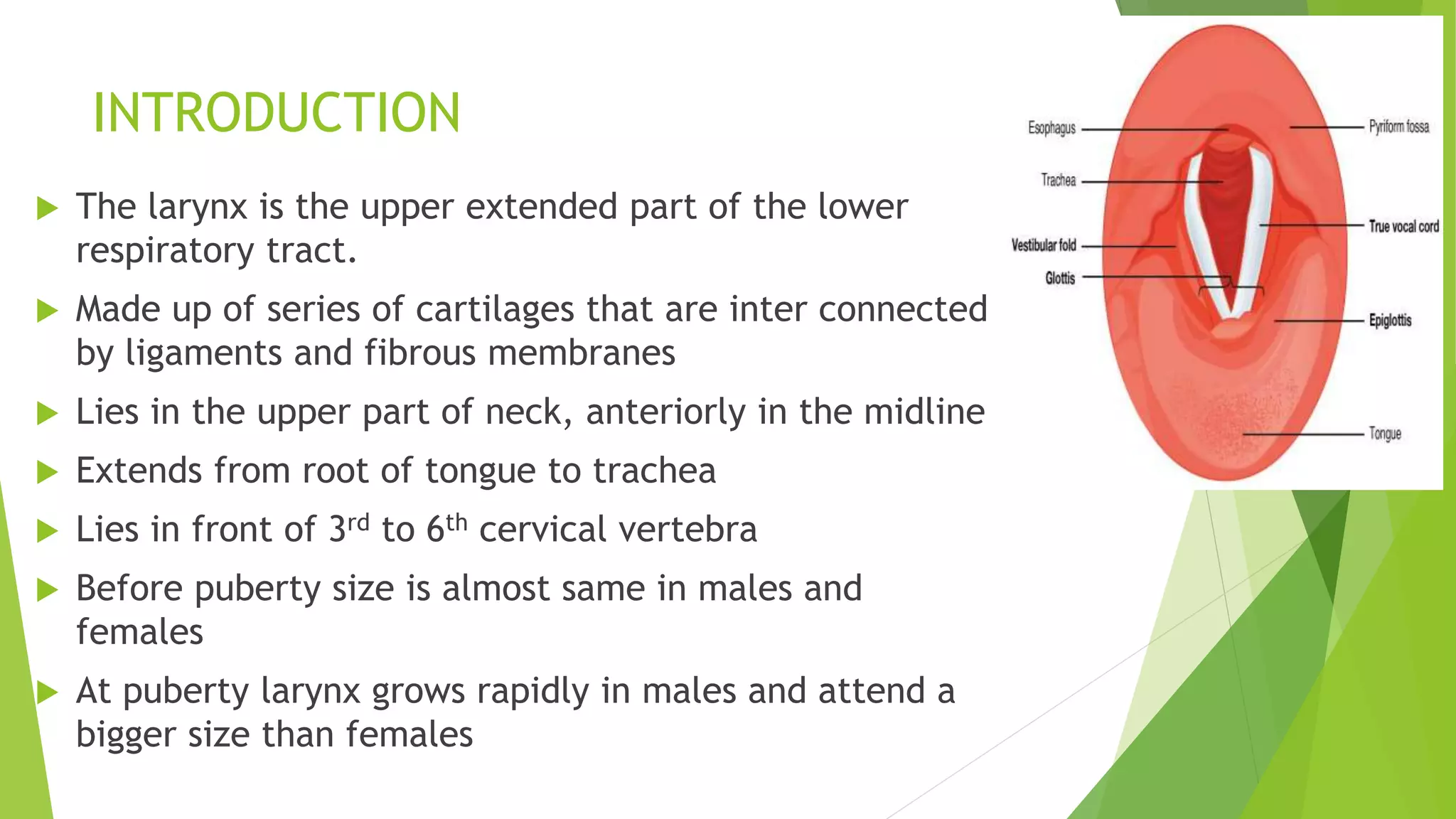
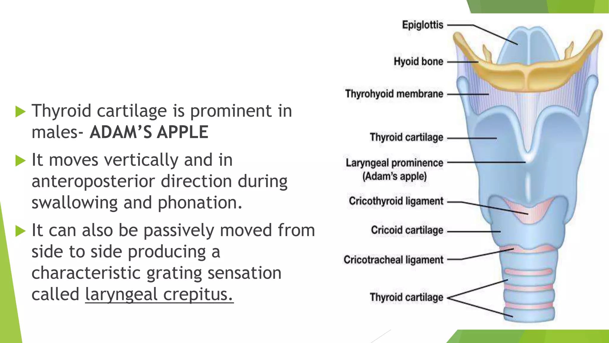
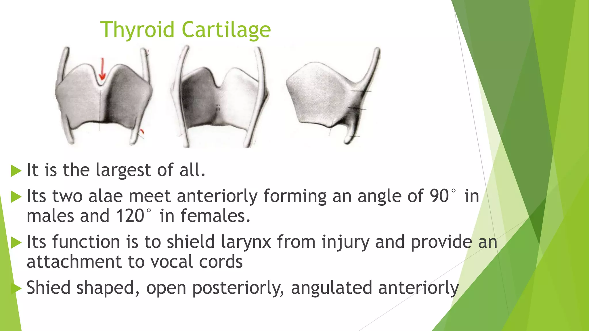
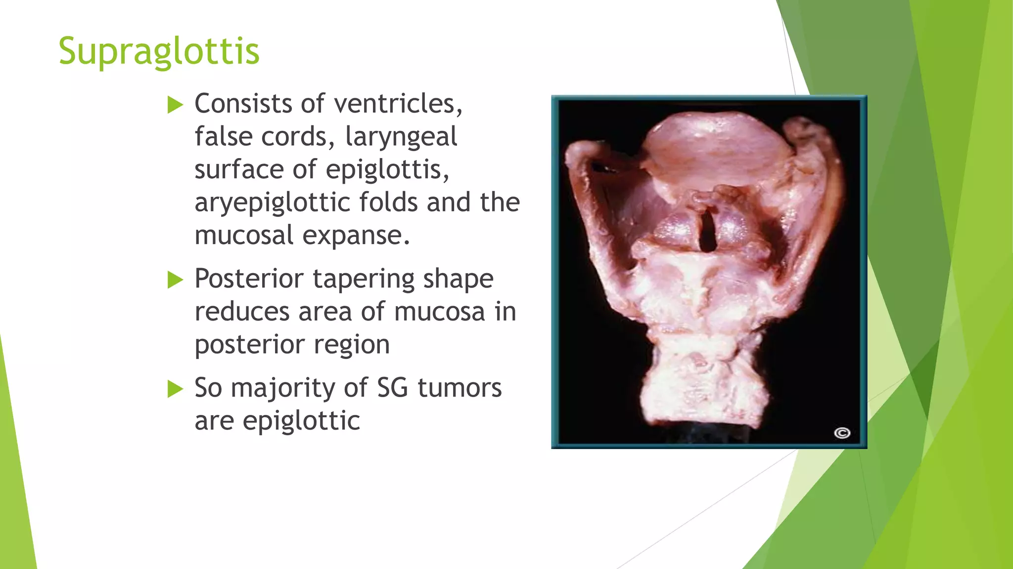
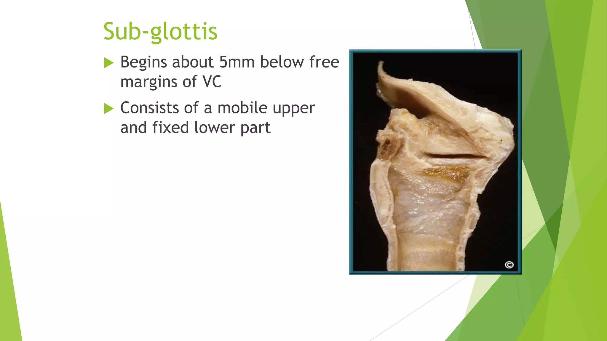
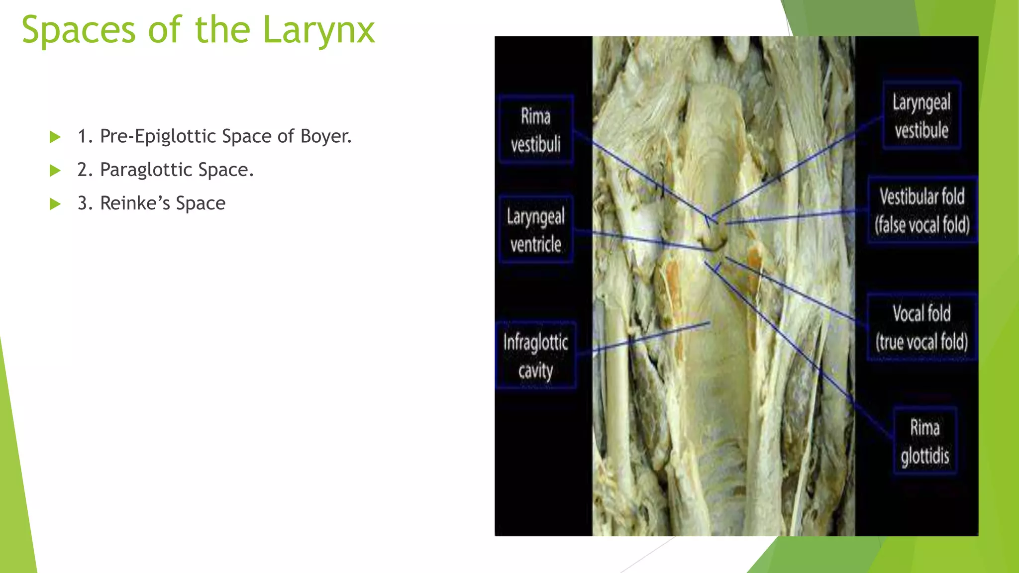
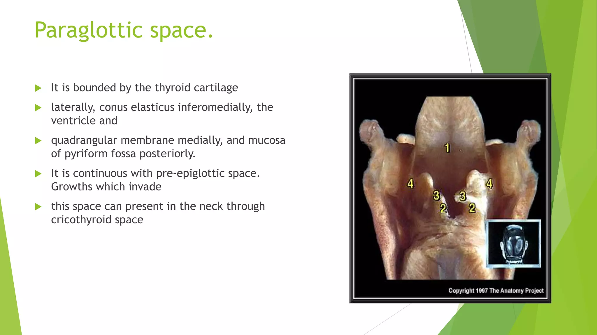
ANANTOMY AND
PHYSIOLOGY OF
LARYNX
DR. SANKALP KESHARI (MS)
ASSISTANT PROFESSOR
DEPT. OF ENT & HNS
MLN MEDICAL COLLEGE , PRAYAGRAJ
INTRODUCTION
 The larynx is the upper extended part of the lower
respiratory tract.
 Made up of series of cartilages that are inter connected
by ligaments and fibrous membranes
 Lies in the upper part of neck, anteriorly in the midline
 Extends from root of tongue to trachea
 Lies in front of 3rd to 6th cervical vertebra
 Before puberty size is almost same in males and
females
 At puberty larynx grows rapidly in males and attend a
bigger size than females
 Thyroid cartilage is prominent in
males- ADAM’S APPLE
 It moves vertically and in
anteroposterior direction during
swallowing and phonation.
 It can also be passively moved from
side to side producing a
characteristic grating sensation
called laryngeal crepitus.
PHYSIOLOGY OF LARYNX
 1. Protection of lower airways
 2. Phonation
 3. Respiration
 4. Fixation of the chest.
Laryngeal framework.
LARYNGEAL CARTILAGES
Unpaired: Thyroid, cricoid and epiglottis.
Paired: Arytenoid, corniculate and cuneiform
Thyroid Cartilage
 It is the largest of all.
 Its two alae meet anteriorly forming an angle of 90° in
males and 120° in females.
 Its function is to shield larynx from injury and provide an
attachment to vocal cords
 Shied shaped, open posteriorly, angulated anteriorly
Cricoid Cartilage
 Signet ring shaped
 Stronger than thyroid
cartilage.
 Lamina – 2 to 3 cm
from above
downwards,
considerably broader
than anterior arch.
 Important from structural & functional point of view
Base for entire larynx
Support to arytenoid
Attachment to intrinsic muscles
Only part of cartilagenous framework that
forms continuous 360 degree ring
Once injured or strictured , difficult to
resect while preserving laryngeal function
 3. Epiglottis. It is a leaf-like, yellow, elastic cartilage
forming anterior wall of laryngeal inlet.
 A stalk-like process of epiglottis (petiole) attaches
the epiglottis to the thyroid angle just above the
attachment of vocal cords.
 Anterior surface of epiglottis is separated from
thyrohyoid membrane and upper part of thyroid
cartilage by a potential space filled with fat— the
pre-epiglottic space.
 4. Arytenoid cartilages. They are paired.
Each arytenoid cartilage is pyramidal in
shape. It has a base which articulates with
cricoid cartilage.
 5. Corniculate cartilages (of Santorini) (Corn
= horn). They are paired. Each articulates
with the apex of arytenoid cartilage as if
forming its horn.
 6. Cuneiform cartilages (of Wrisberg). They
are rod shaped. Each is situated in
aryepiglottic fold in front of corniculate
cartilage and provides passive supports to the
fold.
 Thyroid, cricoid and most of the arytenoid
cartilages are hyalin cartilages
 epiglottis, corniculate, cuneiform and tip of
arytenoid near the corniculate cartilage are
elastic fibrocartilage.
Supraglottis
 Consists of ventricles,
false cords, laryngeal
surface of epiglottis,
aryepiglottic folds and the
mucosal expanse.
 Posterior tapering shape
reduces area of mucosa in
posterior region
 So majority of SG tumors
are epiglottic
Glottis
 Consists of true cords,
anterior commissure and
posterior commissure
 Narrow triangular space
between the true cords is
called rima glottis
 Anterior 2/3 is membranous
 Posterior third consists of
vocal processes of
arytenoids
 Posterior 1/3 of cords and
covering mucosa are called
posterior commissure
Sub-glottis
 Begins about 5mm below free
margins of VC
 Consists of a mobile upper
and fixed lower part
Mucosa
 Mucosa of glottic and Supraglottic regions
is stratified squamous epithelium.
 Mucosa of ventricles and sub-glottic
regions is pseudo-stratified ciliated
epithelium
 Supra and sub glottic regions particularly
ventricles are rich in submucosal mucous
or minor salivary glands while glottis is
not.
LARYNGEAL JOINTS
 Cricothyroid Joint. It is a synovial joint. Each is formed by
the inferior cornua of thyroid cartilage with a facet on the
cricoid cartilage.
 Cricoid cartilage rotates at these joints on a transverse axis
which passes transversely through these joints.
 Cricoarytenoid Joint. It is a synovial joint surrounded by
capsular ligament. It is formed between the base of arytenoid
and a facet on the upper border of cricoid lamina.
 Two types of movements occur:
 (i) rotatory
 (ii) gliding
LARYNGEAL MEMBRANES
 1. Extrinsic membranes and ligaments
 (a) Thyrohyoid membrane
 (b) Cricotracheal membrane
 (c) Hyoepiglottic ligament
 2. Intrinsic membranes and ligaments
 (a) Cricovocal membrane. It is a triangular fibroelastic membrane.
 Its upper border is free and stretches between middle of thyroid angle to
the vocal process of arytenoid and forms the vocal ligament
 Its lower border attaches to the arch of cricoid cartilage.
 thus, with its fellow on the opposite side, forms conus elasticus where
subglottic foreign bodies sometimes get impacted.
 b) Quadrangular membrane. It lies deep to mucosa of
aryepiglottic folds and is not well-defined.
 (c) Cricothyroid ligament. The anterior part of cricothyroid
membrane is thickened to form the ligament and its lateral
part forms the cricovocal membrane.
 (d) Thyroepiglottic ligament. It attaches epiglottis to thyroid
cartilage.
CAVITY OF THE LARYNX
 Inlet of Larynx. It is an oblique opening bounded
anteriorly by free margin of epiglottis; on the sides, by aryepiglottic folds and
posteriorly by interarytenoid fold
Vestibule. It extends from laryngeal inlet to vestibular folds.
anterior wall is formed by posterior surface of epiglottis; sides by the aryepiglottic
folds and posterior wall by mucous membrane over the anterior surface of arytenoids.
 Glottis (Rima Glottidis).
 It is the elongated space between vocal cords anteriorly, and vocal
processes and base of arytenoids posteriorly
 glottis is about 24 mm in men and 16 mm in women. It is the narrowest part
of laryngeal cavity.
 Anterior two-thirds of glottis is also called phonatory glottis as it is
concerned with phonation
 posterior one-third called respiratory glottis.
Spaces of the Larynx
 1. Pre-Epiglottic Space of Boyer.
 2. Paraglottic Space.
 3. Reinke’s Space
Pre-Epiglottic Space
 Bound sup by hyo-
epiglottic ligament, ant
by thyrohyoid memb. &
thyroid cartilage and
posteriorly by epiglottis
 Filled with fat and
areolar tissue
 Continuous with para-
glottic space
 Cx of laryngeal surface
of epiglottis readily
spread to PreEpiSpace
Paraglottic space.
 It is bounded by the thyroid cartilage
 laterally, conus elasticus inferomedially, the
ventricle and
 quadrangular membrane medially, and mucosa
of pyriform fossa posteriorly.
 It is continuous with pre-epiglottic space.
Growths which invade
 this space can present in the neck through
cricothyroid space
Reinke’s Space
 Mucosa over the vocal ligament
loosely attached to ligaments
 Thus there is a submucosal
space along most of the length
of truer VC
PAEDIATRIC LARYNX
 1. Infant’s larynx is positioned high in the neck level of glottis being
opposite to C3 or C4 at rest and reaches C1 or C2 during swallowing.
 This high position allows the epiglottis to meet soft palate and make a
nasopharyngeal channel for nasal breathing during suckling.
 2. Laryngeal cartilages are soft and collapse easily. Epiglottis is omega
shaped
 3. Thyroid cartilage in an infant is flat. It also overlaps the cricoid
cartilage and is in turn overlapped by the hyoid bone.
 4. Infant’s larynx is small and conical.
 5. Submucosal tissues of infant’s larynx are comparatively loose and easily
undergo oedematous change with trauma or inflammation leading to
obstruction
 Infant’s larynx shows two spurts in growth: first 3 years of life, larynx
grows in width and length
 The second spurt in growth occurs during adolescence when the thyroid
angle develops.
 In childhood, vocal cord is 6 mm in females and 8 mm in males. It
increases to 15–19 mm in adult female and 17–23 in adult male.
THANKYOU

larynx anatomy.pptx

  • 1.
    ANANTOMY AND PHYSIOLOGY OF LARYNX DR.SANKALP KESHARI (MS) ASSISTANT PROFESSOR DEPT. OF ENT & HNS MLN MEDICAL COLLEGE , PRAYAGRAJ
  • 2.
    INTRODUCTION  The larynxis the upper extended part of the lower respiratory tract.  Made up of series of cartilages that are inter connected by ligaments and fibrous membranes  Lies in the upper part of neck, anteriorly in the midline  Extends from root of tongue to trachea  Lies in front of 3rd to 6th cervical vertebra  Before puberty size is almost same in males and females  At puberty larynx grows rapidly in males and attend a bigger size than females
  • 3.
     Thyroid cartilageis prominent in males- ADAM’S APPLE  It moves vertically and in anteroposterior direction during swallowing and phonation.  It can also be passively moved from side to side producing a characteristic grating sensation called laryngeal crepitus.
  • 4.
    PHYSIOLOGY OF LARYNX 1. Protection of lower airways  2. Phonation  3. Respiration  4. Fixation of the chest.
  • 5.
  • 6.
    LARYNGEAL CARTILAGES Unpaired: Thyroid,cricoid and epiglottis. Paired: Arytenoid, corniculate and cuneiform
  • 7.
    Thyroid Cartilage  Itis the largest of all.  Its two alae meet anteriorly forming an angle of 90° in males and 120° in females.  Its function is to shield larynx from injury and provide an attachment to vocal cords  Shied shaped, open posteriorly, angulated anteriorly
  • 8.
    Cricoid Cartilage  Signetring shaped  Stronger than thyroid cartilage.  Lamina – 2 to 3 cm from above downwards, considerably broader than anterior arch.
  • 9.
     Important fromstructural & functional point of view Base for entire larynx Support to arytenoid Attachment to intrinsic muscles Only part of cartilagenous framework that forms continuous 360 degree ring Once injured or strictured , difficult to resect while preserving laryngeal function
  • 10.
     3. Epiglottis.It is a leaf-like, yellow, elastic cartilage forming anterior wall of laryngeal inlet.  A stalk-like process of epiglottis (petiole) attaches the epiglottis to the thyroid angle just above the attachment of vocal cords.  Anterior surface of epiglottis is separated from thyrohyoid membrane and upper part of thyroid cartilage by a potential space filled with fat— the pre-epiglottic space.
  • 11.
     4. Arytenoidcartilages. They are paired. Each arytenoid cartilage is pyramidal in shape. It has a base which articulates with cricoid cartilage.  5. Corniculate cartilages (of Santorini) (Corn = horn). They are paired. Each articulates with the apex of arytenoid cartilage as if forming its horn.  6. Cuneiform cartilages (of Wrisberg). They are rod shaped. Each is situated in aryepiglottic fold in front of corniculate cartilage and provides passive supports to the fold.
  • 12.
     Thyroid, cricoidand most of the arytenoid cartilages are hyalin cartilages  epiglottis, corniculate, cuneiform and tip of arytenoid near the corniculate cartilage are elastic fibrocartilage.
  • 14.
    Supraglottis  Consists ofventricles, false cords, laryngeal surface of epiglottis, aryepiglottic folds and the mucosal expanse.  Posterior tapering shape reduces area of mucosa in posterior region  So majority of SG tumors are epiglottic
  • 15.
    Glottis  Consists oftrue cords, anterior commissure and posterior commissure  Narrow triangular space between the true cords is called rima glottis  Anterior 2/3 is membranous  Posterior third consists of vocal processes of arytenoids  Posterior 1/3 of cords and covering mucosa are called posterior commissure
  • 16.
    Sub-glottis  Begins about5mm below free margins of VC  Consists of a mobile upper and fixed lower part
  • 17.
    Mucosa  Mucosa ofglottic and Supraglottic regions is stratified squamous epithelium.  Mucosa of ventricles and sub-glottic regions is pseudo-stratified ciliated epithelium  Supra and sub glottic regions particularly ventricles are rich in submucosal mucous or minor salivary glands while glottis is not.
  • 18.
    LARYNGEAL JOINTS  CricothyroidJoint. It is a synovial joint. Each is formed by the inferior cornua of thyroid cartilage with a facet on the cricoid cartilage.  Cricoid cartilage rotates at these joints on a transverse axis which passes transversely through these joints.  Cricoarytenoid Joint. It is a synovial joint surrounded by capsular ligament. It is formed between the base of arytenoid and a facet on the upper border of cricoid lamina.  Two types of movements occur:  (i) rotatory  (ii) gliding
  • 20.
    LARYNGEAL MEMBRANES  1.Extrinsic membranes and ligaments  (a) Thyrohyoid membrane  (b) Cricotracheal membrane  (c) Hyoepiglottic ligament
  • 21.
     2. Intrinsicmembranes and ligaments  (a) Cricovocal membrane. It is a triangular fibroelastic membrane.  Its upper border is free and stretches between middle of thyroid angle to the vocal process of arytenoid and forms the vocal ligament  Its lower border attaches to the arch of cricoid cartilage.  thus, with its fellow on the opposite side, forms conus elasticus where subglottic foreign bodies sometimes get impacted.
  • 23.
     b) Quadrangularmembrane. It lies deep to mucosa of aryepiglottic folds and is not well-defined.  (c) Cricothyroid ligament. The anterior part of cricothyroid membrane is thickened to form the ligament and its lateral part forms the cricovocal membrane.  (d) Thyroepiglottic ligament. It attaches epiglottis to thyroid cartilage.
  • 24.
    CAVITY OF THELARYNX  Inlet of Larynx. It is an oblique opening bounded anteriorly by free margin of epiglottis; on the sides, by aryepiglottic folds and posteriorly by interarytenoid fold Vestibule. It extends from laryngeal inlet to vestibular folds. anterior wall is formed by posterior surface of epiglottis; sides by the aryepiglottic folds and posterior wall by mucous membrane over the anterior surface of arytenoids.
  • 26.
     Glottis (RimaGlottidis).  It is the elongated space between vocal cords anteriorly, and vocal processes and base of arytenoids posteriorly  glottis is about 24 mm in men and 16 mm in women. It is the narrowest part of laryngeal cavity.  Anterior two-thirds of glottis is also called phonatory glottis as it is concerned with phonation  posterior one-third called respiratory glottis.
  • 27.
    Spaces of theLarynx  1. Pre-Epiglottic Space of Boyer.  2. Paraglottic Space.  3. Reinke’s Space
  • 28.
    Pre-Epiglottic Space  Boundsup by hyo- epiglottic ligament, ant by thyrohyoid memb. & thyroid cartilage and posteriorly by epiglottis  Filled with fat and areolar tissue  Continuous with para- glottic space  Cx of laryngeal surface of epiglottis readily spread to PreEpiSpace
  • 29.
    Paraglottic space.  Itis bounded by the thyroid cartilage  laterally, conus elasticus inferomedially, the ventricle and  quadrangular membrane medially, and mucosa of pyriform fossa posteriorly.  It is continuous with pre-epiglottic space. Growths which invade  this space can present in the neck through cricothyroid space
  • 31.
    Reinke’s Space  Mucosaover the vocal ligament loosely attached to ligaments  Thus there is a submucosal space along most of the length of truer VC
  • 32.
    PAEDIATRIC LARYNX  1.Infant’s larynx is positioned high in the neck level of glottis being opposite to C3 or C4 at rest and reaches C1 or C2 during swallowing.  This high position allows the epiglottis to meet soft palate and make a nasopharyngeal channel for nasal breathing during suckling.  2. Laryngeal cartilages are soft and collapse easily. Epiglottis is omega shaped  3. Thyroid cartilage in an infant is flat. It also overlaps the cricoid cartilage and is in turn overlapped by the hyoid bone.  4. Infant’s larynx is small and conical.  5. Submucosal tissues of infant’s larynx are comparatively loose and easily undergo oedematous change with trauma or inflammation leading to obstruction
  • 33.
     Infant’s larynxshows two spurts in growth: first 3 years of life, larynx grows in width and length  The second spurt in growth occurs during adolescence when the thyroid angle develops.  In childhood, vocal cord is 6 mm in females and 8 mm in males. It increases to 15–19 mm in adult female and 17–23 in adult male.
  • 34.