Downloaded 342 times


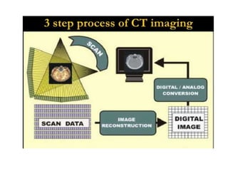


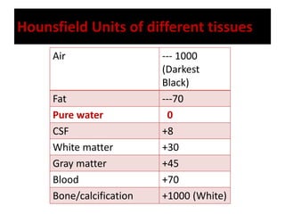
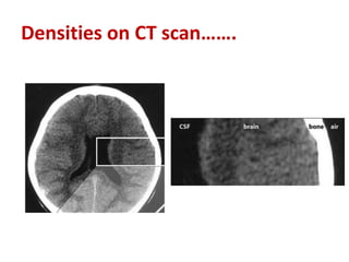


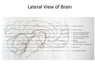


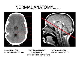
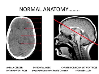

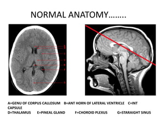
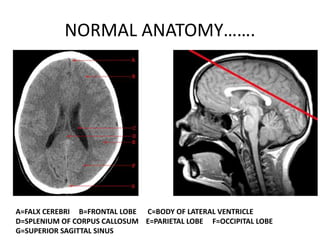


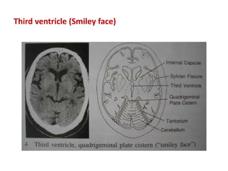
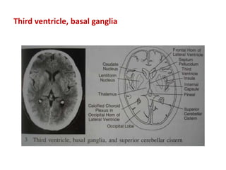


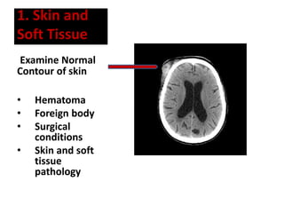
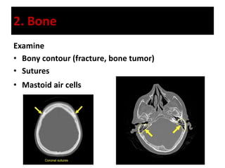


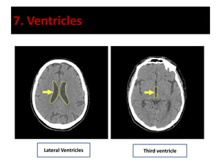
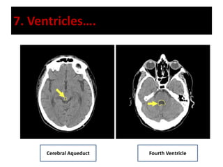
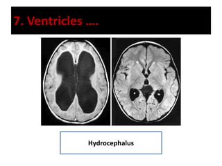
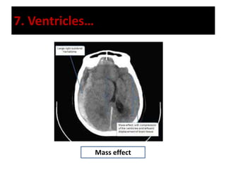


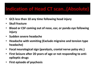

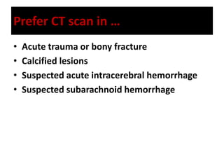
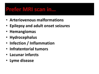
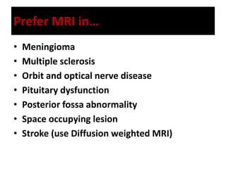


The document provides a comprehensive overview of interpreting head CT scans, detailing the basic principles, normal neuroanatomy, and descriptions of typical findings. It covers techniques for identifying structures, evaluating densities, and recognizing abnormalities, alongside the historical context of CT technology. Additionally, it discusses the appropriate usage of CT versus MRI in various clinical scenarios.


