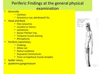
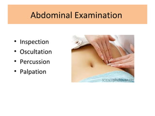
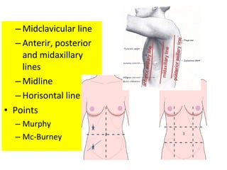
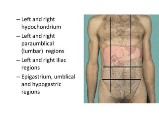
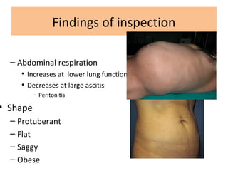
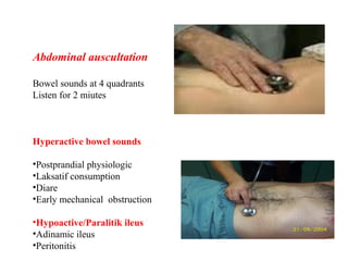
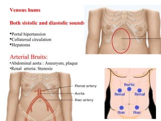
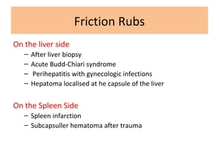
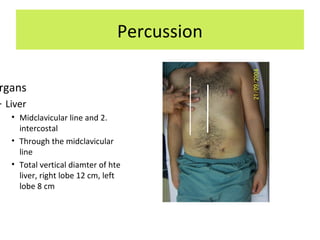
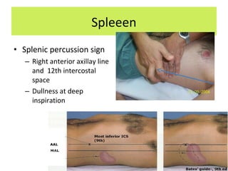
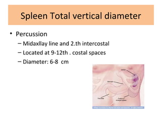
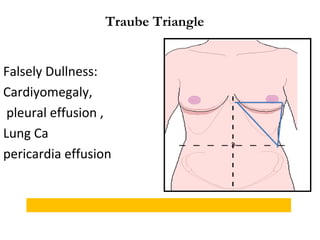
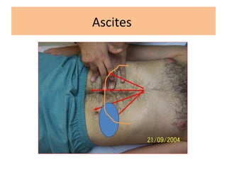
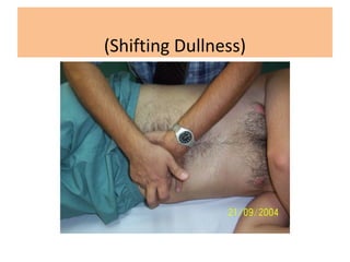
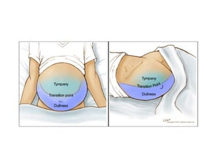
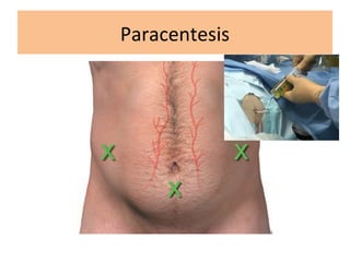
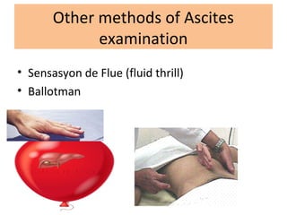
Abdominal Examination
Prof. Dr. Osman Cavit Özdoğan
Gastroenterology
Aproach to GIS Symptoms
Gi Symptoms
Localizaiton
• Upper Gi
• Lower Gi
• Other
Cardinal Symptoms
• Heartburn
• Dyspesia
• Dysphagia
• Non-cardiac chest pain
• Nausea
• Diarea
• Constipation
• Abdominal pain
• Weight loss
• Gi bleeding
Upper GI
• Dysphagia
• İntermittant
• Progressive
• Malignite
• Orofarengeal
• Özofageal
• Odynofagia
• Reflax
• Pill özofagitis
• Reflux Sypmtoms
– Heartburn
– Chest pain
– Regurgitation
• Hoarseness
• Hiccups
• Refractory cough
• Bad taste
• Belching
• Halitosis
Regurgitation
• effortless return of esophageal or gastric
contents into the hypopharynx…
– Esophageal regurgitation
• Achalasia
• Esophageal tumor
– Gastric regurgitation
• GERD
Abdominal Distention
• increased abdominal girth…
– Fat
– Flatus
– Fetus
– Fluid
• Hematemesis
–Fresh blood
–Coffe ground
• Melena
–Appereance
–Smell
Globus
• “Stuck of moutfull at the farenx”
• GI related causes:
– GERD (oropharyngeal reflux)
– Achalasia
• Globus hystericus
Lower GI
• Abdominal Pain
• Visceral pain
• Pariatel pain
• Reflecting pain : Acute Cholecystitis
• Localization
– Epigastric
– Other quadrants
– Diffuse abdomen
– Lower abdomen
• Related to meals or defecation
Stool patern
• Frequency
• Volume
• Shape
• Goat stool
• Slimming
• Color
– Black
– Red
Constipation
• an inability to have spontaneous complete
and releiving bowel movements…
– Slow colonic transit type
• Frequency<2 per week
• Consistency: Hard
• Colonic dysfunction
– Difficult evacuation type
• Frequency>2 per week
• Consistency: Soft
• Anorectal dysfunction
Hematochesia
• Fresh red
• Purple brown
• Small driblets
• Diffuse
Diarrhea
• a decrease in stool consistency due to increased
water content…
– Acute Diarrhea
• Viral
• Bacterial (E. coli, etc.)
– Chronic Diarrhea
• IBS
• IBD (CD and UC)
• Infectious (Amoeba, Tbc.)
• Antibiotic associated
• Others
Steatorrhea
• presence of excess fat in feces…
• Causes
– Chronic pancreatitis
– Celiac disease
– Postgastrectomy
– Cholestasis (esp. PSC)
– Giardiasis
– Crohn’s disease
Fecal Incontinence
• involuntary defication…
• Causes:
– Accidental injury to the rectum/anus
– Diabetes Mellitus
– Rectoanal tumors
– Fecal impaction
Anal Symptoms
• Pruritis
• Pain
• Tenesmus
• Hard Defecation
Hepatitis and liver failure
• Jaundice
• Fatique
• Weight loss or gain
• Loss of apetite
• Cirrhosis
– History of jaundice
– Abdominal distention
– Change in consinousness
Asterixis
• Synonyms: Liver flap, Flepping tremor
• Causes:
– Hepatic: Liver failure→ Hepatic Encephalopathy
– Renal: Renal failure → Uremic syndrome
– Pulmonary: Severe respiratory insufficiency
Why Abdominal Examination?
• Treatment should be urgent or elective
– GI Bleeding? Perforation? Acute abdomen?
– Peptic ulcer activation? Or Acute pancreatitis?
• Findings in physical examination targets to the
diagnosis
• Differential diagnosis?
• How do we decide the lab. Tests for diagnosis
• Does the patient need any intervention
immediately?
Be carefull!!
• Introduce yourself before examination
• Outpatient clinic should not be crowded
– Only one person with the patient, close relative
• Be calm and cool
– Do not shout, be angry or talk with sarcastic
words
• Always have the records on the files
• Always give detailed information for the
diagnosis & the treatment
General Physical Examination
• Always before the abdominal
examination
• Upper part of the patient’s body
should be naked
• Outpatient room should be warm
• Be at the right side of the patient
• All your belongings should be ready
– White coat
– Light source
– ruler
– Pencil, notebook etc.
• Enogh day light
• Hands should be warm and clean
Periferic Findings at the general physical
examination
• Generally
• Cachexia
• Orianted or not, dehidrated? Etc.
• Head and Neck
• Pale conjuctiva
• Jaundice or icterus
• xantelazma
• Kayser-Fleisher ring
• Temporal muscle wasting
• Rhinophyma
• Periferic extremities
• Clubbing
• White nail
• Palmar erythema
• Dupuytren Contractures
• Tenar ve hypotenal muscle atrophia
• Spider nevus,
• pyoderma gangrenozum
Glossitis
• inflammation of the tongue…
• GI related causes
– Pernisious anemia
– Pemphigus vulgaris
– Iron deficiency anemia
Angular Stomatitis
• an inflammatory lesion at the labial
commissure…
• GI related causes:
– Celiac disease
– Crohn’s disease
– Plummer-Vinson syndrome
Fe, B vit. deficiency
Aphtous ulcers
Abdominal Examination
• Inspection
• Oscultation
• Percussion
• Palpation
– Midclavicular line
– Anterir, posterior
and midaxillary
lines
– Midline
– Horisontal line
• Points
– Murphy
– Mc-Burney
– Left and right
hypochondrium
– Left and right
paraumblical
(lumbar) regions
– Left and right iliac
regions
– Epigastrium, umblical
and hypogastric
regions
Findings of inspection
– Abdominal respiration
• Increases at lower lung function
• Decreases at large ascitis
– Peritonitis
• Shape
– Protuberant
– Flat
– Saggy
– Obese
• You should define the lesion and its location when
you find a lesion.
– Diastasis recti
– Hernias
• İncisisonal
• Umblical
• ınguinal
• Others
– Scars
Collateral veins ve Caput medusa
– moles
– Stria
– Dilated veins
– Dilated intestinal loops
– Cullen’s sign
• Acute pankreatitis
• Abdominal trauma
• Ektopic pregnancy
– Grey Turner sign
• Acute pankreatitis
– 24-48 hr
– High mortalitiy
– Sister Mary Joseph nodule:
• Skin metastasis of gastric ca
– Pultations
• Aortic anuerysm
– Eccyhmosis
– Cafe au lait sign
Abdominal auscultation
Bowel sounds at 4 quadrants
Listen for 2 miutes
Hyperactive bowel sounds
•Postprandial physiologic
•Laksatif consumption
•Diare
•Early mechanical obstruction
•Hypoactive/Paralitik ileus
•Adinamic ileus
•Peritonitis
Venous hums
Both sistolic and diastolic sounds
Portal hipertansion
Collatersal circulation
Hepatoma
Arterial Bruits:
•Abdominal aorta : Aneurysm, plaque
•Renal arteria: Stenosis
Friction Rubs
On the liver side
– After liver biopsy
– Acute Budd-Chiari syndrome
– Perihepatitis with gynecologic infections
– Hepatoma localised at he capsule of the liver
On the Spleen Side
– Spleen infarction
– Subcapsuller hematoma after trauma
Percussion
rgans
– Liver
• Midclavicular line and 2.
intercostal
• Through the midclavicular
line
• Total vertical diamter of hte
liver, right lobe 12 cm, left
lobe 8 cm
Spleeen
• Splenic percussion sign
– Right anterior axillay line
and 12th intercostal
space
– Dullness at deep
inspiration
Spleen Total vertical diameter
• Percussion
– Midaxllay line and 2.th intercostal
– Located at 9-12th . costal spaces
– Diameter: 6-8 cm
Traube Triangle
Falsely Dullness:
Cardiyomegaly,
pleural effusion ,
Lung Ca
pericardia effusion
Ascites
(Shifting Dullness)
Paracentesis
Other methods of Ascites
examination
• Sensasyon de Flue (fluid thrill)
• Ballotman
Palpation
• Superficial Palpation:
– How is the situation at
the abdomen?
– Are there any sensitive
sides
– Any mass
– Ant dull
– Localizations
Deep palpation:
– Sensitive sides
• Patient has defence ?
• Muscular defense
• Abdominal guarding
– automically
• Rebound
• Rigidity
– Mass lesions
• Steely, smooth,
subcutan, deep, with
pain, or painless
Liver Palpation
• Diameter
– Midclavicular 6-12 cm right lobe
– 4-8 cm left lobe
– Use midsternal line for left lobe
• Steely, fibrotic, smooth
• Any pain
• Nodüler, irregüler
Karaciğer palpasyonu
Hook Yöntemi
Large Hepatomegaly
• Amiloidosis
• Malign infiltration
• Polycystic liver
• Congestive Heart Failure
– Hepatojuguler reflax
• Faty liver
Spleen Palpation
Large Splenomegaly
• KML infiltration
• Non-Hogkin lymphoma
• Kala azar Disease (Leismaniasis)
• Agnogenic Myeloid Metaplasia
• Murphy’s sign: Tenderness over the gall
bladder
• Palpable gall bladde: Courvoisier gallbladder
Pankreas Ca at the head of it
Kidney Palpation
Conclusion
• Repeat again and again
• Spend more time at internal medicine ward
• Performphysical examinaitonto your parents
• My kid is a doctor, oh ı am so happy
• Of course ask any time you want
• Look at B.Bates
Unutmamak lazım ki doktorun çok hastası olabilir,
ancak size gelen hastanın tek doktoru var o da
sizsiniz.

Abdominal Examination

  • 1.
    Abdominal Examination Prof. Dr.Osman Cavit Özdoğan Gastroenterology
  • 2.
  • 3.
    Gi Symptoms Localizaiton • UpperGi • Lower Gi • Other Cardinal Symptoms • Heartburn • Dyspesia • Dysphagia • Non-cardiac chest pain • Nausea • Diarea • Constipation • Abdominal pain • Weight loss • Gi bleeding
  • 4.
    Upper GI • Dysphagia •İntermittant • Progressive • Malignite • Orofarengeal • Özofageal • Odynofagia • Reflax • Pill özofagitis • Reflux Sypmtoms – Heartburn – Chest pain – Regurgitation
  • 5.
    • Hoarseness • Hiccups •Refractory cough • Bad taste • Belching • Halitosis
  • 6.
    Regurgitation • effortless returnof esophageal or gastric contents into the hypopharynx… – Esophageal regurgitation • Achalasia • Esophageal tumor – Gastric regurgitation • GERD
  • 7.
    Abdominal Distention • increasedabdominal girth… – Fat – Flatus – Fetus – Fluid
  • 8.
    • Hematemesis –Fresh blood –Coffeground • Melena –Appereance –Smell
  • 9.
    Globus • “Stuck ofmoutfull at the farenx” • GI related causes: – GERD (oropharyngeal reflux) – Achalasia • Globus hystericus
  • 10.
    Lower GI • AbdominalPain • Visceral pain • Pariatel pain • Reflecting pain : Acute Cholecystitis • Localization – Epigastric – Other quadrants – Diffuse abdomen – Lower abdomen • Related to meals or defecation
  • 11.
    Stool patern • Frequency •Volume • Shape • Goat stool • Slimming • Color – Black – Red
  • 12.
    Constipation • an inabilityto have spontaneous complete and releiving bowel movements… – Slow colonic transit type • Frequency<2 per week • Consistency: Hard • Colonic dysfunction – Difficult evacuation type • Frequency>2 per week • Consistency: Soft • Anorectal dysfunction
  • 13.
    Hematochesia • Fresh red •Purple brown • Small driblets • Diffuse
  • 14.
    Diarrhea • a decreasein stool consistency due to increased water content… – Acute Diarrhea • Viral • Bacterial (E. coli, etc.) – Chronic Diarrhea • IBS • IBD (CD and UC) • Infectious (Amoeba, Tbc.) • Antibiotic associated • Others
  • 15.
    Steatorrhea • presence ofexcess fat in feces… • Causes – Chronic pancreatitis – Celiac disease – Postgastrectomy – Cholestasis (esp. PSC) – Giardiasis – Crohn’s disease
  • 16.
    Fecal Incontinence • involuntarydefication… • Causes: – Accidental injury to the rectum/anus – Diabetes Mellitus – Rectoanal tumors – Fecal impaction
  • 17.
    Anal Symptoms • Pruritis •Pain • Tenesmus • Hard Defecation
  • 18.
    Hepatitis and liverfailure • Jaundice • Fatique • Weight loss or gain • Loss of apetite • Cirrhosis – History of jaundice – Abdominal distention – Change in consinousness
  • 19.
    Asterixis • Synonyms: Liverflap, Flepping tremor • Causes: – Hepatic: Liver failure→ Hepatic Encephalopathy – Renal: Renal failure → Uremic syndrome – Pulmonary: Severe respiratory insufficiency
  • 20.
    Why Abdominal Examination? •Treatment should be urgent or elective – GI Bleeding? Perforation? Acute abdomen? – Peptic ulcer activation? Or Acute pancreatitis? • Findings in physical examination targets to the diagnosis • Differential diagnosis? • How do we decide the lab. Tests for diagnosis • Does the patient need any intervention immediately?
  • 21.
    Be carefull!! • Introduceyourself before examination • Outpatient clinic should not be crowded – Only one person with the patient, close relative • Be calm and cool – Do not shout, be angry or talk with sarcastic words • Always have the records on the files • Always give detailed information for the diagnosis & the treatment
  • 22.
    General Physical Examination •Always before the abdominal examination • Upper part of the patient’s body should be naked • Outpatient room should be warm • Be at the right side of the patient • All your belongings should be ready – White coat – Light source – ruler – Pencil, notebook etc. • Enogh day light • Hands should be warm and clean
  • 23.
    Periferic Findings atthe general physical examination • Generally • Cachexia • Orianted or not, dehidrated? Etc. • Head and Neck • Pale conjuctiva • Jaundice or icterus • xantelazma • Kayser-Fleisher ring • Temporal muscle wasting • Rhinophyma • Periferic extremities • Clubbing • White nail • Palmar erythema • Dupuytren Contractures • Tenar ve hypotenal muscle atrophia • Spider nevus, • pyoderma gangrenozum
  • 26.
    Glossitis • inflammation ofthe tongue… • GI related causes – Pernisious anemia – Pemphigus vulgaris – Iron deficiency anemia
  • 27.
    Angular Stomatitis • aninflammatory lesion at the labial commissure… • GI related causes: – Celiac disease – Crohn’s disease – Plummer-Vinson syndrome Fe, B vit. deficiency
  • 28.
  • 32.
    Abdominal Examination • Inspection •Oscultation • Percussion • Palpation
  • 33.
    – Midclavicular line –Anterir, posterior and midaxillary lines – Midline – Horisontal line • Points – Murphy – Mc-Burney
  • 34.
    – Left andright hypochondrium – Left and right paraumblical (lumbar) regions – Left and right iliac regions – Epigastrium, umblical and hypogastric regions
  • 35.
    Findings of inspection –Abdominal respiration • Increases at lower lung function • Decreases at large ascitis – Peritonitis • Shape – Protuberant – Flat – Saggy – Obese
  • 36.
    • You shoulddefine the lesion and its location when you find a lesion. – Diastasis recti – Hernias • İncisisonal • Umblical • ınguinal • Others – Scars
  • 37.
    Collateral veins veCaput medusa
  • 38.
    – moles – Stria –Dilated veins – Dilated intestinal loops – Cullen’s sign • Acute pankreatitis • Abdominal trauma • Ektopic pregnancy – Grey Turner sign • Acute pankreatitis – 24-48 hr – High mortalitiy
  • 39.
    – Sister MaryJoseph nodule: • Skin metastasis of gastric ca – Pultations • Aortic anuerysm – Eccyhmosis – Cafe au lait sign
  • 42.
    Abdominal auscultation Bowel soundsat 4 quadrants Listen for 2 miutes Hyperactive bowel sounds •Postprandial physiologic •Laksatif consumption •Diare •Early mechanical obstruction •Hypoactive/Paralitik ileus •Adinamic ileus •Peritonitis
  • 43.
    Venous hums Both sistolicand diastolic sounds Portal hipertansion Collatersal circulation Hepatoma Arterial Bruits: •Abdominal aorta : Aneurysm, plaque •Renal arteria: Stenosis
  • 44.
    Friction Rubs On theliver side – After liver biopsy – Acute Budd-Chiari syndrome – Perihepatitis with gynecologic infections – Hepatoma localised at he capsule of the liver On the Spleen Side – Spleen infarction – Subcapsuller hematoma after trauma
  • 45.
    Percussion rgans – Liver • Midclavicularline and 2. intercostal • Through the midclavicular line • Total vertical diamter of hte liver, right lobe 12 cm, left lobe 8 cm
  • 46.
    Spleeen • Splenic percussionsign – Right anterior axillay line and 12th intercostal space – Dullness at deep inspiration
  • 47.
    Spleen Total verticaldiameter • Percussion – Midaxllay line and 2.th intercostal – Located at 9-12th . costal spaces – Diameter: 6-8 cm
  • 48.
    Traube Triangle Falsely Dullness: Cardiyomegaly, pleuraleffusion , Lung Ca pericardia effusion
  • 49.
  • 50.
  • 52.
  • 53.
    Other methods ofAscites examination • Sensasyon de Flue (fluid thrill) • Ballotman
  • 54.
    Palpation • Superficial Palpation: –How is the situation at the abdomen? – Are there any sensitive sides – Any mass – Ant dull – Localizations
  • 55.
    Deep palpation: – Sensitivesides • Patient has defence ? • Muscular defense • Abdominal guarding – automically • Rebound • Rigidity – Mass lesions • Steely, smooth, subcutan, deep, with pain, or painless
  • 56.
    Liver Palpation • Diameter –Midclavicular 6-12 cm right lobe – 4-8 cm left lobe – Use midsternal line for left lobe • Steely, fibrotic, smooth • Any pain • Nodüler, irregüler
  • 57.
  • 58.
    Large Hepatomegaly • Amiloidosis •Malign infiltration • Polycystic liver • Congestive Heart Failure – Hepatojuguler reflax • Faty liver
  • 59.
  • 60.
    Large Splenomegaly • KMLinfiltration • Non-Hogkin lymphoma • Kala azar Disease (Leismaniasis) • Agnogenic Myeloid Metaplasia
  • 61.
    • Murphy’s sign:Tenderness over the gall bladder • Palpable gall bladde: Courvoisier gallbladder Pankreas Ca at the head of it
  • 62.
  • 63.
    Conclusion • Repeat againand again • Spend more time at internal medicine ward • Performphysical examinaitonto your parents • My kid is a doctor, oh ı am so happy • Of course ask any time you want • Look at B.Bates
  • 64.
    Unutmamak lazım kidoktorun çok hastası olabilir, ancak size gelen hastanın tek doktoru var o da sizsiniz.

Editor's Notes

  • #7 Regurgitation should be distinguished from vomiting, the term applied to the ejection of gastroduodenal content preceded by nausea and accompanied by the abdominal muscular activity. Esophageal regurgitation is rarely acidic, on the other hand gastric regurgitation is frequently acidic.
  • #8 Abdominal distention refers to increased abdominal girth—the result of increased intra-abdominal pressure forcing the abdominal wall outward. Distention may be mild or severe, depending on the amount of pressure. It may be localized or diffuse and may occur gradually or suddenly. Acute abdominal distention may signal life-threatening peritonitis or acute bowel obstruction. Abdominal distention may result from fat, flatus, a fetus (pregnancy or intra-abdominal mass [ectopic pregnancy]), or fluid. Fluid and gas are normally present in the GI tract but not in the peritoneal cavity. However, if fluid and gas can&amp;apos;t pass freely through the GI tract, abdominal distention occurs. In the peritoneal cavity, distention may reflect acute bleeding, accumulation of ascitic fluid, or air from perforation of an abdominal organ. Abdominal distention doesn&amp;apos;t always signal pathology. For example, in anxious patients or those with digestive distress, localized distention in the left upper quadrant can result from aerophagia —the unconscious swallowing of air. Generalized distention can result from ingestion of fruits or vegetables with large quantities of unabsorbable carbohydrates, such as legumes, or from abnormal food fermentation by microbes. Don&amp;apos;t forget to rule out pregnancy in all females with abdominal distention.
  • #10 The globus sensation, a sense that something is lodged continuously in the throat, must be differentiated from dysphagia or odynophagia before embarking on an unnecessary investigation . The globus sensation typically is sensed midline at the laryngeal level, but can lateralize in as many as 20% of patients. The sensation classically is reported as a “lump in the throat,” but feeling that a foreign body, sharp object, or food particle is lodged also is a compatible description. Most notably, globus sensation does not interfere with swallowing; although one in five patients with globus sensation notes something abnormal during food swallows, the original symptom abates during the process. Globus may accompany a variety of disorders that cause dysphagia, such as GERD and distal esophageal motility disorders. Furthermore, up to 45% of the general population may have intermittent symptoms resembling globus. This symptom most often reflects a functional gastrointestinal disorder. In some instances globus is associated with substantial anxiety; that anxiety is sometimes thought to be etiological (globus hystericus).
  • #13 Patients consider themselves to be constipated when they pass stools more infrequently, require more effort for passage (“straining”), or experience more pain or discomfort during passage than they think appropriate. Difficult evacuation of feces, especially when the consistency of stools is softer than normal, is more likely to be caused by disorders of the pelvic floor or anorectum than by slow colonic transit.
  • #15 A decrease in stool consistency or fluidity and stools that cause urgency or abdominal discomfort are more likely to be termed diarrhea by patients than increases in frequency alone.
  • #20 A bilateral, coarse movement, asterixis is characterized by sudden relaxation of muscle groups holding a sustained posture. This elicited sign is most commonly observed in the wrists and fingers, but may also appear during any sustained voluntary action. Typically, it signals hepatic, renal, or pulmonary disease.
  • #28 Angular cheilitis (also called cheilosis or angular stomatitis) is an inflammatory lesion at the labial commissure, or corner of the mouth, and often occurs bilaterally. The condition manifests as deep cracks or splits. In severe cases, the splits can bleed when the mouth is opened and shallow ulcers or a crust may form.
  • #62 Ne - zönkkk