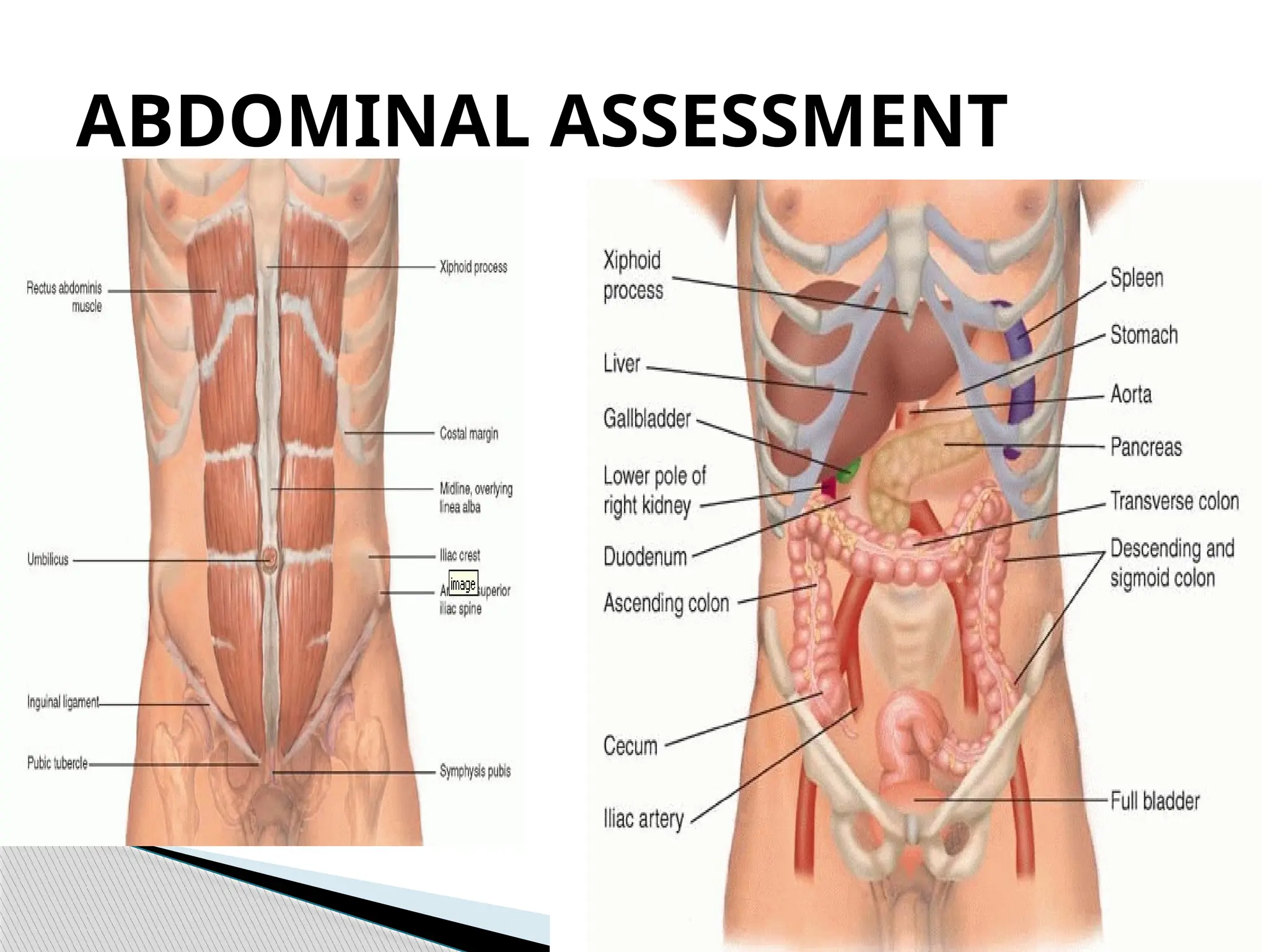
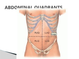
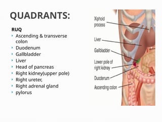
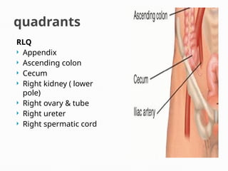
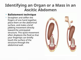
ABDOMINAL ASSESSMENT
ABDOMINAL QUADRANTS
QUADRANTS:
RUQ
 Ascending & transverse
colon
 Duodenum
 Gallbladder
 Liver
 Head of pancreas
 Right kidney(upper pole)
 Right ureter,
 Right adrenal gland
 pylorus
quadrants
RLQ
 Appendix
 Ascending colon
 Cecum
 Right kidney ( lower
pole)
 Right ovary & tube
 Right ureter
 Right spermatic cord
Quadrants:
 LUQ
 Left adrenal gland
 Left kidney
 Left ureter
 Body & tail of pancreas
 Spleen
 Stomach
 Transverse &
descending colon
 LLQ
 Left kidney (lower pole)
 Left ovary
 Left ureter
 Descending & sigmoid
colon
 Left spermatic cord
 Midline:
 Bladder
 Uterus
 Prostate gland
9 abdominal regions:
 RUQ
Pancreatitis
Hepatomegaly
Hepatitis
Duodenal ulcer
Cholecystitis
 RLQ
 Appendicitis
 Ectopic pregnancy
 Renal calculi
 Ovarian cyst
 EPIGASTRIUM
GERD
AAA
 PERIUMBILICAL
Intestinal Obstruction
Peptic Ulcer
 LUQ
Gastric ulcer
MI
Splenic enlargement
& rupture
 LLQ
Ectopic pregnancy
Ovarian cyst
Hernia
PID
Etiologies of Abdominal pain:
anatomical regions where they are
perceived
 Check that the patient has an empty bladder.
 Make the patient comfortable in the supine position, with a
pillow under the head and perhaps another under the knees.
Slide your hand under the low back to see if the patient is
relaxed and lying flat on the table.
 Ask the patient to keep the arms at the sides or folded across
the chest. If the arms are above the head, the abdominal wall
stretches and tightens, making palpation difficult. Move the
gown to below the nipple line, and the drape to the level of the
symphysis pubis.
 Before you begin palpation, ask the patient to point to any areas
of pain so you can examine these areas last.
TECHNIQUES OF
EXAMINATION
 Warm your hands and stethoscope. To warm your hands,
rub them together or place them under hot water. You can
also palpate through the patient's gown to absorb warmth
from the patient's body before exposing the abdomen.
 Approach the patient calmly and avoid quick, unexpected
movements. Watch the patient's face for any signs of pain
or discomfort. Make sure you avoid long fingernails.
 Distract the patient if necessary with conversation or
questions. If the patient is frightened or ticklish, begin
palpation with the patient's hand under yours. After a few
moments, slip your hand underneath to palpate directly.
TECHNIQUES OF
EXAMINATION
 Abdominal skin may be paler than the
general skin tone because this skin is
seldom exposed to natural elements.
1. Observe coloration of the skin.
 Purple discoloration at the flanks ( Grey Turner
sign)- indicates bleeding within the abdominal
wall due to trauma to kidneys, pancreas or
duodenum.
 Pale,taut skin= ascites
 Redness= inflammation
 Bruises & local discoloration
Abnormalities:
 Scattered veins may be visible.
 Dilated superficial capillaries without a
pattern may be seen in older clients. They
are visible in sunlight.
2. Note vascularity of the abdomen.
 Dilated veins- cirrhosis of liver, portal HPN,
ascites
 Spider angioma- dilated arterioles & capillaries
with a central star seen in liver dse.
Abnormalities:
 Old, silvery white striae or stretch marks
from past pregnancies or weight gain are
normal.
3. Note striae.
 Dark bluish pink striae- Cushing’s syndrome
 Striae may be caused by ascites, which
stretches skin as a result from liver failure or
liver disease.
Abnormalities:
 Ask about source of scar & use centimeter ruler
to measure scar’s length.
 Document the location by quadrant, length,
characteristics.
 Pale, smooth, minimally raised old scars.
4. Inspect scars.
 Nonhealing scars, redness, inflammation.
 Deep, irregular scars results from burns.
 Keloidsresult from trauma or surgery.
Abnormalities:
 Abdomen is free of lesions or rashes.
 Flat or raised brown moles.
5. Assess lesions & rashes.
 Changes in moles( size, color, & border
symmetry)
 Any bleeding moles or petechiae are abnormal.
Abnormalities:
 Note the color of umbilical area.
 Assess contour of umbilicus.
 Umbilicus is midline at lateral line.
 Umbilical skin tones are similar to
surrounding abdominal skin tones or even
pinkish.
 It is inverted or protruding no more than
0.5cm & is round or conical.
6. Inspect umbilicus.
 Cullen’s sign- bluish or purple discoloration around
umbilicus that indicates intra abdominal bleeding.
 Deviated umbilicus may be caused by pressure
from mass, enlarged organs, fluid or scar.
 Everted umbilicus is seen in abdominal distention.
 Large everted umbilicus suggests umbilical hernia.
Abnormalities:
 Look across the abdomen at eye level from
client’s side, from behind client’s head, & from
the foot of the bed.
 Measure abdominal girth as indicated.
 Abdomen should be evenly rounded.
7. Inspect abdominal contour.
Normal contour of abdomen
 Generalized protuberant/ distended abdomen
-due to obesity, air or fluid accumulation
 Distention below the umbilicus
-due to full bladder, uterine enlargement, or
ovarian tumor
Distention of the upper abdomen- massess of
pancreas or gastric dilatation
Abnormalities:
 Look at client’s abdomen as she lies in a relaxed
supine position.
 To assess abdomen for herniation or
differentiate mass, ask pt. to raise head.
 Abdomen is symmetric.
 Abdomen does not bulge when pt.raises
head.
8. Assess abdominal
symmetry.
 Asymmetry- organ enlargement, large
massess, hernia, bowel obstruction
 Hernia is seen as bulging in the abdominal
wall.
 Diastasis recti appear as bulging between a
vertical midline separation of the abdominis
muscles.
Abnormalities:
 Observe aortic pulsations & peristaltic waves.
 Abdominal respiratory movement may be
seen esp. in male patients.
 A slight pulsation of abdominal aorta, which
is visible in the epigastrium, extends full
length in thin people.
 Peristaltic waves are not seen through
although visible in very thin people.
9. Inspect abdominal movement
when patient breathes.
 Diminished abdominal respiration or change in
thoracic breathing in male pts. Reflect
peritoneal irritation.
 AAA-vigorous, wide, exaggerated pulsations
 IO- peristaltic waves are increased & progress
in ripple-like fashion from LUQ to RUQ.
Abnormalities:
 Use diaphragm of steth & make sure it is warm before
you place on pt’s abdomen.
 Apply slight pressure on the tender abdomen.
 Begin in the RLQ, RUQ, LUQ, LLQ.
 Listen for up to 5mins in each of the quadrants.
 BORBORYGMI-a hyperactive bowel sounds that can
be heard as loud, prolonged gurgling sounds.
 Usually high pitched sounds & occur 5-30 times
mins per minute
 Results fr. The movement of air & fluid thru the GIT.
10. Auscultate bowel sounds.
 Hypoactive bowel sounds- diminished bowel
motility. ( Abdominal surgery)
 Hyperactive bowel sounds- increased bowel
motility. ( diarrhea)
 Decreased/absent bowel sounds-absence of
bowel motility referral!!
 Peritonitis or paralytic ileus
Abnormalities:
 Use bell of steth to listen for bruits ( low-
pitched, murmurlike sound) over the aorta,
renal, iliac, & femoral arteries.
 Bruits are not normally heard over
abdominal aorta, renal, iliac or femoral
arteries.
11. Auscultate vascular
sounds.
 Bruit with both systolic & diastolic components
occur when blood flow in an artery is turbulent
or obstructed indicating aneurysm or arterial
stenosis.
Abnormalities:
 Listen over R & L lower rib cage with the
diaphragm of the steth.
 No friction rub over spleen & liver is present.
12. Auscultate friction rub over liver
& spleen.
 FRICTION RUB- high-pitched, rough, grating
sound produced when large surface area of the
liver/ spleen rubs the peritoneum.
 Hepatic abscess- a friction rub heard over the
right costal area.
 Splenic infarction-friction rub at the anterior
axillary line in the lower left costal area.
Abnormalities:
 Light &systematically percuss all quadrants.
 Tympany predominates over the abdomen
because air in the stomach & intestines.
 Dullness is heard over the liver ,spleen &
nonevacuated descending colon.
13. Percuss for tone.
 Hyperresonance/ accentuated tympany-
gaseous distended
 Enlarged area of dullness- enlarged liver or
spleen
 Abnormal dullness- distended bladder, large
masses or ascites
Abnormalities:
 To assess for tenderness in difficult to palpate
structures.
 Place left hand flat against lower R ribcage.
 Use ulnar side of R fist to strike L hand.
 NO tenderness is elicited.
 Tenderness over the liver may be associated
w/ inflammation or infection ( hepatitis/
cholecystitis)
14. Perform blunt percussion on the
liver.
 Perform blunt percussion on the kidneys at the
costovertebral angles over the 12th
rib.
 No tenderness or pain is elicited.
 Tenderness or sharp pain suggests kidney
infection, renal calculi.
 Use fingertips & begin palpation in a nontender
quadrant.
 Compress to a depth of 1cm in a dipping
motion
 Then gently lift fingers & move to the next area.
 Abdomen is nontender & soft. No guarding.
15. Perform light palpation.
 Involuntary reflex guarding reflects peritoneal
irritation.
 Ride sided guarding due to cholecystitis.
Abnormalities:
 Use palmar surface of the fingers, compress to
a maximum depth of 5-6cm.
 Perform bimanual palpation if resistance is
encountered.
 Normal tenderness is possible over xiphoid
process, aorta, cecum, sigmoid colon, &
ovaries with deep palpation.
16. Deeply palpate all quadrants to delineate
abdominal organs & detect subtle masses.
 Severe tenderness or pain- trauma, peritonitis,
infection, tumors, or enlarged organs.
Abnormalities:
 Note location, size (cm), shape, consistency,
demarcation, pulsability, tenderness, mobility.
 No palpable masses are present.
 A mass due to tumor, cyst, abscess, enlarged
organ.
17. Palpate masses.
 Umbilicus & surrounding area are free of
swellings, bulges, or masses.
 Soft center of the umbilicus= herniation.
 Hard nodule = metastatic node from GI Ca.
18. Palpate umbilicus & surrounding area for
swellings, bulges or masses.
 Use thumb & 1st
finger or 2 hands to palpate
deeply in the epigastrium.
 Asses pulsation of abdominal aorta.
 Normal aorta is approximately 2.5-3.0cm
wide w/moderately strong & regular pulse.
 AAA- wide, bounding pulse may be felt.
19. Palpate aorta.
 Stand at the right side & place L hand under
client’s back at the level of the 11th
-12th
ribs.
 Lay R hand parallel to the R costal margin.
 Ask client to inhale then compress upward &
inward w/ fingers.
 Liver not palpable. Palpable to thin
patient.It should be firm, smooth, & even.
 Mild tenderness is normal,
20. Palpate liver.
 Cancer- hard, firm liver
 Nodularity- tumors, metastatic Ca, late cirrhosis
 Tenderness- CHF, acute hepatitis
 Enlarged liver- more than 1-3cm below costal
margin( hepatitis, tumors, cirrhosis)
Abnormalities:
 Stand to the R of the pt’s chest
 Curl/ hook fingers of both hands over the edge
of R costal margin.
 Ask pt. to take deep breath & gently pull inward
& upward with fingers.
Hooking technique for liver
palpation.
Hooking technique
 Stand at R side, reach over the abdomen w/ left
arm & place hand under posterior rib.
 Pull up gently.
 Place R hand below the L costal margin w/
fingers pointing toward the client’s head.
 Ask pt. to inhale & press inward & upward as
you support w/ other hand.
 The spleen is seldom palpable at the L costal
margin.
21. Palpate spleen.
 Palpable spleen suggests enlargement w/c may
result from trauma, chronic blood disorders &
cancers.
 Tenderness accompanied by peritoneal
inflammation or capsular stretching is assoc. w/
splenic enlargement.
Abnormalities:
 Support R posterior flank w/ L hand & place R
hand in the RUQ just below the costal margin
at the MCL.
 Ask pt to inhale.
 Compress fingers deeply during peak
inspiration.
 Ask pt. to exhale & hold breath briefly.
 Gradually release pressure on R hand.
 You feel it slip beneath the fingers
22. Palpate kidneys.
 Kidneys are not normally palpable.
 Sometimes the lower pole of the R kidney
may be palpable by the capture method
because of its lower position,
 It is firm, smooth & rounded.
 Enlarged kidney- cyst, tumor, hydronephrosis
Abnormalities:
 Begin at symphysis pubis & move upward to
estimate bladder borders.
 Bladder is not palpable.
 Distended bladder is smooth, round, & firm
mass extending as far as the umbilicus.
 Dull percussion tones.
23. Palpate urinary bladder.
Special abdominal tests:
Assessing Possible Ascites
 A protuberant abdomen with
bulging flanks suggests the
possibility of ascitic fluid. Because
ascitic fluid characteristically sinks
with gravity, whereas gas-filled
loops of bowel float to the top,
percussion gives a dull note in
dependent areas of the abdomen.
 Look for such a pattern by
percussing outward in several
directions from the central area of
tympany.
 Map the border between
tympany and dullness.
 Ascites from increased hydrostatic pressure in
cirrhosis, congestive heart failure, constrictive
pericarditis, or inferior vena cava or hepatic
vein obstruction;
 from decreased osmotic pressure in nephrotic
syndrome, malnutrition. Also in ovarian cancer.
EXAMPLES OF ABNORMALITIES
2 possible techniques to confirm
ascites:
 1. Test for shifting
dullness.
 After mapping the borders of
tympany and dullness, ask the
patient to turn onto one side.
 Percuss and mark the
borders again.
 In a person without ascites,
the borders between tympany
and dullness usually stay
relatively constant.
 2. Test for a fluid
wave.
 Ask the patient or an assistant
to press the edges of both
hands firmly down the midline
of the abdomen.
 This pressure helps to stop the
transmission of a wave through
fat.
 While you tap one flank sharply
with your fingertips, feel on the
opposite flank for an impulse
transmitted through the fluid.
Unfortunately, this sign is often
negative until ascites is obvious,
and it is sometimes positive in
people without ascites.
Identifying an Organ or a Mass in an
Ascitic Abdomen
 Ballotement technique
 Straighten and stiffen the
fingers of one hand together,
place them on the abdominal
surface, and make a brief
jabbing movement directly
toward the anticipated
structure. This quick movement
often displaces the fluid so that
your fingertips can briefly
touch the surface of the
structure through the
abdominal wall.
Assessing Possible
Appendicitis
 Ask the patient to point to
where the pain began and
where it is now. Ask the patient
to cough. Determine whether
and where pain results.
 EXAMPLES OF
ABNORMALITIES
The pain of appendicitis
classically begins near the
umbilicus, then shifts to the
right lower quadrant, where
coughing increases it.
 Search carefully for an
area of local
tenderness.
 EXAMPLES OF
ABNORMALITIES
 Localized tenderness
anywhere in the right
lower quadrant, even in
the right flank, may
indicate appendicitis.
 Check for Rovsing's sign and for referred
rebound tenderness.
 Press deeply and evenly in the left lower
quadrant. Then quickly withdraw your fingers.
 No rebound tenderness is present.
 Pain in the right lower quadrant during left-
sided pressure suggests appendicitis (a
positive Rovsing's sign).
 So does right lower quadrant pain on quick
withdrawal (referred rebound tenderness).
EXAMPLES OF ABNORMALITIES
 Psoas sign.
 Place your hand just above the patient's right
knee and ask the patient to raise that thigh
against your hand.
 Alternatively, ask the patient to turn onto the
left side. Then extend the patient's right leg at
the hip.
 Flexion of the leg at the hip makes the psoas
muscle contract; extension stretches it.
 No abdominal pain is present.
 Increased abdominal pain on either maneuver
constitutes a positive psoas sign, suggesting
irritation of the psoas muscle by an
inflamed appendix.
EXAMPLES OF ABNORMALITIES
 Obturator sign.
 Flex the patient's right thigh at the hip, with the
knee bent, and rotate the leg internally at the
hip.
 This maneuver stretches the internal obturator
muscle.
 No abdominal pain is present.
 Right hypogastric pain constitutes a positive
obturator sign, suggesting irritation of the
obturator muscle by an inflamed appendix.
EXAMPLES OF ABNORMALITIES
 Hypersensitivity test
 Stroke the abdomen w/ a sharp object(tongue
blade) or
 Grasp a fold of the skin with your thumb &
index finger & quickly let go.
 Do this several times along abdominal wall
 The client feels no pain & no exaggerated
sensation.
 Pain or exaggerated sensation felt in the
RLQ is positive for skin hypersentivity test &
may indicate appendicitis.
Abnormalities:
 When right upper quadrant pain and tenderness
suggest acute cholecystitis, look for Murphy's sign.
 Hook your left thumb or the fingers of your right hand
under the costal margin at the point where the lateral
border of the rectus muscle intersects with the costal
margin.
 Alternatively, if the liver is enlarged, hook your thumb or
fingers under the liver edge at a comparable point
below.
 Ask the patient to take a deep breath. Watch the
patient's breathing and note the degree of tenderness.
Assessing Possible Acute
Cholecystitis
A sharp increase in tenderness with a sudden
stop in inspiratory effort constitutes a
positive Murphy's sign of acute cholecystitis.
Hepatic tenderness may also increase with this
maneuver but is usually less well localized.
EXAMPLES OF ABNORMALITIES
 Abdomen is protuberant with active bowel
sounds. It is soft and nontender; no palpable
masses or hepatosplenomegaly. Liver span is 7
cm in the right midclavicular line; edge is
smooth and palpable 1 cm below the right
costal margin. Spleen and kidneys not felt. No
costovertebral angle (CVA) tenderness.”
RECORDING YOUR FINDINGS
 Abdomen is flat. No bowel sounds heard. It is
firm and boardlike, with increased tenderness,
guarding, and rebound in the right
midquadrant. Liver percusses to 7 cm in the
midclavicular line; edge not felt. Spleen and
kidneys not felt. No palpable masses. No CVA
tenderness.
RECORDING YOUR FINDINGS
Daghang
salamat!!!
THANK
YOU!!!

ABDOMINAL ASSESSMENT it is useful pro.pptx

  • 1.
  • 3.
  • 4.
    QUADRANTS: RUQ  Ascending &transverse colon  Duodenum  Gallbladder  Liver  Head of pancreas  Right kidney(upper pole)  Right ureter,  Right adrenal gland  pylorus
  • 5.
    quadrants RLQ  Appendix  Ascendingcolon  Cecum  Right kidney ( lower pole)  Right ovary & tube  Right ureter  Right spermatic cord
  • 6.
    Quadrants:  LUQ  Leftadrenal gland  Left kidney  Left ureter  Body & tail of pancreas  Spleen  Stomach  Transverse & descending colon
  • 7.
     LLQ  Leftkidney (lower pole)  Left ovary  Left ureter  Descending & sigmoid colon  Left spermatic cord  Midline:  Bladder  Uterus  Prostate gland
  • 8.
  • 9.
     RUQ Pancreatitis Hepatomegaly Hepatitis Duodenal ulcer Cholecystitis RLQ  Appendicitis  Ectopic pregnancy  Renal calculi  Ovarian cyst  EPIGASTRIUM GERD AAA  PERIUMBILICAL Intestinal Obstruction Peptic Ulcer  LUQ Gastric ulcer MI Splenic enlargement & rupture  LLQ Ectopic pregnancy Ovarian cyst Hernia PID Etiologies of Abdominal pain: anatomical regions where they are perceived
  • 10.
     Check thatthe patient has an empty bladder.  Make the patient comfortable in the supine position, with a pillow under the head and perhaps another under the knees. Slide your hand under the low back to see if the patient is relaxed and lying flat on the table.  Ask the patient to keep the arms at the sides or folded across the chest. If the arms are above the head, the abdominal wall stretches and tightens, making palpation difficult. Move the gown to below the nipple line, and the drape to the level of the symphysis pubis.  Before you begin palpation, ask the patient to point to any areas of pain so you can examine these areas last. TECHNIQUES OF EXAMINATION
  • 11.
     Warm yourhands and stethoscope. To warm your hands, rub them together or place them under hot water. You can also palpate through the patient's gown to absorb warmth from the patient's body before exposing the abdomen.  Approach the patient calmly and avoid quick, unexpected movements. Watch the patient's face for any signs of pain or discomfort. Make sure you avoid long fingernails.  Distract the patient if necessary with conversation or questions. If the patient is frightened or ticklish, begin palpation with the patient's hand under yours. After a few moments, slip your hand underneath to palpate directly. TECHNIQUES OF EXAMINATION
  • 12.
     Abdominal skinmay be paler than the general skin tone because this skin is seldom exposed to natural elements. 1. Observe coloration of the skin.
  • 13.
     Purple discolorationat the flanks ( Grey Turner sign)- indicates bleeding within the abdominal wall due to trauma to kidneys, pancreas or duodenum.  Pale,taut skin= ascites  Redness= inflammation  Bruises & local discoloration Abnormalities:
  • 14.
     Scattered veinsmay be visible.  Dilated superficial capillaries without a pattern may be seen in older clients. They are visible in sunlight. 2. Note vascularity of the abdomen.
  • 15.
     Dilated veins-cirrhosis of liver, portal HPN, ascites  Spider angioma- dilated arterioles & capillaries with a central star seen in liver dse. Abnormalities:
  • 16.
     Old, silverywhite striae or stretch marks from past pregnancies or weight gain are normal. 3. Note striae.
  • 17.
     Dark bluishpink striae- Cushing’s syndrome  Striae may be caused by ascites, which stretches skin as a result from liver failure or liver disease. Abnormalities:
  • 18.
     Ask aboutsource of scar & use centimeter ruler to measure scar’s length.  Document the location by quadrant, length, characteristics.  Pale, smooth, minimally raised old scars. 4. Inspect scars.
  • 19.
     Nonhealing scars,redness, inflammation.  Deep, irregular scars results from burns.  Keloidsresult from trauma or surgery. Abnormalities:
  • 20.
     Abdomen isfree of lesions or rashes.  Flat or raised brown moles. 5. Assess lesions & rashes.
  • 21.
     Changes inmoles( size, color, & border symmetry)  Any bleeding moles or petechiae are abnormal. Abnormalities:
  • 22.
     Note thecolor of umbilical area.  Assess contour of umbilicus.  Umbilicus is midline at lateral line.  Umbilical skin tones are similar to surrounding abdominal skin tones or even pinkish.  It is inverted or protruding no more than 0.5cm & is round or conical. 6. Inspect umbilicus.
  • 23.
     Cullen’s sign-bluish or purple discoloration around umbilicus that indicates intra abdominal bleeding.  Deviated umbilicus may be caused by pressure from mass, enlarged organs, fluid or scar.  Everted umbilicus is seen in abdominal distention.  Large everted umbilicus suggests umbilical hernia. Abnormalities:
  • 24.
     Look acrossthe abdomen at eye level from client’s side, from behind client’s head, & from the foot of the bed.  Measure abdominal girth as indicated.  Abdomen should be evenly rounded. 7. Inspect abdominal contour.
  • 25.
  • 26.
     Generalized protuberant/distended abdomen -due to obesity, air or fluid accumulation  Distention below the umbilicus -due to full bladder, uterine enlargement, or ovarian tumor Distention of the upper abdomen- massess of pancreas or gastric dilatation Abnormalities:
  • 27.
     Look atclient’s abdomen as she lies in a relaxed supine position.  To assess abdomen for herniation or differentiate mass, ask pt. to raise head.  Abdomen is symmetric.  Abdomen does not bulge when pt.raises head. 8. Assess abdominal symmetry.
  • 28.
     Asymmetry- organenlargement, large massess, hernia, bowel obstruction  Hernia is seen as bulging in the abdominal wall.  Diastasis recti appear as bulging between a vertical midline separation of the abdominis muscles. Abnormalities:
  • 29.
     Observe aorticpulsations & peristaltic waves.  Abdominal respiratory movement may be seen esp. in male patients.  A slight pulsation of abdominal aorta, which is visible in the epigastrium, extends full length in thin people.  Peristaltic waves are not seen through although visible in very thin people. 9. Inspect abdominal movement when patient breathes.
  • 30.
     Diminished abdominalrespiration or change in thoracic breathing in male pts. Reflect peritoneal irritation.  AAA-vigorous, wide, exaggerated pulsations  IO- peristaltic waves are increased & progress in ripple-like fashion from LUQ to RUQ. Abnormalities:
  • 31.
     Use diaphragmof steth & make sure it is warm before you place on pt’s abdomen.  Apply slight pressure on the tender abdomen.  Begin in the RLQ, RUQ, LUQ, LLQ.  Listen for up to 5mins in each of the quadrants.  BORBORYGMI-a hyperactive bowel sounds that can be heard as loud, prolonged gurgling sounds.  Usually high pitched sounds & occur 5-30 times mins per minute  Results fr. The movement of air & fluid thru the GIT. 10. Auscultate bowel sounds.
  • 32.
     Hypoactive bowelsounds- diminished bowel motility. ( Abdominal surgery)  Hyperactive bowel sounds- increased bowel motility. ( diarrhea)  Decreased/absent bowel sounds-absence of bowel motility referral!!  Peritonitis or paralytic ileus Abnormalities:
  • 33.
     Use bellof steth to listen for bruits ( low- pitched, murmurlike sound) over the aorta, renal, iliac, & femoral arteries.  Bruits are not normally heard over abdominal aorta, renal, iliac or femoral arteries. 11. Auscultate vascular sounds.
  • 35.
     Bruit withboth systolic & diastolic components occur when blood flow in an artery is turbulent or obstructed indicating aneurysm or arterial stenosis. Abnormalities:
  • 36.
     Listen overR & L lower rib cage with the diaphragm of the steth.  No friction rub over spleen & liver is present. 12. Auscultate friction rub over liver & spleen.
  • 37.
     FRICTION RUB-high-pitched, rough, grating sound produced when large surface area of the liver/ spleen rubs the peritoneum.  Hepatic abscess- a friction rub heard over the right costal area.  Splenic infarction-friction rub at the anterior axillary line in the lower left costal area. Abnormalities:
  • 38.
     Light &systematicallypercuss all quadrants.  Tympany predominates over the abdomen because air in the stomach & intestines.  Dullness is heard over the liver ,spleen & nonevacuated descending colon. 13. Percuss for tone.
  • 39.
     Hyperresonance/ accentuatedtympany- gaseous distended  Enlarged area of dullness- enlarged liver or spleen  Abnormal dullness- distended bladder, large masses or ascites Abnormalities:
  • 40.
     To assessfor tenderness in difficult to palpate structures.  Place left hand flat against lower R ribcage.  Use ulnar side of R fist to strike L hand.  NO tenderness is elicited.  Tenderness over the liver may be associated w/ inflammation or infection ( hepatitis/ cholecystitis) 14. Perform blunt percussion on the liver.
  • 41.
     Perform bluntpercussion on the kidneys at the costovertebral angles over the 12th rib.  No tenderness or pain is elicited.  Tenderness or sharp pain suggests kidney infection, renal calculi.
  • 42.
     Use fingertips& begin palpation in a nontender quadrant.  Compress to a depth of 1cm in a dipping motion  Then gently lift fingers & move to the next area.  Abdomen is nontender & soft. No guarding. 15. Perform light palpation.
  • 43.
     Involuntary reflexguarding reflects peritoneal irritation.  Ride sided guarding due to cholecystitis. Abnormalities:
  • 44.
     Use palmarsurface of the fingers, compress to a maximum depth of 5-6cm.  Perform bimanual palpation if resistance is encountered.  Normal tenderness is possible over xiphoid process, aorta, cecum, sigmoid colon, & ovaries with deep palpation. 16. Deeply palpate all quadrants to delineate abdominal organs & detect subtle masses.
  • 45.
     Severe tendernessor pain- trauma, peritonitis, infection, tumors, or enlarged organs. Abnormalities:
  • 46.
     Note location,size (cm), shape, consistency, demarcation, pulsability, tenderness, mobility.  No palpable masses are present.  A mass due to tumor, cyst, abscess, enlarged organ. 17. Palpate masses.
  • 47.
     Umbilicus &surrounding area are free of swellings, bulges, or masses.  Soft center of the umbilicus= herniation.  Hard nodule = metastatic node from GI Ca. 18. Palpate umbilicus & surrounding area for swellings, bulges or masses.
  • 48.
     Use thumb& 1st finger or 2 hands to palpate deeply in the epigastrium.  Asses pulsation of abdominal aorta.  Normal aorta is approximately 2.5-3.0cm wide w/moderately strong & regular pulse.  AAA- wide, bounding pulse may be felt. 19. Palpate aorta.
  • 49.
     Stand atthe right side & place L hand under client’s back at the level of the 11th -12th ribs.  Lay R hand parallel to the R costal margin.  Ask client to inhale then compress upward & inward w/ fingers.  Liver not palpable. Palpable to thin patient.It should be firm, smooth, & even.  Mild tenderness is normal, 20. Palpate liver.
  • 51.
     Cancer- hard,firm liver  Nodularity- tumors, metastatic Ca, late cirrhosis  Tenderness- CHF, acute hepatitis  Enlarged liver- more than 1-3cm below costal margin( hepatitis, tumors, cirrhosis) Abnormalities:
  • 52.
     Stand tothe R of the pt’s chest  Curl/ hook fingers of both hands over the edge of R costal margin.  Ask pt. to take deep breath & gently pull inward & upward with fingers. Hooking technique for liver palpation.
  • 53.
  • 54.
     Stand atR side, reach over the abdomen w/ left arm & place hand under posterior rib.  Pull up gently.  Place R hand below the L costal margin w/ fingers pointing toward the client’s head.  Ask pt. to inhale & press inward & upward as you support w/ other hand.  The spleen is seldom palpable at the L costal margin. 21. Palpate spleen.
  • 55.
     Palpable spleensuggests enlargement w/c may result from trauma, chronic blood disorders & cancers.  Tenderness accompanied by peritoneal inflammation or capsular stretching is assoc. w/ splenic enlargement. Abnormalities:
  • 56.
     Support Rposterior flank w/ L hand & place R hand in the RUQ just below the costal margin at the MCL.  Ask pt to inhale.  Compress fingers deeply during peak inspiration.  Ask pt. to exhale & hold breath briefly.  Gradually release pressure on R hand.  You feel it slip beneath the fingers 22. Palpate kidneys.
  • 57.
     Kidneys arenot normally palpable.  Sometimes the lower pole of the R kidney may be palpable by the capture method because of its lower position,  It is firm, smooth & rounded.
  • 58.
     Enlarged kidney-cyst, tumor, hydronephrosis Abnormalities:
  • 59.
     Begin atsymphysis pubis & move upward to estimate bladder borders.  Bladder is not palpable.  Distended bladder is smooth, round, & firm mass extending as far as the umbilicus.  Dull percussion tones. 23. Palpate urinary bladder.
  • 60.
  • 61.
    Assessing Possible Ascites A protuberant abdomen with bulging flanks suggests the possibility of ascitic fluid. Because ascitic fluid characteristically sinks with gravity, whereas gas-filled loops of bowel float to the top, percussion gives a dull note in dependent areas of the abdomen.  Look for such a pattern by percussing outward in several directions from the central area of tympany.  Map the border between tympany and dullness.
  • 62.
     Ascites fromincreased hydrostatic pressure in cirrhosis, congestive heart failure, constrictive pericarditis, or inferior vena cava or hepatic vein obstruction;  from decreased osmotic pressure in nephrotic syndrome, malnutrition. Also in ovarian cancer. EXAMPLES OF ABNORMALITIES
  • 63.
    2 possible techniquesto confirm ascites:  1. Test for shifting dullness.  After mapping the borders of tympany and dullness, ask the patient to turn onto one side.  Percuss and mark the borders again.  In a person without ascites, the borders between tympany and dullness usually stay relatively constant.
  • 64.
     2. Testfor a fluid wave.  Ask the patient or an assistant to press the edges of both hands firmly down the midline of the abdomen.  This pressure helps to stop the transmission of a wave through fat.  While you tap one flank sharply with your fingertips, feel on the opposite flank for an impulse transmitted through the fluid. Unfortunately, this sign is often negative until ascites is obvious, and it is sometimes positive in people without ascites.
  • 65.
    Identifying an Organor a Mass in an Ascitic Abdomen  Ballotement technique  Straighten and stiffen the fingers of one hand together, place them on the abdominal surface, and make a brief jabbing movement directly toward the anticipated structure. This quick movement often displaces the fluid so that your fingertips can briefly touch the surface of the structure through the abdominal wall.
  • 66.
    Assessing Possible Appendicitis  Askthe patient to point to where the pain began and where it is now. Ask the patient to cough. Determine whether and where pain results.  EXAMPLES OF ABNORMALITIES The pain of appendicitis classically begins near the umbilicus, then shifts to the right lower quadrant, where coughing increases it.  Search carefully for an area of local tenderness.  EXAMPLES OF ABNORMALITIES  Localized tenderness anywhere in the right lower quadrant, even in the right flank, may indicate appendicitis.
  • 67.
     Check forRovsing's sign and for referred rebound tenderness.  Press deeply and evenly in the left lower quadrant. Then quickly withdraw your fingers.  No rebound tenderness is present.
  • 68.
     Pain inthe right lower quadrant during left- sided pressure suggests appendicitis (a positive Rovsing's sign).  So does right lower quadrant pain on quick withdrawal (referred rebound tenderness). EXAMPLES OF ABNORMALITIES
  • 69.
     Psoas sign. Place your hand just above the patient's right knee and ask the patient to raise that thigh against your hand.  Alternatively, ask the patient to turn onto the left side. Then extend the patient's right leg at the hip.  Flexion of the leg at the hip makes the psoas muscle contract; extension stretches it.  No abdominal pain is present.
  • 70.
     Increased abdominalpain on either maneuver constitutes a positive psoas sign, suggesting irritation of the psoas muscle by an inflamed appendix. EXAMPLES OF ABNORMALITIES
  • 71.
     Obturator sign. Flex the patient's right thigh at the hip, with the knee bent, and rotate the leg internally at the hip.  This maneuver stretches the internal obturator muscle.  No abdominal pain is present.
  • 72.
     Right hypogastricpain constitutes a positive obturator sign, suggesting irritation of the obturator muscle by an inflamed appendix. EXAMPLES OF ABNORMALITIES
  • 73.
     Hypersensitivity test Stroke the abdomen w/ a sharp object(tongue blade) or  Grasp a fold of the skin with your thumb & index finger & quickly let go.  Do this several times along abdominal wall  The client feels no pain & no exaggerated sensation.
  • 74.
     Pain orexaggerated sensation felt in the RLQ is positive for skin hypersentivity test & may indicate appendicitis. Abnormalities:
  • 75.
     When rightupper quadrant pain and tenderness suggest acute cholecystitis, look for Murphy's sign.  Hook your left thumb or the fingers of your right hand under the costal margin at the point where the lateral border of the rectus muscle intersects with the costal margin.  Alternatively, if the liver is enlarged, hook your thumb or fingers under the liver edge at a comparable point below.  Ask the patient to take a deep breath. Watch the patient's breathing and note the degree of tenderness. Assessing Possible Acute Cholecystitis
  • 76.
    A sharp increasein tenderness with a sudden stop in inspiratory effort constitutes a positive Murphy's sign of acute cholecystitis. Hepatic tenderness may also increase with this maneuver but is usually less well localized. EXAMPLES OF ABNORMALITIES
  • 77.
     Abdomen isprotuberant with active bowel sounds. It is soft and nontender; no palpable masses or hepatosplenomegaly. Liver span is 7 cm in the right midclavicular line; edge is smooth and palpable 1 cm below the right costal margin. Spleen and kidneys not felt. No costovertebral angle (CVA) tenderness.” RECORDING YOUR FINDINGS
  • 78.
     Abdomen isflat. No bowel sounds heard. It is firm and boardlike, with increased tenderness, guarding, and rebound in the right midquadrant. Liver percusses to 7 cm in the midclavicular line; edge not felt. Spleen and kidneys not felt. No palpable masses. No CVA tenderness. RECORDING YOUR FINDINGS
  • 79.
  • 80.