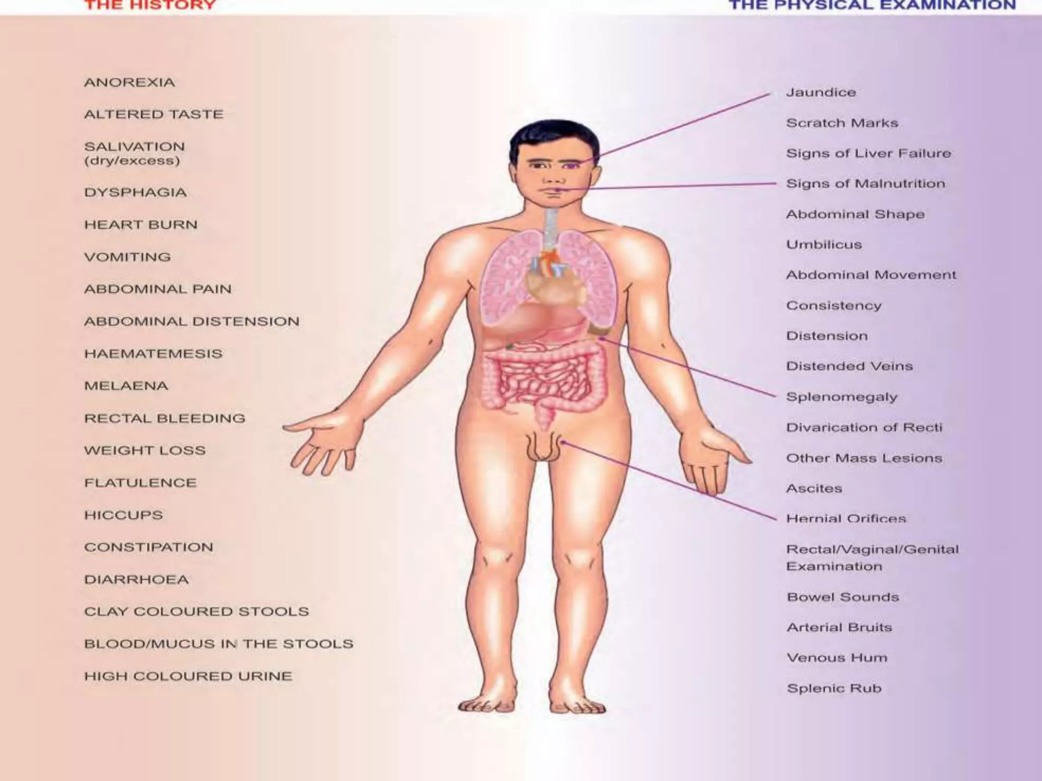
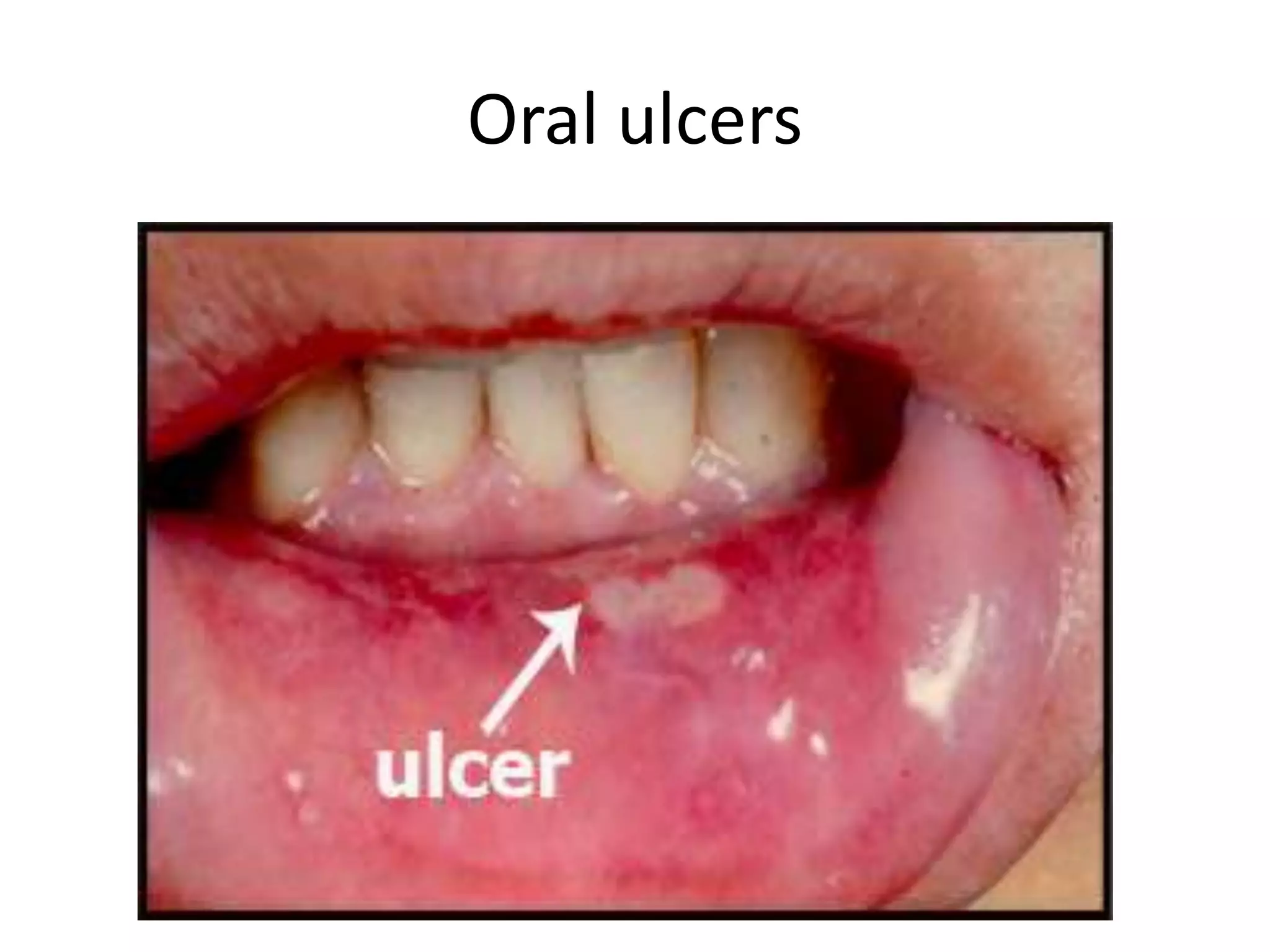
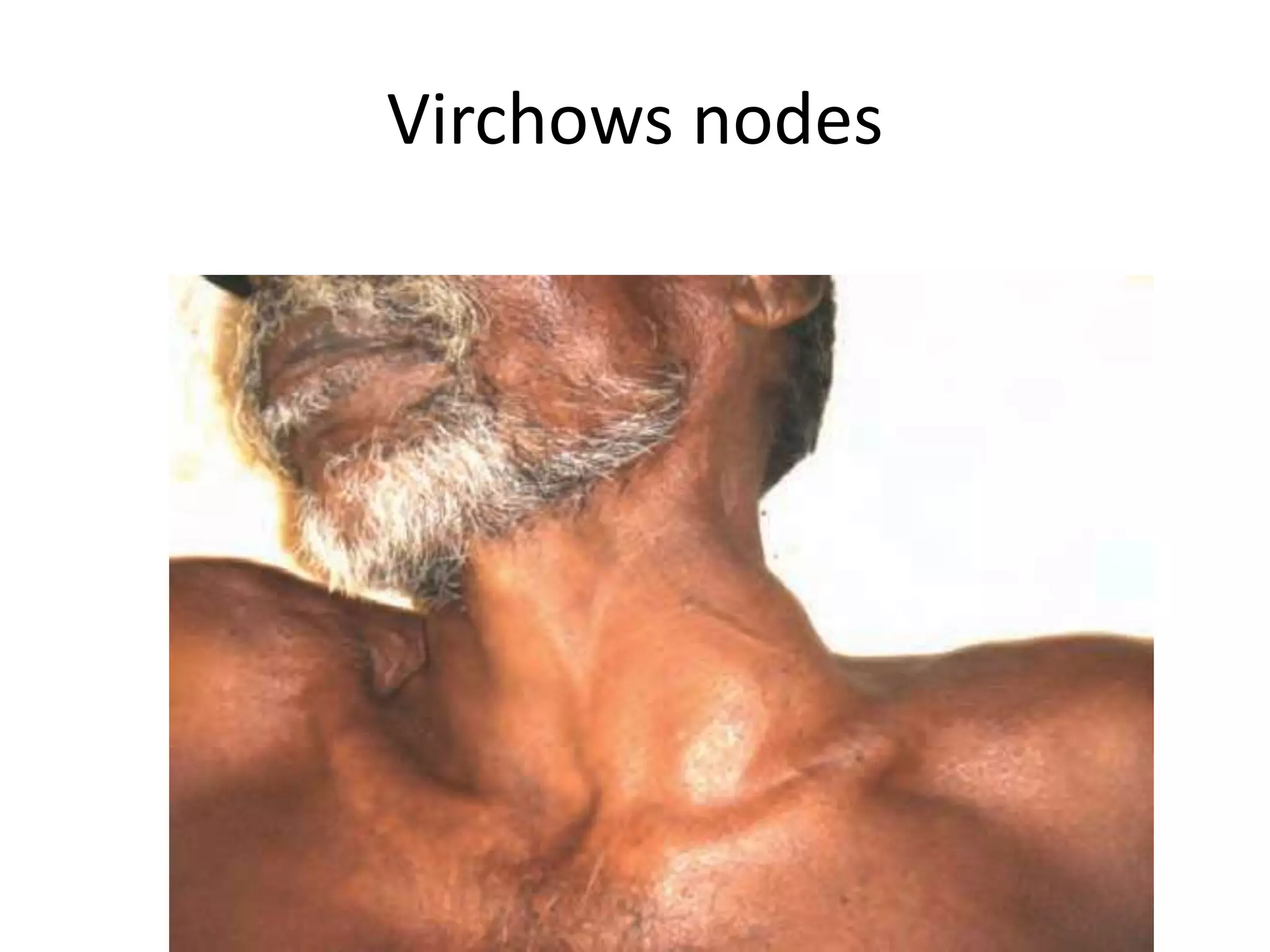
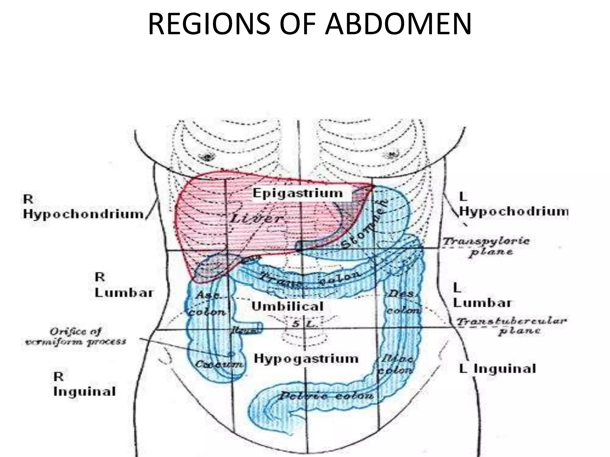
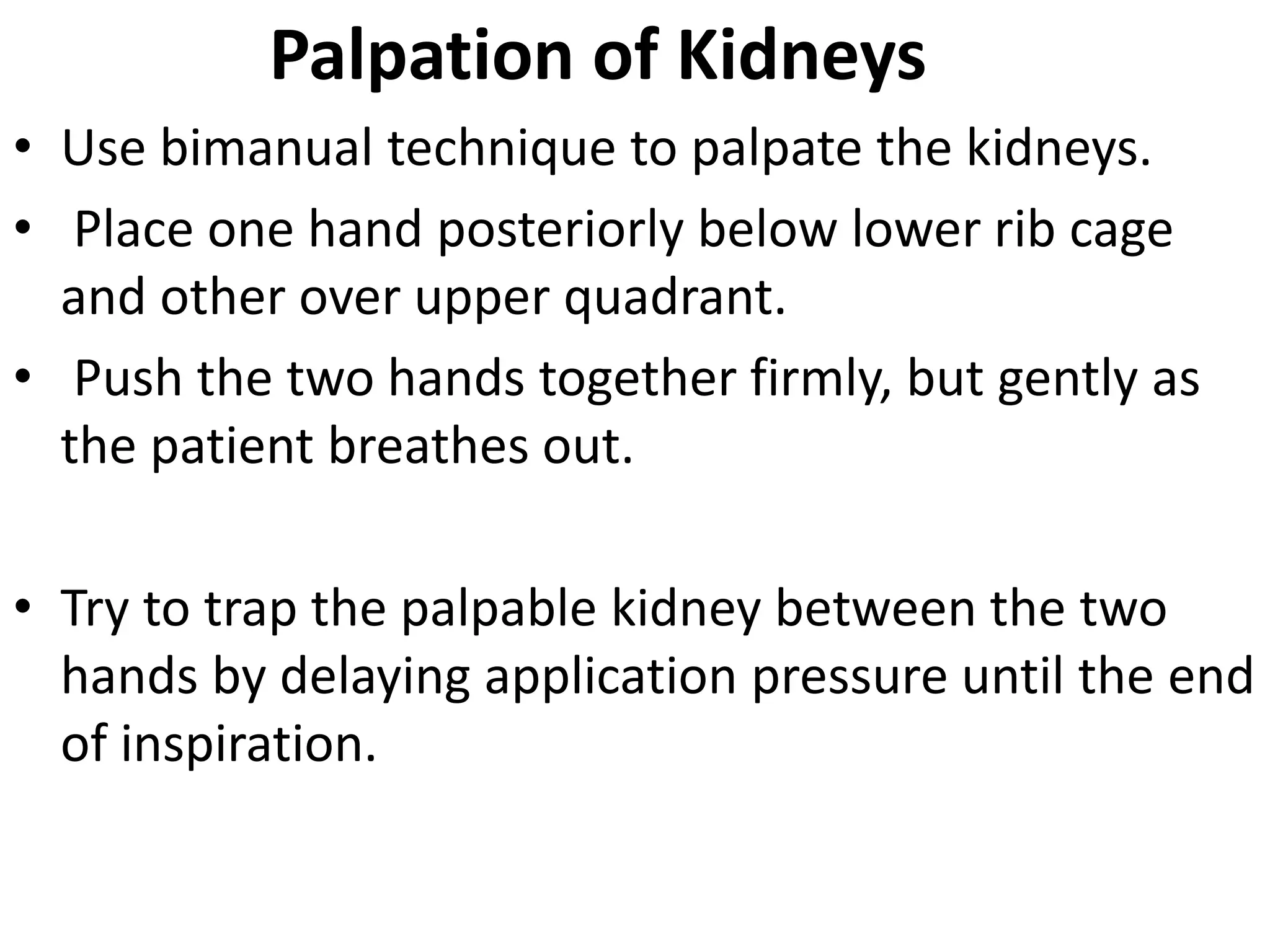
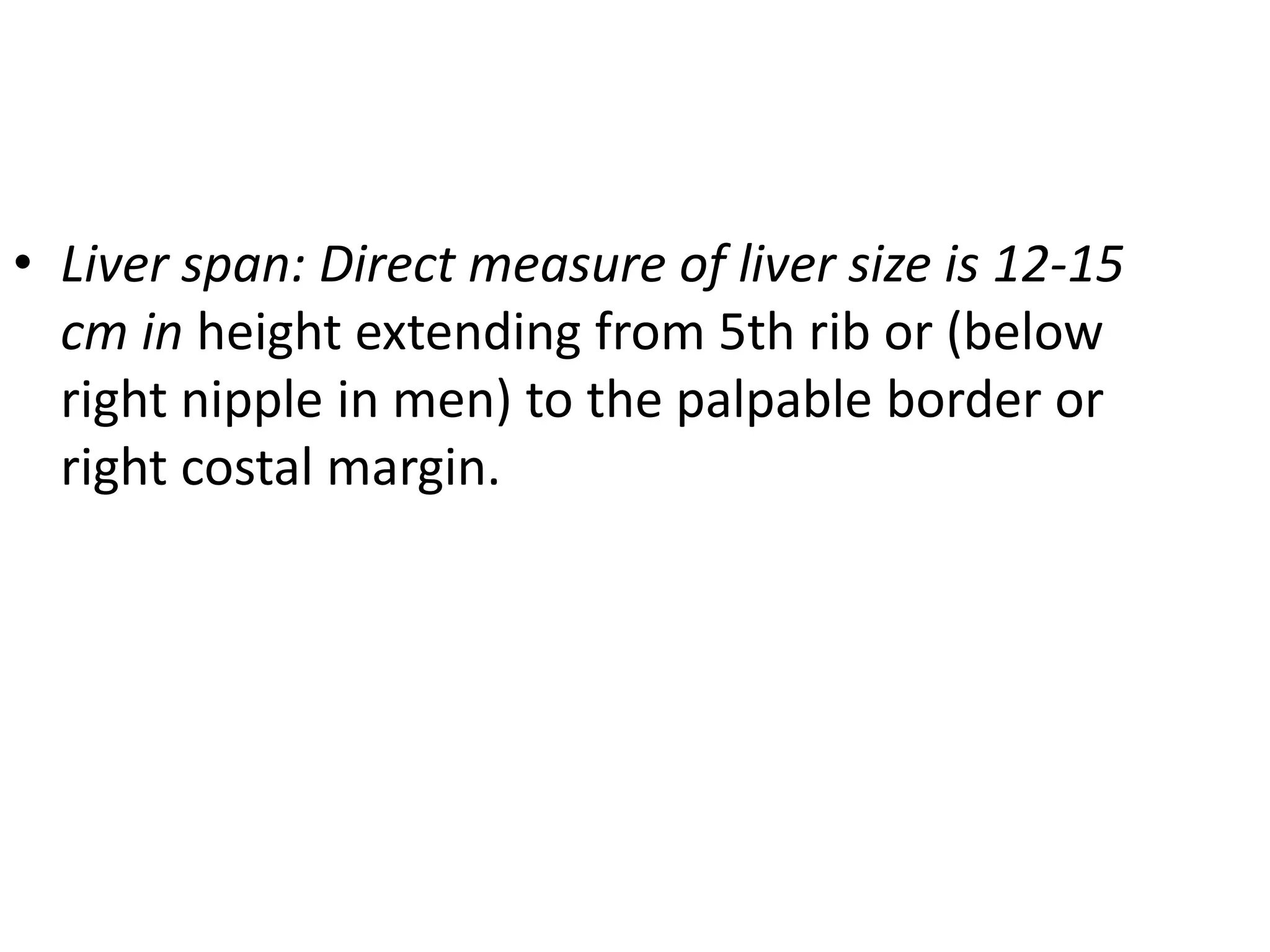
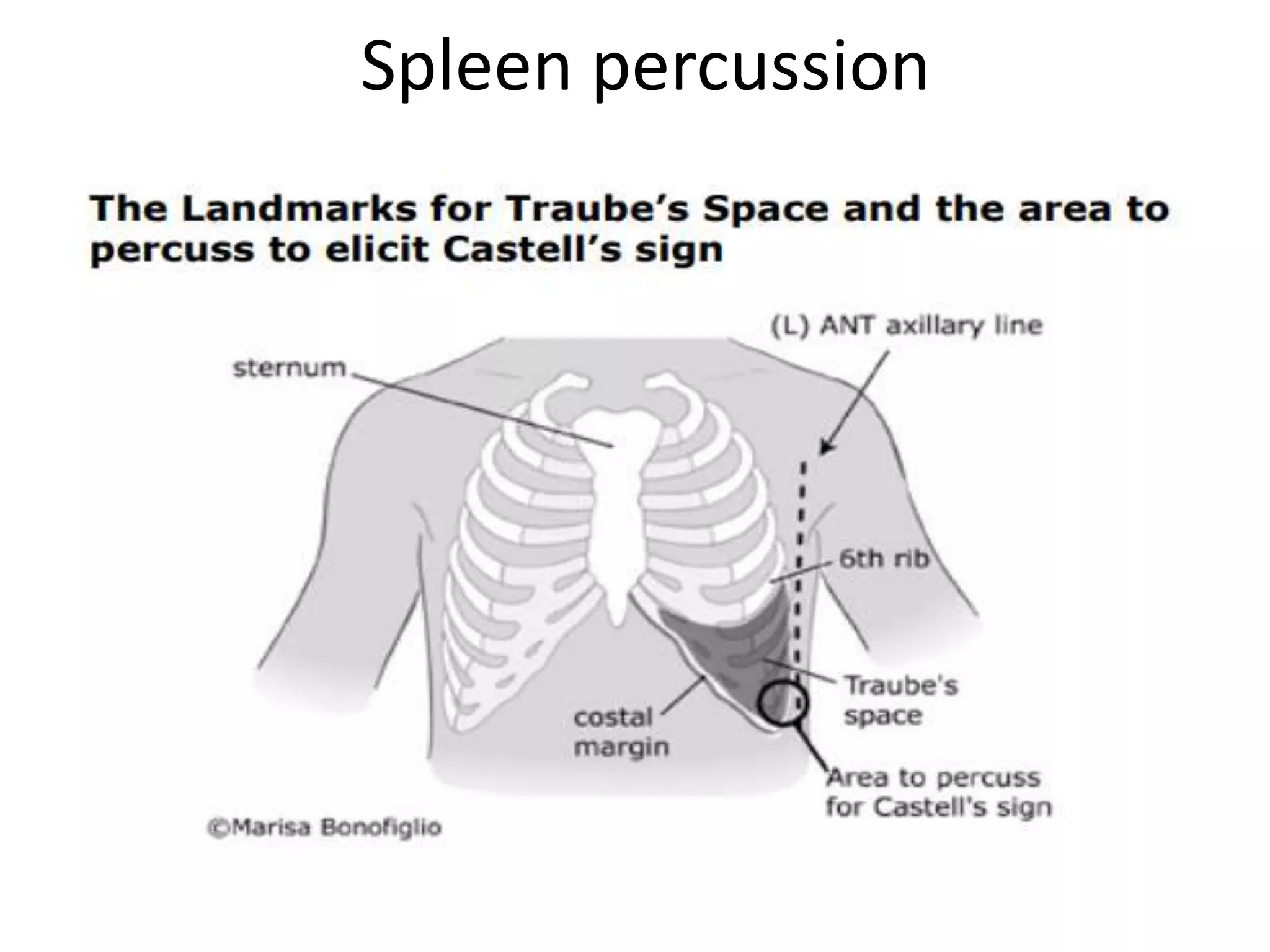
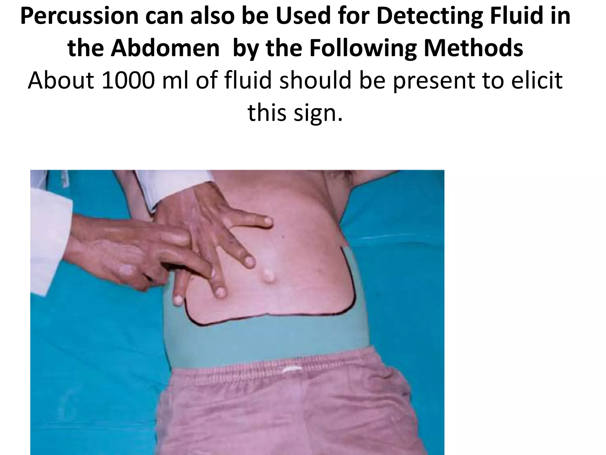
This document provides an overview of techniques for examining the abdomen through inspection, palpation, percussion, and auscultation. Key points covered include assessing the shape and movements of the abdomen, palpating the liver, gallbladder, spleen and kidneys, using percussion to define organ boundaries, and listening for bowel sounds, succussion splash, bruits, venous hum, and friction rubs over the abdomen. The document serves as a guide for medical students to perform a thorough physical examination of the abdomen.
Brief Presentation on clinical examination of Respiratory System with Report of Normal case
references:
macleod's clinical examination 13th edition
hutchinson clinical methods
R Alagappan - Manual of Practical Medicine, 4th Edition
Brief Presentation on clinical examination of Respiratory System with Report of Normal case
references:
macleod's clinical examination 13th edition
hutchinson clinical methods
R Alagappan - Manual of Practical Medicine, 4th Edition
basics about chronic liver disease for a pediatrician. fast and easy guide to common causes of chronic liver diseases in children
Please leave a comment if you like it..
Brief Presentation on clinical examination of Cardio Vascular System with Report of Normal case
references:
macleod's clinical examination 13th edition
hutchinson clinical methods
Examination of Swelling in a patient is always a task for MBBS students. This PPT provides the students, how to elicit a history & also the easy way to examine a swelling.
The abdominal examination consists of four basic components: inspection, palpation, percussion, and auscultation. It is important to begin with the general examination of the abdomen with the patient in a completely supine position. The presence of any of the following signs may indicate specific disorders.
INTUSSUSCEPTION (ISS)
DEFINITION
It is telescoping or invagination of one portion (segment) of bowel into the adjacent segment.
TYPES
1. Antegrade: Most common.
2. Retrograde: Rare (jejunogastric in gastrojejunostomy stoma).
This presentation explains in detail the definition, pathophysiology, signs & symptoms, management, and prognosis of intestinal obstruction, ileus, and volvulus.
Similar to Per abdomen examination - Clinical Methods - Abdomen (20)
- Video recording of this lecture in English language: https://youtu.be/kqbnxVAZs-0
- Video recording of this lecture in Arabic language: https://youtu.be/SINlygW1Mpc
- Link to download the book free: https://nephrotube.blogspot.com/p/nephrotube-nephrology-books.html
- Link to NephroTube website: www.NephroTube.com
- Link to NephroTube social media accounts: https://nephrotube.blogspot.com/p/join-nephrotube-on-social-media.html
Title: Sense of Smell
Presenter: Dr. Faiza, Assistant Professor of Physiology
Qualifications:
MBBS (Best Graduate, AIMC Lahore)
FCPS Physiology
ICMT, CHPE, DHPE (STMU)
MPH (GC University, Faisalabad)
MBA (Virtual University of Pakistan)
Learning Objectives:
Describe the primary categories of smells and the concept of odor blindness.
Explain the structure and location of the olfactory membrane and mucosa, including the types and roles of cells involved in olfaction.
Describe the pathway and mechanisms of olfactory signal transmission from the olfactory receptors to the brain.
Illustrate the biochemical cascade triggered by odorant binding to olfactory receptors, including the role of G-proteins and second messengers in generating an action potential.
Identify different types of olfactory disorders such as anosmia, hyposmia, hyperosmia, and dysosmia, including their potential causes.
Key Topics:
Olfactory Genes:
3% of the human genome accounts for olfactory genes.
400 genes for odorant receptors.
Olfactory Membrane:
Located in the superior part of the nasal cavity.
Medially: Folds downward along the superior septum.
Laterally: Folds over the superior turbinate and upper surface of the middle turbinate.
Total surface area: 5-10 square centimeters.
Olfactory Mucosa:
Olfactory Cells: Bipolar nerve cells derived from the CNS (100 million), with 4-25 olfactory cilia per cell.
Sustentacular Cells: Produce mucus and maintain ionic and molecular environment.
Basal Cells: Replace worn-out olfactory cells with an average lifespan of 1-2 months.
Bowman’s Gland: Secretes mucus.
Stimulation of Olfactory Cells:
Odorant dissolves in mucus and attaches to receptors on olfactory cilia.
Involves a cascade effect through G-proteins and second messengers, leading to depolarization and action potential generation in the olfactory nerve.
Quality of a Good Odorant:
Small (3-20 Carbon atoms), volatile, water-soluble, and lipid-soluble.
Facilitated by odorant-binding proteins in mucus.
Membrane Potential and Action Potential:
Resting membrane potential: -55mV.
Action potential frequency in the olfactory nerve increases with odorant strength.
Adaptation Towards the Sense of Smell:
Rapid adaptation within the first second, with further slow adaptation.
Psychological adaptation greater than receptor adaptation, involving feedback inhibition from the central nervous system.
Primary Sensations of Smell:
Camphoraceous, Musky, Floral, Pepperminty, Ethereal, Pungent, Putrid.
Odor Detection Threshold:
Examples: Hydrogen sulfide (0.0005 ppm), Methyl-mercaptan (0.002 ppm).
Some toxic substances are odorless at lethal concentrations.
Characteristics of Smell:
Odor blindness for single substances due to lack of appropriate receptor protein.
Behavioral and emotional influences of smell.
Transmission of Olfactory Signals:
From olfactory cells to glomeruli in the olfactory bulb, involving lateral inhibition.
Primitive, less old, and new olfactory systems with different path
Muktapishti is a traditional Ayurvedic preparation made from Shoditha Mukta (Purified Pearl), is believed to help regulate thyroid function and reduce symptoms of hyperthyroidism due to its cooling and balancing properties. Clinical evidence on its efficacy remains limited, necessitating further research to validate its therapeutic benefits.
Title: Sense of Taste
Presenter: Dr. Faiza, Assistant Professor of Physiology
Qualifications:
MBBS (Best Graduate, AIMC Lahore)
FCPS Physiology
ICMT, CHPE, DHPE (STMU)
MPH (GC University, Faisalabad)
MBA (Virtual University of Pakistan)
Learning Objectives:
Describe the structure and function of taste buds.
Describe the relationship between the taste threshold and taste index of common substances.
Explain the chemical basis and signal transduction of taste perception for each type of primary taste sensation.
Recognize different abnormalities of taste perception and their causes.
Key Topics:
Significance of Taste Sensation:
Differentiation between pleasant and harmful food
Influence on behavior
Selection of food based on metabolic needs
Receptors of Taste:
Taste buds on the tongue
Influence of sense of smell, texture of food, and pain stimulation (e.g., by pepper)
Primary and Secondary Taste Sensations:
Primary taste sensations: Sweet, Sour, Salty, Bitter, Umami
Chemical basis and signal transduction mechanisms for each taste
Taste Threshold and Index:
Taste threshold values for Sweet (sucrose), Salty (NaCl), Sour (HCl), and Bitter (Quinine)
Taste index relationship: Inversely proportional to taste threshold
Taste Blindness:
Inability to taste certain substances, particularly thiourea compounds
Example: Phenylthiocarbamide
Structure and Function of Taste Buds:
Composition: Epithelial cells, Sustentacular/Supporting cells, Taste cells, Basal cells
Features: Taste pores, Taste hairs/microvilli, and Taste nerve fibers
Location of Taste Buds:
Found in papillae of the tongue (Fungiform, Circumvallate, Foliate)
Also present on the palate, tonsillar pillars, epiglottis, and proximal esophagus
Mechanism of Taste Stimulation:
Interaction of taste substances with receptors on microvilli
Signal transduction pathways for Umami, Sweet, Bitter, Sour, and Salty tastes
Taste Sensitivity and Adaptation:
Decrease in sensitivity with age
Rapid adaptation of taste sensation
Role of Saliva in Taste:
Dissolution of tastants to reach receptors
Washing away the stimulus
Taste Preferences and Aversions:
Mechanisms behind taste preference and aversion
Influence of receptors and neural pathways
Impact of Sensory Nerve Damage:
Degeneration of taste buds if the sensory nerve fiber is cut
Abnormalities of Taste Detection:
Conditions: Ageusia, Hypogeusia, Dysgeusia (parageusia)
Causes: Nerve damage, neurological disorders, infections, poor oral hygiene, adverse drug effects, deficiencies, aging, tobacco use, altered neurotransmitter levels
Neurotransmitters and Taste Threshold:
Effects of serotonin (5-HT) and norepinephrine (NE) on taste sensitivity
Supertasters:
25% of the population with heightened sensitivity to taste, especially bitterness
Increased number of fungiform papillae
Local Advanced Lung Cancer: Artificial Intelligence, Synergetics, Complex Sys...Oleg Kshivets
Overall life span (LS) was 1671.7±1721.6 days and cumulative 5YS reached 62.4%, 10 years – 50.4%, 20 years – 44.6%. 94 LCP lived more than 5 years without cancer (LS=2958.6±1723.6 days), 22 – more than 10 years (LS=5571±1841.8 days). 67 LCP died because of LC (LS=471.9±344 days). AT significantly improved 5YS (68% vs. 53.7%) (P=0.028 by log-rank test). Cox modeling displayed that 5YS of LCP significantly depended on: N0-N12, T3-4, blood cell circuit, cell ratio factors (ratio between cancer cells-CC and blood cells subpopulations), LC cell dynamics, recalcification time, heparin tolerance, prothrombin index, protein, AT, procedure type (P=0.000-0.031). Neural networks, genetic algorithm selection and bootstrap simulation revealed relationships between 5YS and N0-12 (rank=1), thrombocytes/CC (rank=2), segmented neutrophils/CC (3), eosinophils/CC (4), erythrocytes/CC (5), healthy cells/CC (6), lymphocytes/CC (7), stick neutrophils/CC (8), leucocytes/CC (9), monocytes/CC (10). Correct prediction of 5YS was 100% by neural networks computing (error=0.000; area under ROC curve=1.0).
Flu Vaccine Alert in Bangalore Karnatakaaddon Scans
As flu season approaches, health officials in Bangalore, Karnataka, are urging residents to get their flu vaccinations. The seasonal flu, while common, can lead to severe health complications, particularly for vulnerable populations such as young children, the elderly, and those with underlying health conditions.
Dr. Vidisha Kumari, a leading epidemiologist in Bangalore, emphasizes the importance of getting vaccinated. "The flu vaccine is our best defense against the influenza virus. It not only protects individuals but also helps prevent the spread of the virus in our communities," he says.
This year, the flu season is expected to coincide with a potential increase in other respiratory illnesses. The Karnataka Health Department has launched an awareness campaign highlighting the significance of flu vaccinations. They have set up multiple vaccination centers across Bangalore, making it convenient for residents to receive their shots.
To encourage widespread vaccination, the government is also collaborating with local schools, workplaces, and community centers to facilitate vaccination drives. Special attention is being given to ensuring that the vaccine is accessible to all, including marginalized communities who may have limited access to healthcare.
Residents are reminded that the flu vaccine is safe and effective. Common side effects are mild and may include soreness at the injection site, mild fever, or muscle aches. These side effects are generally short-lived and far less severe than the flu itself.
Healthcare providers are also stressing the importance of continuing COVID-19 precautions. Wearing masks, practicing good hand hygiene, and maintaining social distancing are still crucial, especially in crowded places.
Protect yourself and your loved ones by getting vaccinated. Together, we can help keep Bangalore healthy and safe this flu season. For more information on vaccination centers and schedules, residents can visit the Karnataka Health Department’s official website or follow their social media pages.
Stay informed, stay safe, and get your flu shot today!
- Video recording of this lecture in English language: https://youtu.be/lK81BzxMqdo
- Video recording of this lecture in Arabic language: https://youtu.be/Ve4P0COk9OI
- Link to download the book free: https://nephrotube.blogspot.com/p/nephrotube-nephrology-books.html
- Link to NephroTube website: www.NephroTube.com
- Link to NephroTube social media accounts: https://nephrotube.blogspot.com/p/join-nephrotube-on-social-media.html
Basavarajeeyam is a Sreshta Sangraha grantha (Compiled book ), written by Neelkanta kotturu Basavaraja Virachita. It contains 25 Prakaranas, First 24 Chapters related to Rogas& 25th to Rasadravyas.
Tom Selleck Health: A Comprehensive Look at the Iconic Actor’s Wellness Journeygreendigital
Tom Selleck, an enduring figure in Hollywood. has captivated audiences for decades with his rugged charm, iconic moustache. and memorable roles in television and film. From his breakout role as Thomas Magnum in Magnum P.I. to his current portrayal of Frank Reagan in Blue Bloods. Selleck's career has spanned over 50 years. But beyond his professional achievements. fans have often been curious about Tom Selleck Health. especially as he has aged in the public eye.
Follow us on: Pinterest
Introduction
Many have been interested in Tom Selleck health. not only because of his enduring presence on screen but also because of the challenges. and lifestyle choices he has faced and made over the years. This article delves into the various aspects of Tom Selleck health. exploring his fitness regimen, diet, mental health. and the challenges he has encountered as he ages. We'll look at how he maintains his well-being. the health issues he has faced, and his approach to ageing .
Early Life and Career
Childhood and Athletic Beginnings
Tom Selleck was born on January 29, 1945, in Detroit, Michigan, and grew up in Sherman Oaks, California. From an early age, he was involved in sports, particularly basketball. which played a significant role in his physical development. His athletic pursuits continued into college. where he attended the University of Southern California (USC) on a basketball scholarship. This early involvement in sports laid a strong foundation for his physical health and disciplined lifestyle.
Transition to Acting
Selleck's transition from an athlete to an actor came with its physical demands. His first significant role in "Magnum P.I." required him to perform various stunts and maintain a fit appearance. This role, which he played from 1980 to 1988. necessitated a rigorous fitness routine to meet the show's demands. setting the stage for his long-term commitment to health and wellness.
Fitness Regimen
Workout Routine
Tom Selleck health and fitness regimen has evolved. adapting to his changing roles and age. During his "Magnum, P.I." days. Selleck's workouts were intense and focused on building and maintaining muscle mass. His routine included weightlifting, cardiovascular exercises. and specific training for the stunts he performed on the show.
Selleck adjusted his fitness routine as he aged to suit his body's needs. Today, his workouts focus on maintaining flexibility, strength, and cardiovascular health. He incorporates low-impact exercises such as swimming, walking, and light weightlifting. This balanced approach helps him stay fit without putting undue strain on his joints and muscles.
Importance of Flexibility and Mobility
In recent years, Selleck has emphasized the importance of flexibility and mobility in his fitness regimen. Understanding the natural decline in muscle mass and joint flexibility with age. he includes stretching and yoga in his routine. These practices help prevent injuries, improve posture, and maintain mobilit
These lecture slides, by Dr Sidra Arshad, offer a quick overview of the physiological basis of a normal electrocardiogram.
Learning objectives:
1. Define an electrocardiogram (ECG) and electrocardiography
2. Describe how dipoles generated by the heart produce the waveforms of the ECG
3. Describe the components of a normal electrocardiogram of a typical bipolar lead (limb II)
4. Differentiate between intervals and segments
5. Enlist some common indications for obtaining an ECG
6. Describe the flow of current around the heart during the cardiac cycle
7. Discuss the placement and polarity of the leads of electrocardiograph
8. Describe the normal electrocardiograms recorded from the limb leads and explain the physiological basis of the different records that are obtained
9. Define mean electrical vector (axis) of the heart and give the normal range
10. Define the mean QRS vector
11. Describe the axes of leads (hexagonal reference system)
12. Comprehend the vectorial analysis of the normal ECG
13. Determine the mean electrical axis of the ventricular QRS and appreciate the mean axis deviation
14. Explain the concepts of current of injury, J point, and their significance
Study Resources:
1. Chapter 11, Guyton and Hall Textbook of Medical Physiology, 14th edition
2. Chapter 9, Human Physiology - From Cells to Systems, Lauralee Sherwood, 9th edition
3. Chapter 29, Ganong’s Review of Medical Physiology, 26th edition
4. Electrocardiogram, StatPearls - https://www.ncbi.nlm.nih.gov/books/NBK549803/
5. ECG in Medical Practice by ABM Abdullah, 4th edition
6. Chapter 3, Cardiology Explained, https://www.ncbi.nlm.nih.gov/books/NBK2214/
7. ECG Basics, http://www.nataliescasebook.com/tag/e-c-g-basics
3. General physical examination
a.Nutritional status
b. Anaemia
c. Finger clubbing: malabsorption, chronic liver
impairment, inflammatory bowel diseases,
hepatoma
d. Leukonychia
e. Koilonychia is suggestive of chronic iron deficiency
f. Lymphadenopathy : In leukaemia or lymphoma
there may be generalised lymphadenopathy
20. Inspection – shape of abdomen
• Generalised fullness or distension
• Localised distension
a. Symmetrical and centered around umbilicus?
b. Asymmetrical—liver or spleen or ovary
• Scaphoid or sunken abdomen is seen in advanced
starvation or malignancy.
21. Umbilicus
• Normal Slightly retracted and inverted
• Everted In umbilical hernia
• Omphalolith - Inspissated desquamated
epithelium & other debris.
• Slit Vertical (pelvic or ovarian tumours)
• Horizontal (cirrhosis of liver with ascites).
22. Movements with respiration
• Normally there is a gentle rise in abdominal wall in
inspiration and a fall during expiration.
• In peritonitis abdomen is still or silent.
24. Skin or Surface of Abdomen
• Striae atrophica or gravidarum: recent change in
abdominal size
• Linea nigra is a pigmentation below umbilicus seen
in pregnancy
• Purple striae: seen in Cushing’s
syndrome
27. • Cullen’s sign:A faint blue discoloration around the
umbilicus may occur as the result of
hemoperitoneum
• Grey-Turner’s sign : a blue-red-purple or green-
brown discoloration of the flanks (Turner’s sign)
reflects tissue catabolism of hemoglobin from
severe necrotizing pancreatitis with hemorrhage.
30. • At the end, always inspect external genitalia
and hernial orifices.
31. Palpation
• Measurements
• Abdominal girth should be measured.
• Measure the distance between lower end of
xiphisternum to umbilicus and from umbilicus to
symphysis pubis
• Spinoumbilical measurement: Normally they are
equidistant.
33. Superficial palpation
Guarding = muscles contract when pressure is applied
- Ridigity = indicates peritoneal inflamation.
involuntary reflex contraction.
• The most extreme form is seen in the board-like
rigidity often associated with a perforated hollow
viscus.
- Rebound = Releasing of pressure causing pain.
34. • Guarding and rigidity, however, may be
localized, as over an acutely inflamed
appendix in the right iliac fossa or diverticulitis
in the left iliac fossa.
• Deep palpation
36. Palpation by Dipping
• This method is used in tense ascites to detect the
presence of hepatic or splenic enlargement.
• Sudden displacement of liquid gives a tapping
sensation over the surface of liver or spleen.
• For eliciting this, place the hand flat on
abdomen and make quick dipping movements.
37. • The site of tenderness is important.
• Tenderness in the epigastrium suggests peptic ulcer;
• in the right hypochondrium, cholecystitis;
• in the left iliac fossa, diverticulitis;
• in the right iliac fossa, appendicitis or Crohn’s
ileitis.
38. Organomegaly
• When an organ is enlarged, assess the following:
a. Edge or border (sharp or rounded)
b. Surface (smooth or nodular)
c. Consistency (soft, firm or hard)
d. Presence of tenderness
e. Movement with respiration.
43. Palpation of Gallbladder
• It is felt as a firm, smooth, rough or globular
swelling with distinct borders, just lateral to the
edge of the rectus abdominis near the tip of ninth
costal cartilage.
• It moves with respiration.
46. GALL BLADDER ENLARGEMENT CAUSES
• In a jaundiced patient with CA of head of the
pancreas or other malignant causes of obstruction
of the common bile duct (below the entry of the
cystic duct), the ducts above the obstruction
become dilated, as does the gallbladder.
• In mucocele of the gallbladder
• In carcinoma of the gallbladder, the gallbladder may
be felt as a stony, hard, irregular swelling, unlike the
firm, regular swelling.
47. Murphy’s sign
• In acute inflammation of the gallbladder (acute
cholecystitis), severe pain is present.
• Not seen in chronic cholecystitis
48. Courvoisier’s law
• In a jaundiced patient, a palpable gallbladder is
likely to be due to extrahepatic obstruction, e.g.
from pancreatic cancer.
• In cholelithiasis, the gallbladder wall is diseased,
thickened, contracted and not palpable due to
repeated cholecystitis.
49. PALPATION OF SPLEEN
• To become palpable, spleen should have enlarged
2-3 times. Direction of enlargement is towards right
iliac fossa.
• Enlargement takes place in a superior and posterior
direction before it becomes palpable subcostally.
50. Palpation of spleen
• Palpate from right iliac fossa to left
hypochondrium
• Wait for one full phase of respiration
• At the height of inspiration, release the pressure
on the examining hand so that the finger tips slip
over the lower pole of spleen, confirming its
presence and surface characteristics.
53. Palpation of Kidneys
• Use bimanual technique to palpate the kidneys.
• Place one hand posteriorly below lower rib cage
and other over upper quadrant.
• Push the two hands together firmly, but gently as
the patient breathes out.
• Try to trap the palpable kidney between the two
hands by delaying application pressure until the end
of inspiration.
54. • Confirm the structure of the kidney, by pushing
the kidney between the two hands (ballotting)
and by assessing its degree of movement
during respiration.
• Assess the size, surface and consistency of a
palpable kidney.
• Examine the left kidney from either side.
57. Palpation of Urinary Bladder
• Normally it is not palpable.It is palpable as a
smooth, firm, regular, oval shaped swelling in
suprapubic region and its dome may reach as far
as the umbilicus.
• Its lower border cannot be felt. It is
symmetrically placed in suprapubic region
beneath the umbilicus, which is dull on
percussion.
58. Percussion
• Defining Boundaries
• Liver
• In normal liver, upper border is at 5th inter costal
space where note is dull;
• This extends down to the lower border found at or
just below right subcostal margin.
• Percussion below the right costal margin is useful in
hepatomegaly. Ask the patient to breathe in deeply
as you percuss, lightly keeping the fingers parallel to
the rib margin.
59. • Liver span: Direct measure of liver size is 12-15
cm in height extending from 5th rib or (below
right nipple in men) to the palpable border or
right costal margin.
61. Percussion by Nixon’s method
• Right lateral decubitus , begin percussion
midway along the Left costal margin ,
• proceed in a line perpendicular to the Left
costal margin .
• if the upper limit of dullness extends >8 cm
above the Left costal margin, this indicates
possible splenomegaly
62.
63.
64. Percussion can also be Used for Detecting Fluid in
the Abdomen by the Following Methods
About 1000 ml of fluid should be present to elicit
this sign.
65.
66. Fluid thrill
This is felt when there is a large amount of fluid
under tension, i.e. > 2000 ml.
68. • Chronic ascites is associated with
• 1. Umbilical hernia
• 2. Puncture marks
• 3. Striae
• 4. Divarication of recti.
69. Auscultation
• Bowel Sounds
• Normal motility of the gut creates a characteristic
gurgling sounds every 5-10 seconds.
Bowel sounds are increased in:
1. Simple, acute, mechanical, small bowel obstruction.
Increased bowel sounds with colicky pain is
pathognomonic of small bowel obstruction.
2. Malabsorption
3. Severe GI bleeding
4. Carcinoid syndrome.
70. • Bowel sounds are absent in
a. Paralytic ileus
b. Peritonitis.
71. Succussion Splash
• It is a sound resembling shaking a half filled bottle.
It is heard in:
• 1. Pyloric stenosis
• 2. Advanced intestinal obstruction
• 3. Paralytic ileus (with grossly distended loops of
bowel)
• 4. Normal stomach within 2 hours after a meal.
72. Bruit
• Bruit over aorta can be heard above and to the left of
umbilicus in cases of aortic aneurysm.
• Bruit over mid abdomen is heard in renal artery
stenosis.
• Bruit over common iliac artery can be heard in stenosis
or aneurysm.
Bruit over liver may be heard in:
a. Haemangioma
b. Hepatocellular carcinoma
c. Acute alcoholic hepatitis
d. Hepatic artery aneurysm.
73. Venous Hum
• It is heard between xiphisternum and umbilicus
due to turbulence of blood flow in well-
developed collaterals as a result of portal
hypertension (Cruveilhier- Baumgarten
syndrome).
• It signifies a congenital patent umbilical vein
draining into the portal vein.
74. Friction Rub
• It is heard in perisplenitis or perihepatitis due to
microinfarction and inflammation.
• Splenic rub is heard in the following conditions:
• a. Chronic myeloid leukaemia
• b. Infective endocarditis
• c. Sickle cell anaemia
• d. After biopsy
75. References
• Hutchisons clinical methods
• Mcleods clinical examinations
• S.DAS clinical manual of surgery
• Harrisons principles of internal medicine ,19th
edition