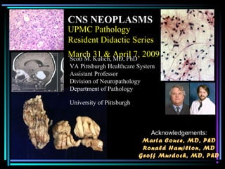
1. UPMC Pathology
Resident Didactic Series
March 31 & April 7, 2009
CNS NEOPLASMS
Scott M. Kulich, MD, PhD
VA Pittsburgh Healthcare System
Assistant Professor
Division of Neuropathology
Department of Pathology
University of Pittsburgh
Acknowledgements:
Marta Couce, MD, PhD
Ronald Hamilton, MD
Geoff Murdoch, MD, PhD
6. NEURORADIOLOGY FOR
PATHOLOGISTSQuestion: Who cares?
Answer: You will when your
favorite neurosurgeon hands
you a piece of tissue the size
of a grain of salt and tells you
he needs you to tell him if he
can go ahead and stick Gliadel
chemotherapeutic wafers in the
patient’s brain
7. NEURORADIOLOGY FOR
PATHOLOGISTSQuestion: Who cares?
Neuroradiology = Gross pathology
Answer: You will when your
favorite neurosurgeon hands
you a piece of tissue the size
of a grain of salt and tells you
he needs you to tell him if he
can go ahead and stick Gliadel
chemotherapeutic wafers in the
patient’s brain
8. NEURORADIOLOGY FOR
PATHOLOGISTS
• Two main imaging techniques
– Computerized tomography (CT)
• 3D X-rays
• White areas = areas that absorb or “attenuate”
the passage of x-ray beam (acute
hematoma, bone, calcium = hyperdense/
attenuating)
• Black areas = areas that do not absorb or
“attenuate” the passage of x-ray beam (fat, air,
CSF, edema = hypodense/ attenuating)
Neuroradiology
for
10. NEURORADIOLOGY FOR
PATHOLOGISTS
• Magnetic resonance imaging (MRI)
• Not ionizing radiation but magnetic field to
excite protons which emit “signal” upon
relaxation
• Image appearance dependent upon time
interval between each excitation and time
interval between each collection
• Two basic “weights” of images based upon TE
and TR
– T1: Short TE and TR
» T1 is the one…that looks like a brain
– T2 :Long TE and TR
11. NEURORADIOLOGY FOR
PATHOLOGISTS
• Magnetic resonance imaging (MRI)
• Not ionizing radiation but magnetic field to
excite protons which emit “signal” upon
relaxation
• Image appearance dependent upon time
interval between each excitation and time
interval between each collection
• Two basic “weights” of images based upon TE
and TR
– T1: Short TE and TR
» T1 is the one…that looks like a brain
– T2 :Long TE and TR
12. NEURORADIOLOGY FOR
PATHOLOGISTS
• Magnetic resonance imaging (MRI)
• Not ionizing radiation but magnetic field to
excite protons which emit “signal” upon
relaxation
• Image appearance dependent upon time
interval between each excitation and time
interval between each collection
• Two basic “weights” of images based upon TE
and TR
– T1: Short TE and TR
» T1 is the one…that looks like a brain
– T2 :Long TE and TR
15. NEURORADIOLOGY FOR
PATHOLOGISTS
• Important info to glean from neuroimaging
– Age
– Location, location, location
– Multicentricity
– Bilateral hemisphere involvement
– Architecture
– Contrast enhancement
– Interaction with surrounding tissue
27. NEURORADIOLOGY FOR
PATHOLOGISTS
• Contrast enhancement
– Breached blood-brain barrier
– Seen with neoplasms but can be seen with other
conditions (e.g. infectious, demyelinating, …)
– Pattern of enhancement often helpful
• Homogeneous versus non-homogeneous
– Lymphoma, hemangiopericytoma, meningioma
– GBM, mets, abscesses
• Patchy versus circumferential ( i.e. ring enhancement)
28. NEURORADIOLOGY FOR
PATHOLOGISTS
• Contrast enhancement
– Breached blood-brain barrier
– Seen with neoplasms but can be seen with other
conditions (e.g. infectious, demyelinating, …)
– Pattern of enhancement often helpful
• Homogeneous versus non-homogeneous
– Lymphoma, hemangiopericytoma, meningioma
– GBM, mets, abscesses
• Patchy versus circumferential ( i.e. ring enhancement)
35. Approach to intraoperative consults
• Review of imaging and history
• Questions for surgeon
– What do you NEED to know?
– Can you get more tissue if necessary?
• Specimen preparation
– Intraoperative cytology vs frozen sections
• touch and smear preparations
36. Approach to intraoperative consults
• Review of imaging and history
• Questions for surgeon
– What do you NEED to know?
– Can you get more tissue if necessary?
• Specimen preparation
– Intraoperative cytology vs frozen sections
• touch and smear preparations
37. Approach to intraoperative consults
• Review of imaging and history
• Questions for surgeon
– What do you NEED to know?
– Can you get more tissue if necessary?
• Specimen preparation
– Intraoperative cytology vs frozen sections
• touch and smear preparations
42. A “wiley” approach to intraoperative
consults
• Abnormal versus normal
• Reactive versus neoplastic
• Primary versus metastatic
• Grade of lesion
• Does diagnosis correlate with clinical
and imaging data?
43. A “wiley” approach to intraoperative
consults
• Abnormal versus normal
• Reactive versus neoplastic
• Primary versus metastatic
• Grade of lesion
• Does diagnosis correlate with clinical
and imaging data?
44. A “wiley” approach to intraoperative
consults
• Abnormal versus normal
• Reactive versus neoplastic
• Primary versus metastatic
• Grade of lesion
• Does diagnosis correlate with clinical
and imaging data?
45. A “wiley” approach to intraoperative
consults
• Abnormal versus normal
• Reactive versus neoplastic
• Primary versus metastatic
• Grade of lesion
• Does diagnosis correlate with clinical
and imaging data?
46. A “wiley” approach to intraoperative
consults
• Abnormal versus normal
• Reactive versus neoplastic
• Primary versus metastatic
• Grade of lesion
• Does diagnosis correlate with clinical
and imaging data?