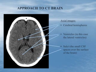
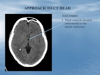
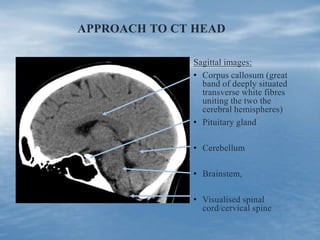
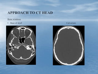
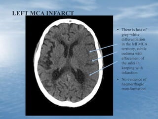
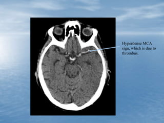
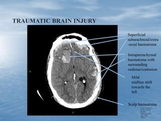
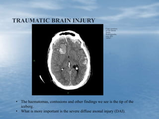
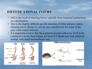
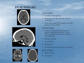
APPROACH TO CT HEAD
Gerrie Potgieter
Radiologist - I-MED
Associate lecturer - University of Notre Dame
INTRODUCTION
• As in clinical medicine (inspection, palpation,
percussion, auscultation) it is important to have a
systematic approach to any imaging.
• Today we will review a suggested approach to a
CT Head.
• This is my proposed system for non-radiology
clinicians.
INTRODUCTION
• All doctors should have a basic approach to interpreting a
CT Head.
• I am very certain that every person sitting here today will
have to review an urgent CT Head at some stage in their
career, due to a report not being available due to a technical
issue.
• We will review some basic anatomy and a suggested
approach.
• I am going to show some images and then discuss the
cases.
APPROACH TO CT BRAIN
Axial images:
• Cerebral hemispheres
• Ventricles (in this case
the lateral ventricles)
• Sulci (the small CSF
spaces over the surface
of the brain)
APPROACH TO CT HEAD
Axial images:
• Third ventricle (located
inferomedial to the
lateral ventricles)
APPROACH TO CT HEAD
Sagittal images:
• Corpus callosum (great
band of deeply situated
transverse white fibres
uniting the two the
cerebral hemispheres)
• Pituitary gland
• Cerebellum
• Brainstem,
• Visualised spinal
cord/cervical spine
APPROACH TO CT HEAD
Bone windows
• Base of skull Calvarium
CASE 1
• 65 year old male with sudden onset
right sided weakness.
LEFT MCA INFARCT
• There is loss of
grey-white
differentiation
in the left MCA
territory, subtle
oedema with
effacement of
the sulci in
keeping with
infarction.
• No evidence of
haemorrhagic
transformation.
Hyperdense MCA
sign, which is due to
thrombus.
CASE 2
• 47 year old male in high speed
motorcycle accident with severe head
injuries.
• GCS 7
TRAUMATIC BRAIN INJURY
Case courtesy
of Dr Derek
Smith,
Radiopaedia.
org, rID:
36667
Superficial
subarachnoid/extra
-axial haematoma
Intraparenchymal
haematoma with
surrounding
oedema/contusion
Scalp haematoma
Mild
midline shift
towards the
left
TRAUMATIC BRAIN INJURY
Case courtesy
of Dr Derek
Smth,
Radiopaedia.
org, rID:
36667
• The haematomas, contusions and other findings we see is the tip of the
iceberg.
• What is more important is the severe diffuse axonal injury (DAI).
DIFFUSE AXONAL INJURY
• DAI is the result of shearing forces, typically from rotational acceleration
or a deceleration.
• Due to the slightly different specific densities of white and grey matter,
shearing due to change in velocity has a predilection for axons at the
grey-white matter junction.
• It is important to know that these patients present with a low GCS in the
context of a severe head trauma and these CT Heads may look relatively
normal, with small haemorrhages only.
CASE 3
• 27 year old male
• Sudden onset severe headache in gym.
• High density/ acute
Intraventricular
haemorrhage
Subarachnoid haemorrhage
Subarachnoid
haemorrhage
Dilated temporal
horns, in keeping
with early
hydrocephalus.
Small
saccular/berry
aneurysm
Small
saccular/berry
aneurysm
CASE 4
• 28 year old boxer
• 4 week history of headaches.
BILATERAL SUBDURAL HAEMATOMAS
The change in
density is due to
the layering effect
from the blood
products.
The lateral
ventricles show
near complete
effacement.
CASE 5
• 71 year old male.
• 50 pack year smoking history.
• Presents with headaches and personality
changes.
METASTASIS
Case courtesy of Dr
David Cuete,
Radiopaedia.org,
rID: 22895
Right frontal
cortically based
enhancing lesion,
with surrounding
low density, in
keeping with a
metastasis.
CEREBRAL ABSCESS
Case courtesy of
A.Prof Frank Gaillard,
Radiopaedia.org, rID:
19468
Right posterior frontal rim
enhancing lesion
This is different from a metastasis
in that rim enhancement is usually
only seen in established infection.
In cerebral infection we usually get
four stages:
• early cerebritis
• late cerebritis
• early abscess/encapsulation - may
occur 10 days after infection
• late abscess/encapsulation - may
occur more than 14 days after
infection
IN SUMMARY
Axial images:
• Cerebral hemispheres
• Ventricles (in this case the lateral
ventricles)
• Sulci (the small CSF spaces over the
surface of the brain)
Sagittal images:
• Corpus callosum (great band of
deeply situated transverse white
fibres uniting the two the cerebral
hemispheres)
• Pituitary gland
• Cerebellum
• Brainstem,
• Visualised spinal cord/cervical spine
Bone windows
• Base of skull
• Calvarium
THANK YOU
References:
• Radiopaedia
• UpToDate

Blood or brain? Head CT updates

  • 1.
    APPROACH TO CTHEAD Gerrie Potgieter Radiologist - I-MED Associate lecturer - University of Notre Dame
  • 2.
    INTRODUCTION • As inclinical medicine (inspection, palpation, percussion, auscultation) it is important to have a systematic approach to any imaging. • Today we will review a suggested approach to a CT Head. • This is my proposed system for non-radiology clinicians.
  • 3.
    INTRODUCTION • All doctorsshould have a basic approach to interpreting a CT Head. • I am very certain that every person sitting here today will have to review an urgent CT Head at some stage in their career, due to a report not being available due to a technical issue. • We will review some basic anatomy and a suggested approach. • I am going to show some images and then discuss the cases.
  • 4.
    APPROACH TO CTBRAIN Axial images: • Cerebral hemispheres • Ventricles (in this case the lateral ventricles) • Sulci (the small CSF spaces over the surface of the brain)
  • 5.
    APPROACH TO CTHEAD Axial images: • Third ventricle (located inferomedial to the lateral ventricles)
  • 6.
    APPROACH TO CTHEAD Sagittal images: • Corpus callosum (great band of deeply situated transverse white fibres uniting the two the cerebral hemispheres) • Pituitary gland • Cerebellum • Brainstem, • Visualised spinal cord/cervical spine
  • 7.
    APPROACH TO CTHEAD Bone windows • Base of skull Calvarium
  • 8.
    CASE 1 • 65year old male with sudden onset right sided weakness.
  • 9.
    LEFT MCA INFARCT •There is loss of grey-white differentiation in the left MCA territory, subtle oedema with effacement of the sulci in keeping with infarction. • No evidence of haemorrhagic transformation.
  • 10.
    Hyperdense MCA sign, whichis due to thrombus.
  • 11.
    CASE 2 • 47year old male in high speed motorcycle accident with severe head injuries. • GCS 7
  • 12.
    TRAUMATIC BRAIN INJURY Casecourtesy of Dr Derek Smith, Radiopaedia. org, rID: 36667 Superficial subarachnoid/extra -axial haematoma Intraparenchymal haematoma with surrounding oedema/contusion Scalp haematoma Mild midline shift towards the left
  • 13.
    TRAUMATIC BRAIN INJURY Casecourtesy of Dr Derek Smth, Radiopaedia. org, rID: 36667 • The haematomas, contusions and other findings we see is the tip of the iceberg. • What is more important is the severe diffuse axonal injury (DAI).
  • 14.
    DIFFUSE AXONAL INJURY •DAI is the result of shearing forces, typically from rotational acceleration or a deceleration. • Due to the slightly different specific densities of white and grey matter, shearing due to change in velocity has a predilection for axons at the grey-white matter junction. • It is important to know that these patients present with a low GCS in the context of a severe head trauma and these CT Heads may look relatively normal, with small haemorrhages only.
  • 15.
    CASE 3 • 27year old male • Sudden onset severe headache in gym.
  • 16.
    • High density/acute Intraventricular haemorrhage Subarachnoid haemorrhage
  • 17.
    Subarachnoid haemorrhage Dilated temporal horns, inkeeping with early hydrocephalus.
  • 18.
  • 19.
  • 20.
    CASE 4 • 28year old boxer • 4 week history of headaches.
  • 21.
    BILATERAL SUBDURAL HAEMATOMAS Thechange in density is due to the layering effect from the blood products. The lateral ventricles show near complete effacement.
  • 22.
    CASE 5 • 71year old male. • 50 pack year smoking history. • Presents with headaches and personality changes.
  • 23.
    METASTASIS Case courtesy ofDr David Cuete, Radiopaedia.org, rID: 22895 Right frontal cortically based enhancing lesion, with surrounding low density, in keeping with a metastasis.
  • 24.
    CEREBRAL ABSCESS Case courtesyof A.Prof Frank Gaillard, Radiopaedia.org, rID: 19468 Right posterior frontal rim enhancing lesion This is different from a metastasis in that rim enhancement is usually only seen in established infection. In cerebral infection we usually get four stages: • early cerebritis • late cerebritis • early abscess/encapsulation - may occur 10 days after infection • late abscess/encapsulation - may occur more than 14 days after infection
  • 25.
    IN SUMMARY Axial images: •Cerebral hemispheres • Ventricles (in this case the lateral ventricles) • Sulci (the small CSF spaces over the surface of the brain) Sagittal images: • Corpus callosum (great band of deeply situated transverse white fibres uniting the two the cerebral hemispheres) • Pituitary gland • Cerebellum • Brainstem, • Visualised spinal cord/cervical spine Bone windows • Base of skull • Calvarium
  • 26.

Editor's Notes

  • #22 Who votes the abnormality is on the right? On the left? Both sides?