Download as PPSX, PPTX


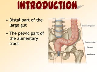
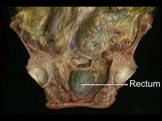
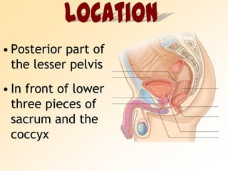
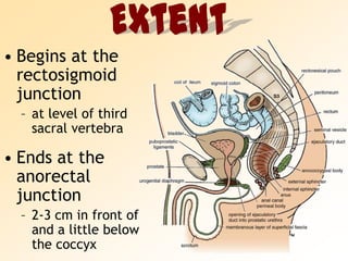

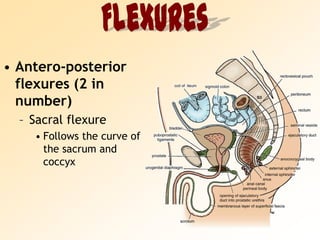
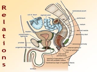
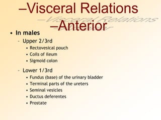

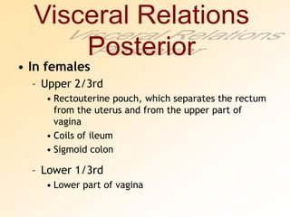
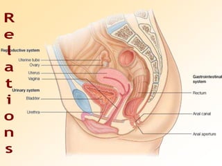
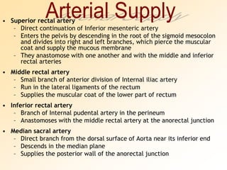
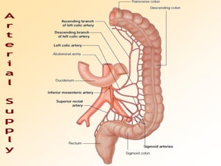
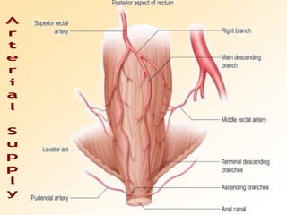
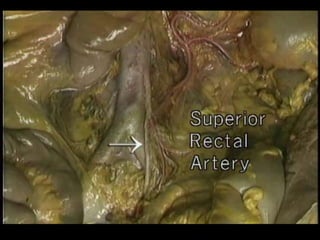
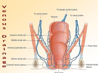

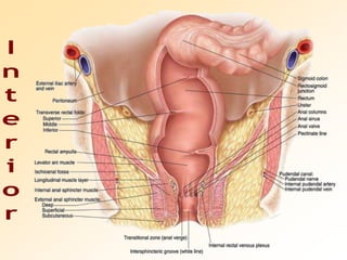
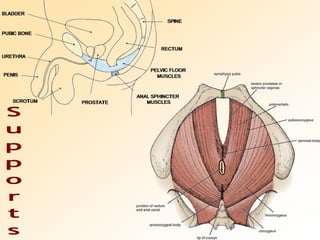
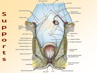


1) The rectum is the distal part of the large intestine located in the pelvic cavity. It extends from the rectosigmoid junction to the anal canal. 2) It has several flexures including anterior-posterior sacral and perineal flexures. It also has lateral flexures that correspond to transverse rectal folds. 3) The rectum receives its blood supply from the superior, middle, and inferior rectal arteries and drains into the superior, middle, and inferior rectal veins.


![CTEV [ clubfoot] DR ARUN LAL ,DR MOHAMED ASHRAF travancore medical college k...](https://cdn.slidesharecdn.com/ss_thumbnails/ctevclubfootdrarunlaldrmohamedashraftravancoremedicalcollegekollamkeralaindia-260208063247-18fc466c-thumbnail.jpg?width=640&height=640&fit=bounds)
