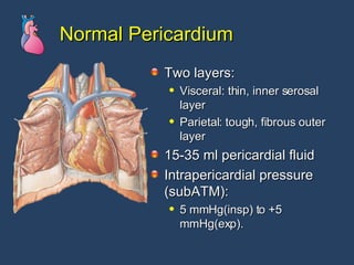
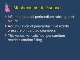
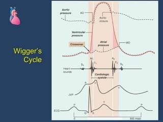
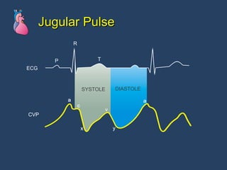
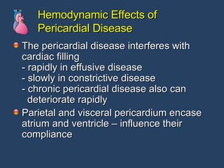
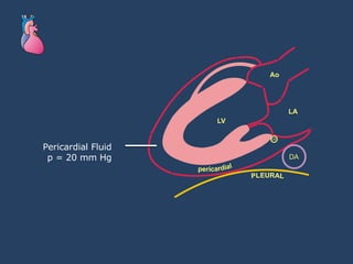
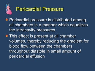
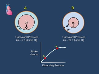
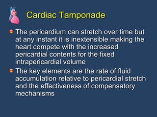
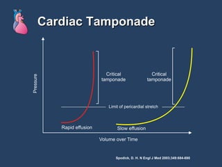
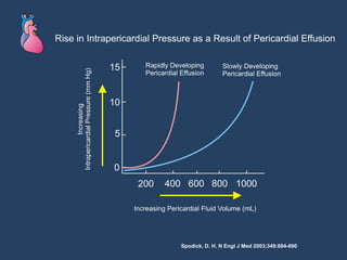
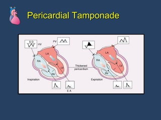
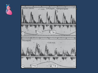
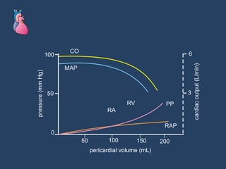
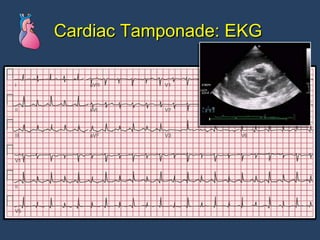
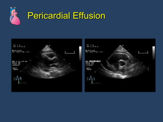
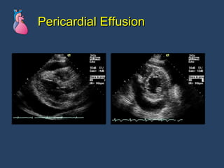
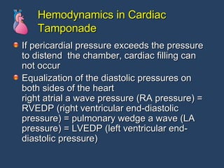
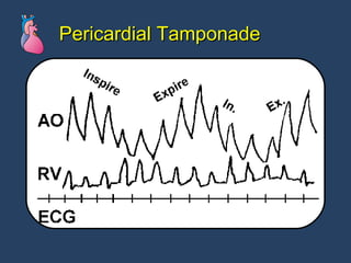
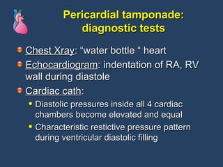
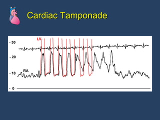
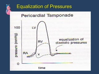
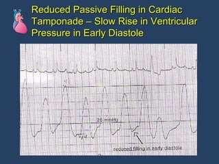
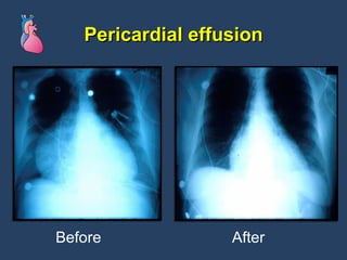
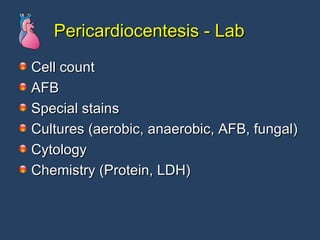
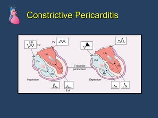
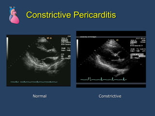
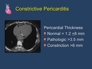
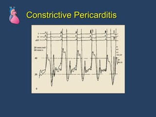
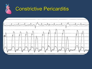
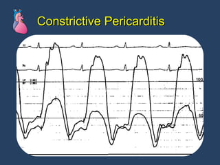
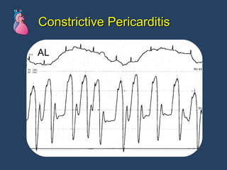
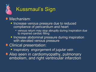
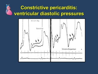
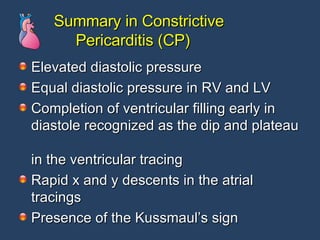
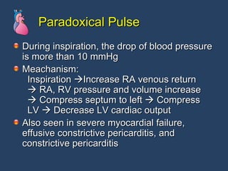
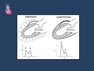
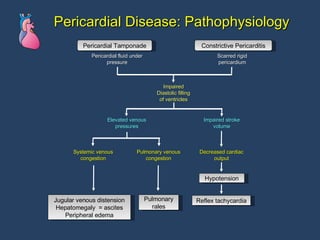
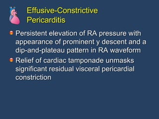
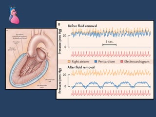
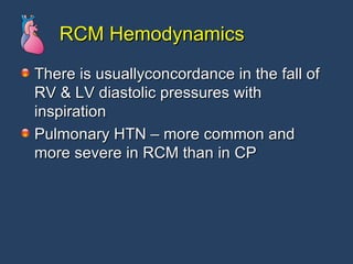
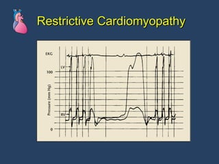
Pericardial Diseases
Normal Pericardium Two layers:  Visceral: thin, inner serosal layer Parietal: tough, fibrous outer layer 15-35 ml pericardial fluid Intrapericardial pressure (subATM):  5 mmHg(insp) to +5 mmHg(exp).
Mechanisms of Disease Inflamed parietal pericardium rubs against pleura. Accumulation of pericardial fluid exerts pressure on cardiac chambers Thickened, +/- calcified  pericardium restricts cardiac filling
Pericarditis
Pericarditis: Pathologic Types Serous Serofibrinous Purulent Hemorrhagic
Acute Pericarditis Infectious viral tuberculosis pyogenic bacterial
Acute Pericarditis Non-infectious Post-myocardial infarction Uremia Neoplastic disease Radiation induced Connective tissue diseases Drug induced
Acute Pericarditis: Clinical Findings Chest pain Pleuritic, positional, may mimic MI Fever, tachycardia, dyspnea Pericardial friction rub 3 component “scratchy” sound Abnormal ECG
Acute Pericarditis: EKG
Purulent Pericarditis Common seen in patients with empyema, mediastinitis, endocarditis, burn, and post- pericardiodectomy Diagnosis: ECG, echocardiography, Gallium67 scan with SPECT, Gallium67 and Tc99 scan  Primary purulent pericarditis is rare, even in immunocompromised host
Acute Pericarditis or Ischemia ? Pericarditis: fever, CPK and ESR elevation, pleuritic pain and friction rub, concave ST elevation in all leads except V1 and aVR, PR segment depression AMI or Prizmental’s angina: Convex ST elevation in regional leads, series evolutionary change in ECG, Q wave
Pericardial Effusion
Pericardial Effusion: Mechanisms Inflammatory Non-inflammatory: Increased capillary permeability Increased capillary pressure decreased plasma oncotic pressure lymphatic obstruction
Pericardial Effusions: Symptoms Dull chest pain Dysphagia Dyspnea Hoarseness Hiccups DOE Anorexia Fatigue
Pericardial Effusions: Signs Tachycardia,  Hypotension  Signs of shock JVD  venous waves lack the normal early diastolic y-descent.
Wigger’s Cycle
Jugular Pulse
Hemodynamic Effects of Pericardial Disease The pericardial disease interferes with cardiac filling - rapidly in effusive disease - slowly in constrictive disease - chronic pericardial disease also can    deteriorate rapidly Parietal and visceral pericardium encase atrium and ventricle – influence their compliance
Pathophysiology of Cardiac Tamponade
Pericardial Fluid p = 20 mm Hg
Pericardial Pressure Pericardial pressure is distributed among all chambers in a manner which equalizes the intracavity pressures This effect is present at all chamber volumes, thereby reducing the gradient for blood flow between the chambers throughout diastole in small amount of pericardial effusion
Transmural Pressure 25 – 5 = 20 mm Hg Transmural Pressure 25 – 20 = 5 mm Hg Stroke Volume Distending Pressure A B A B 25 5 25 20
Cardiac Tamponade The pericardium can stretch over time but at any instant it is inextensible making the heart compete with the increased pericardial contents for the fixed intrapericardial volume The key elements are the rate of fluid accumulation relative to pericardial stretch and the effectiveness of compensatory mechanisms
Cardiac Tamponade Spodick, D. H. N Engl J Med 2003;349:684-690
Spodick, D. H. N Engl J Med 2003;349:684-690
Pericardial Tamponade
Tamponade Doppler
 
Large Pericardial Effusion: Signs Soft heart sounds Reduced intensity of friction rub Ewart’s sign: Dullness, decreased breath sounds, and egophony over posterior L lung due to compression by large pericardial sac Electrical alternans on ECG
Pathophysio of Acute Cardiac Tamponade The Primary abnormality is rapid or slow compression of all cardiac chambers secondary to increased intrapericardial pressure. The pericardium can stretch over time but at any instant it is inextensible making the heart compete with the increased pericardial contents for the fixed intrapericardial volume. The key elements are the rate of fluid accumulation relative to pericardial stretch and the effectiveness of compensatory mechanisms.  i.e.. Cardiac rupture and stiff pericardium vs. inflammatory effusion with slow increase in size of effusion. The true filling pressure of the heart is the myocardial “transmural pressure” which is intracardiac pressure minus pericardial pressure. During inspiration and expiration, the right heart increases its filling at the expense of the left heart, so that its transmural pressure transiently improves and then reverts during expiration. In florid Tamponade this mechanism cannot compensate for the the reduced stroke volume.
Pericardial Effusion/Tamponade: Diagnostic Tests
Pericardial Effusion: CXR
Cardiac Tamponade: EKG
Pericardial Effusion
Hemodynamics in Cardiac Tamponade If pericardial pressure exceeds the pressure to distend  the chamber, cardiac filling can not occur Equalization of the diastolic pressures on both sides of the heart  right atrial a wave pressure (RA pressure) = RVEDP (right ventricular end-diastolic pressure) = pulmonary wedge a wave (LA pressure) = LVEDP (left ventricular end-diastolic pressure)
Pericardial Tamponade
Absence of y Descent Wave in Cardiac Tamponade Because of equalization of four chambers pressures, no blood flow crosses the atrio-ventricular valve in early diastole (passive ventricular filling, y descent) except a wave (atrial contraction)
Pericardial tamponade: diagnostic tests Chest Xray : “water bottle “ heart Echocardiogram : indentation of RA, RV wall during diastole Cardiac cath :  Diastolic pressures inside all 4 cardiac chambers become elevated and equal Characteristic restictive pressure pattern during ventricular diastolic filling
Cardiac Tamponade
Equalization of Pressures
Reduced Passive Filling in Cardiac Tamponade – Slow Rise in Ventricular Pressure in Early Diastole
Pulsus paradoxus Exaggerated (>10mmHg) cyclic decrease in systolic BP during normal inspiration Inspiration: increased venous return  increased RV volume.  Interventricular septum shifts left,  decreased LV volume  decreased stroke volume  systolic pressure falls.
Pericardial Effusion: Management Pericardiocentesis  Blindly in the case of an emergency With ECHO, fluoroscopy or CT guidance Pericardiotomy: If the heart cannot be reached by a needle/catheter. Indicated in patients with intrapericardial bleeding, clotted hemopericardium. Positive airway pressure should be avoided as it decreases cardiac output.
Diagnostic Consideration CXR: water bottle appearance  ECG: sinus tachycardia, electrical alternans,   T wave abnormality, low voltage in   ECG leads (tamponade)   ST elevation, PR segment depression   (acute pericarditis)  2D echocardiography: best noninvasive diagnostic tool in diagnosis pericardial effusion or tamponade CT or MRI: identify pericardial thickening
Treatment Options Medical treatment: fluid administration Pericardiocentesis Subxiphoid pericardiotomy Complete pericardium removal
Pericardiocentesis
Pericardiocentesis The main pericardial procedure performed in the catheterization laboratory is needle puncture and drainage of the pericardial fluid Diagnostic pericardiocentesis – to evaluate the etiology of pericarditis
Pericardiocentesis: Technique Spodick, D. H. N Engl J Med 2003;349:684-690
Pericardial effusion Before After
Pericardiocentesis - Lab Cell count AFB Special stains Cultures (aerobic, anaerobic, AFB, fungal) Cytology Chemistry (Protein, LDH)
Pericardiocentesis - Lab The diagnostic yield may be increased by retrieval of pericardial tissue by a surgical pericardial biopsy performed via thoracotomy, subxiphoid incision, or thoracoscopy.
Pericardiocentesis: Complication Most likely to be uncomplicated if both anterior and posterior echo-free spaces of at least 10 mm Increased risk of injury in smaller effusion In pts on warfarin, the procedure may be deferred until INR is within normal level; if urgent administration of FFP
Nonhemodynamic Considerations Anticoagulation Management of effusion in renal failure Purulent pericarditis Pericardial effusion following cardiac surgery Acute pericarditis and ischemia
Pericardial Disease after Cardiac Surgery First few hours after surgery : hemopericar- dium or hemomediastinum leads to cardiac tamponade (> 60%) Several weeks after op: postpericardiectomy syndrome with fever, chest pain, and friction rub (10-20%) 6 weeks to years after op: constrictive  pericarditis ( 1%)
Summary in Cardiac Tamponade Elevated diastolic pressure Equal end-diastolic pressure in RV and LV Absence of ventricular filling early in  diastole Absent Y decent in the atrial tracings
Constrictive Pericarditis
Constrictive Pericarditis Symmetrical process in which scarring of both the parietal and visceral pericardial layers constains all cardiac chambers Fibrous thickening, adhesion, calcification of the pericardium.
Constrictive Pericarditis Etiologies Idiopathic -most common cause viral TB Mediastinal radiation therapy Post-surgical
Constrictive Pericarditis Should be considered in any patient with: unexplained jugular venous distension Systemic edema Hepatic congestion dyspnea
Constrictive Pericarditis Should also be considered in the post-op heart surgery patient who has: Unexplained tachycardia Low cardiac output Venous congestion in the first months of surgery
Constrictive Pericarditis: Clinical Findings Fatigue, hypotension, tachycardia Elevated JVP Kausmaul’s sign Pericardial “knock” Ascites, edema
Constrictive Pericarditis: Diagnostics
Constrictive Pericarditis
Constrictive Pericarditis Normal Constrictive
Constrictive Pericarditis Pericardial Thickness Normal = 1.2  + 8 mm Pathologic >3.5 mm Constriction >6 mm
Constrictive Pericarditis - EKG Usually shows reduced voltage and diffuse ST-T wave abnormalities that may be mistaken for ischemic CAD
Constrictive pericarditis: Catheterization & Angiography To confirm the presence of constrictive physiology and assess its severity before consideration of pericardiectomy Assist to differentiate pericardial disease from RCM To exclude major coexisting causes of RA HTN such as severe pulmonary HTN To exclude rare instances of localized constriction
Constrictive pericarditis: Diagnosis Cardiac cath:  Elevated, equalized diastolic pressures Restricted filling pattern in RV  (“dip and plateau”) Prominent “y” descent on RA pressure tracing
Constrictive Pericarditis
Hemodynamics in Constrictive Pericarditis During constriction, pericardium encases the heart like a box, and the heart can only distend to an certain extent then stops The rapid early diastolic filling and abrupt halt gives rise to the classic dip and plateau configuration In the atrial pressure tracing, rapid ventricular filling (passive atrial emptying) resulting in a rapid Y descent with a nadir and  sharp rise in atrial pressure as the ventricle can not expand further
Hemodynamics in Constrictive Pericarditis Similarly, following atrial systole the fall in atrial pressure, or x descent is rapid, with a quick rise in atrial pressure --- M shape in right atrial tracing Because the overall volume of pericardium is fixed, it will result in identical LVEDP and RVEDP once the limitation of chamber enlargement are met
Constrictive Pericarditis
Constrictive Pericarditis Normal
Constrictive Pericarditis  Normal
Constrictive Pericarditis
Kussmaul’s Sign Mechanism: Increase venous pressure due to reduced  compliance of pericardium and heart  venous return may stop abruptly during inspiration due to impaired cardiac filling  Increase abdominal pressure during inspiration  with elevated venous pressure  Clinical presentation: inspiratory  engorgement of jugular vein  Also seen in cardiomyopathy, pulmonary embolism, and right ventricular infarction
Constrictive pericarditis: ventricular diastolic pressures
CP: Treatment Purely palliative Surgical pericardiectomy
Summary in Constrictive    Pericarditis (CP) Elevated diastolic pressure Equal diastolic pressure in RV and LV Completion of ventricular filling early in diastole recognized as the dip and plateau  in the ventricular tracing Rapid x and y descents in the atrial tracings Presence of the Kussmaul’s sign
Paradoxical Pulse During inspiration, the drop of blood pressure is more than 10 mmHg  Meachanism:   Inspiration   Increase RA venous return     RA, RV pressure and volume increase     Compress septum to left    Compress   LV    Decrease LV cardiac output  Also seen in severe myocardial failure, effusive constrictive pericarditis, and  constrictive pericarditis
 
Pericardial Disease: Pathophysiology Pericardial Tamponade Constrictive Pericarditis Jugular venous distension Hepatomegaly  = ascites Peripheral edema Pulmonary rales Reflex tachycardia Hypotension Pericardial fluid under pressure Scarred rigid pericardium Impaired Diastolic filling of ventricles Elevated venous pressures Impaired stroke volume Systemic venous congestion Pulmonary venous congestion Decreased cardiac output
Effusive-Constrictive Pericarditis
Effusive-Constrictive Pericarditis Persistent elevation of RA pressure with appearance of prominent y descent and a dip-and-plateau pattern in RA waveform Relief of cardiac tamponade unmasks significant residual visceral pericardial constriction
 
Effusive-Constrictive Pericarditis Diastolic pressures remain equalized between L & R heart after pericardiocentesis Tx: requires extensive visceral and parietal pericardiectomy Causes: idiopathic, CA, radiation, rheumatoid arthritis, TB
Restrictive Cardiomyopathy
Restrictive Cardiomyopathy Results from pathologic infiltration of the myocardium by a variety of processes  amyloidosis and sarcoidosis glycogen storage diseases, hemochromatosis, endomyocardial fibrosis, and hypereosinophilic syndromes  Results in abnormal diastolic ventricular filling and varying degrees of systolic dysfunction
Restrictive Cardiomyopathy Differentiation between CP and RCM is often difficult but important because on CP can be treated with pericardiectomy. Both disorders manifest impaired diastolic filing and elevated diastolic pressures with Sx of CHF
Restrictive Cardiomyopathy Differentiation from constrictive pericarditis may be difficult because of similar clinical and hemodynamic presentations Clues from history, physical exam, ECG, echo, CT and MR scan, biopsy
Restrictive Cardiomyopathy amyloidosis is most likely to simulate constrictive pericarditis  Digoxin should be avoided in patients with cardiac amyloidosis because of enhanced susceptibility to digoxin toxicity No therapy is known to be effective in reversing the progression of cardiac amyloidosis
Restrictive Cardiomyopathy Echocardiography may reveal thickening of the myocardium and varying degrees of systolic ventricular dysfunction.  Doppler echocardiographic analysis may demonstrate evidence of abnormal diastolic filling patterns and elevated venous pressure The ECG may show conduction system disease or low voltage, in contrast to the increased voltage seen with ventricular hypertrophy
Restrictive Cardiomyopathy: Echocardiographic Features Absence of pericardial adhesion and thickening  Left ventricular mass that is normal or increased; myocardial reflectance increased  Moderate to severe biatrial enlargement  Frequent AV valve regurgitation  Signs of pulmonary hypertension  AV valve excursion on M-mode unaffected by respiration
RCM Hemodynamics RA pressure usually elevated Prominent y descent followed by a rapid rise  with an M or W pattern as is CP Respiratory variation in RA pressure may be lacking Diastolic pressures in LV & RV may be elevated but LVDP > RVDP
RCM Hemodynamics There is usuallyconcordance in the fall of RV & LV diastolic pressures with inspiration Pulmonary HTN – more common and more severe in RCM than in CP
Restrictive Cardiomyopathy
 

Pericardial Dse Cath Lab

  • 1.
  • 2.
    Normal Pericardium Twolayers: Visceral: thin, inner serosal layer Parietal: tough, fibrous outer layer 15-35 ml pericardial fluid Intrapericardial pressure (subATM): 5 mmHg(insp) to +5 mmHg(exp).
  • 3.
    Mechanisms of DiseaseInflamed parietal pericardium rubs against pleura. Accumulation of pericardial fluid exerts pressure on cardiac chambers Thickened, +/- calcified pericardium restricts cardiac filling
  • 4.
  • 5.
    Pericarditis: Pathologic TypesSerous Serofibrinous Purulent Hemorrhagic
  • 6.
    Acute Pericarditis Infectiousviral tuberculosis pyogenic bacterial
  • 7.
    Acute Pericarditis Non-infectiousPost-myocardial infarction Uremia Neoplastic disease Radiation induced Connective tissue diseases Drug induced
  • 8.
    Acute Pericarditis: ClinicalFindings Chest pain Pleuritic, positional, may mimic MI Fever, tachycardia, dyspnea Pericardial friction rub 3 component “scratchy” sound Abnormal ECG
  • 9.
  • 10.
    Purulent Pericarditis Commonseen in patients with empyema, mediastinitis, endocarditis, burn, and post- pericardiodectomy Diagnosis: ECG, echocardiography, Gallium67 scan with SPECT, Gallium67 and Tc99 scan Primary purulent pericarditis is rare, even in immunocompromised host
  • 11.
    Acute Pericarditis orIschemia ? Pericarditis: fever, CPK and ESR elevation, pleuritic pain and friction rub, concave ST elevation in all leads except V1 and aVR, PR segment depression AMI or Prizmental’s angina: Convex ST elevation in regional leads, series evolutionary change in ECG, Q wave
  • 12.
  • 13.
    Pericardial Effusion: MechanismsInflammatory Non-inflammatory: Increased capillary permeability Increased capillary pressure decreased plasma oncotic pressure lymphatic obstruction
  • 14.
    Pericardial Effusions: SymptomsDull chest pain Dysphagia Dyspnea Hoarseness Hiccups DOE Anorexia Fatigue
  • 15.
    Pericardial Effusions: SignsTachycardia, Hypotension Signs of shock JVD venous waves lack the normal early diastolic y-descent.
  • 16.
  • 17.
  • 18.
    Hemodynamic Effects ofPericardial Disease The pericardial disease interferes with cardiac filling - rapidly in effusive disease - slowly in constrictive disease - chronic pericardial disease also can deteriorate rapidly Parietal and visceral pericardium encase atrium and ventricle – influence their compliance
  • 19.
  • 20.
  • 21.
    Pericardial Pressure Pericardialpressure is distributed among all chambers in a manner which equalizes the intracavity pressures This effect is present at all chamber volumes, thereby reducing the gradient for blood flow between the chambers throughout diastole in small amount of pericardial effusion
  • 22.
    Transmural Pressure 25– 5 = 20 mm Hg Transmural Pressure 25 – 20 = 5 mm Hg Stroke Volume Distending Pressure A B A B 25 5 25 20
  • 23.
    Cardiac Tamponade Thepericardium can stretch over time but at any instant it is inextensible making the heart compete with the increased pericardial contents for the fixed intrapericardial volume The key elements are the rate of fluid accumulation relative to pericardial stretch and the effectiveness of compensatory mechanisms
  • 24.
    Cardiac Tamponade Spodick,D. H. N Engl J Med 2003;349:684-690
  • 25.
    Spodick, D. H.N Engl J Med 2003;349:684-690
  • 26.
  • 27.
  • 28.
  • 29.
    Large Pericardial Effusion:Signs Soft heart sounds Reduced intensity of friction rub Ewart’s sign: Dullness, decreased breath sounds, and egophony over posterior L lung due to compression by large pericardial sac Electrical alternans on ECG
  • 30.
    Pathophysio of AcuteCardiac Tamponade The Primary abnormality is rapid or slow compression of all cardiac chambers secondary to increased intrapericardial pressure. The pericardium can stretch over time but at any instant it is inextensible making the heart compete with the increased pericardial contents for the fixed intrapericardial volume. The key elements are the rate of fluid accumulation relative to pericardial stretch and the effectiveness of compensatory mechanisms. i.e.. Cardiac rupture and stiff pericardium vs. inflammatory effusion with slow increase in size of effusion. The true filling pressure of the heart is the myocardial “transmural pressure” which is intracardiac pressure minus pericardial pressure. During inspiration and expiration, the right heart increases its filling at the expense of the left heart, so that its transmural pressure transiently improves and then reverts during expiration. In florid Tamponade this mechanism cannot compensate for the the reduced stroke volume.
  • 31.
  • 32.
  • 33.
  • 34.
  • 35.
  • 36.
    Hemodynamics in CardiacTamponade If pericardial pressure exceeds the pressure to distend the chamber, cardiac filling can not occur Equalization of the diastolic pressures on both sides of the heart right atrial a wave pressure (RA pressure) = RVEDP (right ventricular end-diastolic pressure) = pulmonary wedge a wave (LA pressure) = LVEDP (left ventricular end-diastolic pressure)
  • 37.
  • 38.
    Absence of yDescent Wave in Cardiac Tamponade Because of equalization of four chambers pressures, no blood flow crosses the atrio-ventricular valve in early diastole (passive ventricular filling, y descent) except a wave (atrial contraction)
  • 39.
    Pericardial tamponade: diagnostictests Chest Xray : “water bottle “ heart Echocardiogram : indentation of RA, RV wall during diastole Cardiac cath : Diastolic pressures inside all 4 cardiac chambers become elevated and equal Characteristic restictive pressure pattern during ventricular diastolic filling
  • 40.
  • 41.
  • 42.
    Reduced Passive Fillingin Cardiac Tamponade – Slow Rise in Ventricular Pressure in Early Diastole
  • 43.
    Pulsus paradoxus Exaggerated(>10mmHg) cyclic decrease in systolic BP during normal inspiration Inspiration: increased venous return increased RV volume. Interventricular septum shifts left, decreased LV volume decreased stroke volume systolic pressure falls.
  • 44.
    Pericardial Effusion: ManagementPericardiocentesis Blindly in the case of an emergency With ECHO, fluoroscopy or CT guidance Pericardiotomy: If the heart cannot be reached by a needle/catheter. Indicated in patients with intrapericardial bleeding, clotted hemopericardium. Positive airway pressure should be avoided as it decreases cardiac output.
  • 45.
    Diagnostic Consideration CXR:water bottle appearance ECG: sinus tachycardia, electrical alternans, T wave abnormality, low voltage in ECG leads (tamponade) ST elevation, PR segment depression (acute pericarditis) 2D echocardiography: best noninvasive diagnostic tool in diagnosis pericardial effusion or tamponade CT or MRI: identify pericardial thickening
  • 46.
    Treatment Options Medicaltreatment: fluid administration Pericardiocentesis Subxiphoid pericardiotomy Complete pericardium removal
  • 47.
  • 48.
    Pericardiocentesis The mainpericardial procedure performed in the catheterization laboratory is needle puncture and drainage of the pericardial fluid Diagnostic pericardiocentesis – to evaluate the etiology of pericarditis
  • 49.
    Pericardiocentesis: Technique Spodick,D. H. N Engl J Med 2003;349:684-690
  • 50.
  • 51.
    Pericardiocentesis - LabCell count AFB Special stains Cultures (aerobic, anaerobic, AFB, fungal) Cytology Chemistry (Protein, LDH)
  • 52.
    Pericardiocentesis - LabThe diagnostic yield may be increased by retrieval of pericardial tissue by a surgical pericardial biopsy performed via thoracotomy, subxiphoid incision, or thoracoscopy.
  • 53.
    Pericardiocentesis: Complication Mostlikely to be uncomplicated if both anterior and posterior echo-free spaces of at least 10 mm Increased risk of injury in smaller effusion In pts on warfarin, the procedure may be deferred until INR is within normal level; if urgent administration of FFP
  • 54.
    Nonhemodynamic Considerations AnticoagulationManagement of effusion in renal failure Purulent pericarditis Pericardial effusion following cardiac surgery Acute pericarditis and ischemia
  • 55.
    Pericardial Disease afterCardiac Surgery First few hours after surgery : hemopericar- dium or hemomediastinum leads to cardiac tamponade (> 60%) Several weeks after op: postpericardiectomy syndrome with fever, chest pain, and friction rub (10-20%) 6 weeks to years after op: constrictive pericarditis ( 1%)
  • 56.
    Summary in CardiacTamponade Elevated diastolic pressure Equal end-diastolic pressure in RV and LV Absence of ventricular filling early in diastole Absent Y decent in the atrial tracings
  • 57.
  • 58.
    Constrictive Pericarditis Symmetricalprocess in which scarring of both the parietal and visceral pericardial layers constains all cardiac chambers Fibrous thickening, adhesion, calcification of the pericardium.
  • 59.
    Constrictive Pericarditis EtiologiesIdiopathic -most common cause viral TB Mediastinal radiation therapy Post-surgical
  • 60.
    Constrictive Pericarditis Shouldbe considered in any patient with: unexplained jugular venous distension Systemic edema Hepatic congestion dyspnea
  • 61.
    Constrictive Pericarditis Shouldalso be considered in the post-op heart surgery patient who has: Unexplained tachycardia Low cardiac output Venous congestion in the first months of surgery
  • 62.
    Constrictive Pericarditis: ClinicalFindings Fatigue, hypotension, tachycardia Elevated JVP Kausmaul’s sign Pericardial “knock” Ascites, edema
  • 63.
  • 64.
  • 65.
  • 66.
  • 67.
    Constrictive Pericarditis PericardialThickness Normal = 1.2 + 8 mm Pathologic >3.5 mm Constriction >6 mm
  • 68.
    Constrictive Pericarditis -EKG Usually shows reduced voltage and diffuse ST-T wave abnormalities that may be mistaken for ischemic CAD
  • 69.
    Constrictive pericarditis: Catheterization& Angiography To confirm the presence of constrictive physiology and assess its severity before consideration of pericardiectomy Assist to differentiate pericardial disease from RCM To exclude major coexisting causes of RA HTN such as severe pulmonary HTN To exclude rare instances of localized constriction
  • 70.
    Constrictive pericarditis: DiagnosisCardiac cath: Elevated, equalized diastolic pressures Restricted filling pattern in RV (“dip and plateau”) Prominent “y” descent on RA pressure tracing
  • 71.
  • 72.
    Hemodynamics in ConstrictivePericarditis During constriction, pericardium encases the heart like a box, and the heart can only distend to an certain extent then stops The rapid early diastolic filling and abrupt halt gives rise to the classic dip and plateau configuration In the atrial pressure tracing, rapid ventricular filling (passive atrial emptying) resulting in a rapid Y descent with a nadir and sharp rise in atrial pressure as the ventricle can not expand further
  • 73.
    Hemodynamics in ConstrictivePericarditis Similarly, following atrial systole the fall in atrial pressure, or x descent is rapid, with a quick rise in atrial pressure --- M shape in right atrial tracing Because the overall volume of pericardium is fixed, it will result in identical LVEDP and RVEDP once the limitation of chamber enlargement are met
  • 74.
  • 75.
  • 76.
  • 77.
  • 78.
  • 79.
  • 80.
  • 81.
    Kussmaul’s Sign Mechanism:Increase venous pressure due to reduced compliance of pericardium and heart venous return may stop abruptly during inspiration due to impaired cardiac filling Increase abdominal pressure during inspiration with elevated venous pressure Clinical presentation: inspiratory engorgement of jugular vein Also seen in cardiomyopathy, pulmonary embolism, and right ventricular infarction
  • 82.
  • 83.
    CP: Treatment Purelypalliative Surgical pericardiectomy
  • 84.
    Summary in Constrictive Pericarditis (CP) Elevated diastolic pressure Equal diastolic pressure in RV and LV Completion of ventricular filling early in diastole recognized as the dip and plateau in the ventricular tracing Rapid x and y descents in the atrial tracings Presence of the Kussmaul’s sign
  • 85.
    Paradoxical Pulse Duringinspiration, the drop of blood pressure is more than 10 mmHg Meachanism: Inspiration  Increase RA venous return  RA, RV pressure and volume increase  Compress septum to left  Compress LV  Decrease LV cardiac output Also seen in severe myocardial failure, effusive constrictive pericarditis, and constrictive pericarditis
  • 86.
  • 87.
    Pericardial Disease: PathophysiologyPericardial Tamponade Constrictive Pericarditis Jugular venous distension Hepatomegaly = ascites Peripheral edema Pulmonary rales Reflex tachycardia Hypotension Pericardial fluid under pressure Scarred rigid pericardium Impaired Diastolic filling of ventricles Elevated venous pressures Impaired stroke volume Systemic venous congestion Pulmonary venous congestion Decreased cardiac output
  • 88.
  • 89.
    Effusive-Constrictive Pericarditis Persistentelevation of RA pressure with appearance of prominent y descent and a dip-and-plateau pattern in RA waveform Relief of cardiac tamponade unmasks significant residual visceral pericardial constriction
  • 90.
  • 91.
    Effusive-Constrictive Pericarditis Diastolicpressures remain equalized between L & R heart after pericardiocentesis Tx: requires extensive visceral and parietal pericardiectomy Causes: idiopathic, CA, radiation, rheumatoid arthritis, TB
  • 92.
  • 93.
    Restrictive Cardiomyopathy Resultsfrom pathologic infiltration of the myocardium by a variety of processes amyloidosis and sarcoidosis glycogen storage diseases, hemochromatosis, endomyocardial fibrosis, and hypereosinophilic syndromes Results in abnormal diastolic ventricular filling and varying degrees of systolic dysfunction
  • 94.
    Restrictive Cardiomyopathy Differentiationbetween CP and RCM is often difficult but important because on CP can be treated with pericardiectomy. Both disorders manifest impaired diastolic filing and elevated diastolic pressures with Sx of CHF
  • 95.
    Restrictive Cardiomyopathy Differentiationfrom constrictive pericarditis may be difficult because of similar clinical and hemodynamic presentations Clues from history, physical exam, ECG, echo, CT and MR scan, biopsy
  • 96.
    Restrictive Cardiomyopathy amyloidosisis most likely to simulate constrictive pericarditis Digoxin should be avoided in patients with cardiac amyloidosis because of enhanced susceptibility to digoxin toxicity No therapy is known to be effective in reversing the progression of cardiac amyloidosis
  • 97.
    Restrictive Cardiomyopathy Echocardiographymay reveal thickening of the myocardium and varying degrees of systolic ventricular dysfunction. Doppler echocardiographic analysis may demonstrate evidence of abnormal diastolic filling patterns and elevated venous pressure The ECG may show conduction system disease or low voltage, in contrast to the increased voltage seen with ventricular hypertrophy
  • 98.
    Restrictive Cardiomyopathy: EchocardiographicFeatures Absence of pericardial adhesion and thickening Left ventricular mass that is normal or increased; myocardial reflectance increased Moderate to severe biatrial enlargement Frequent AV valve regurgitation Signs of pulmonary hypertension AV valve excursion on M-mode unaffected by respiration
  • 99.
    RCM Hemodynamics RApressure usually elevated Prominent y descent followed by a rapid rise with an M or W pattern as is CP Respiratory variation in RA pressure may be lacking Diastolic pressures in LV & RV may be elevated but LVDP > RVDP
  • 100.
    RCM Hemodynamics Thereis usuallyconcordance in the fall of RV & LV diastolic pressures with inspiration Pulmonary HTN – more common and more severe in RCM than in CP
  • 101.
  • 102.

Editor's Notes

  • #2 Topic Outline Overview Pericarditis Pericardial effusion Tamponade Pericardiocentesis Constrictive pericarditis Restrictive cardiomyopathy