
Recommended
More Related Content
What's hot
What's hot (20)
Similar to Structure Of DNA & Recent advances in genomes
Similar to Structure Of DNA & Recent advances in genomes (20)
More from habibullah181
More from habibullah181 (20)
Recently uploaded
Recently uploaded (20)
Structure Of DNA & Recent advances in genomes
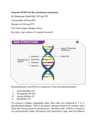
- 1. Structure Of DNA & Recentadvances in genomes By Muhammad HabibUllah 2015-ag-7783 Farooq Zahid 2016-ag-9907 Husnain Ali 2016-ag-5273 UAF Sub Campus Depalpur Okara Bsc Hons. Agri sciences 4th semester Section B Deoxyribonucleic acid (DNA) is composed of four deoxyribonucleotides, i. deoxyadenylate (A), ii. deoxyguany late (G), iii. deoxycytidylate (C) iv. thymidylate (T) To construct a lengthy polypeptide chain, these units are connected by 3′ to 5′ phosphodiesterlinkages. DNA is the genetic substancefound in all creatures, and it binds with histone protein in chromosomes. The helical helix of DNA is formed by two polynucleotide chains. Phosphoric acid, deoxyribose sugar, and four different
- 2. forms ofnitrogenous bases are the major chemical components ofDNA. Phosphoric acid forms an ester link with sugar, a glycosidic connection between sugar and base, and a hydrogen bond between bases. These are the fundamental elements that make up the entire DNA structure. The importance of the nucleotide sequence of DNA cannot be overstated. The genetic information is encoded in a certain base sequence; if the base is changed, the information is changed as well. F. Misher separated DNA from nucleous in 1869, and Altman coined the term nucleic acid in 1889. The suitable DNA double helix model provided by Watson and Crick can explain the gene's specificity.
- 3. DNA's chemical Composition is as follows: There are three primary components in deoxyribonucleic acid. 1. Phosphoric acid (H3PO4) 2. Pentosesugar 3. Nitrogenous Base 1. Phosphoric acid, like phosphate, is found in nature and forms the backboneof DNA molecules, with sugar molecules. It connects the nucleotides byforming an ester phosphatebond between the deoxyribosesugars of two neighbouring nucleotides. This bond connects the 3prime carbon in one nucleotide to the 5prime carbon in the following nucleotide.
- 4. 2. Deoxyribose sugar is a pentose sugar with the molecular formula C5H10O4. Pentosesugar has a pentagonal ring structure with 3 and 5 prime carbonatoms attached to phosphoric acid and 1 prime carbon atom attached to the base. 3. There are two types of nitrogenous base. 1. Purine 2. pyrimidine it is completely fabricated of adenine and guanine with two heterocyclic rings C,H,O, and N atoms.
- 5. The 1prime C of the ribose sugar joins the base through a glycosidic bond in DNA, and this molecule is known as nucleoside. The complete compound is called a nucleotide when 5prime C is connected to phosphoric acid by an ester bond. A strand is formed when two deoxyribonucleotides are linked by a phosphodiester link. DNA contains two strands becauseit is a double helix. The other strand runs in the opposite or antiparallel direction, i.e., if one strand runs from 3 prime to 5 prime end forward, the other runs from 5 prime to 3 prime end backward. A hydrogen connection between two nitrogenous bases connects the two strands. Adenine always creates two bonds with thymine, while cytosine always creates three bonds with the guanine base. NUCLEOSIDE NUCLEOTIDE
- 6. Double Helix structure of DNA The ‘backbones' of DNA molecules are made up of sugar and phosphates in an alternating pattern. The rungs of the ladder are made up of hydrogen-bonded bases. Recent advances in genomes
- 7. There are many recent advances are covered in the genomes. Some important advances are discussed here…. 1. PassengerMutations in More Than 2,500 Cancer Genomes:Overall MolecularFunctionalImpact and Consequences In comparison to a homogeneous background expectation, the percentage of genes in different gene categories influenced by driver (grey band) and probable passenger (faded yellow band) LoFs (dashed black line). Different tumor kinds are represented by the data points in the boxplot. A Kolmogorov Smirnov (KS) testwas used to determine statistical significance between these percentages for each of the cancer cohorts. In comparison to a uniform genomic backdrop, heat map demonstrating enrichment (red color) and depletion (blue color) of motif gain (upper panel) and loss (bottom panel) events induced by putative passenger mutations for distinct TFs. The red-hued TFs are well-known cancer genes. The statistical significance values for several TFs have been given. Gain (positive alteration bias) and loss (negative alteration bias) of motif events found across ETS TF-regulated target genes (on the x axis). The green triangle represents pan-cancer alteration bias, while the coloured circles represent alteration bias for distinct cancer cohorts. The frequency of motif- altering occurrences is shown by the size of the circles. Genes that are differently expressed due to gain of motif events in TFs belonging to the ETS TF family are shown in a Q-Q plot. (E) The presence of significant deletions in the germline and somatic tissues that can engulf or partially erase coding areas and TF binding peaks.
- 8. 2. A cancer rainbow mouse for visualizing the functional genomics of oncogenic clonal expansion Field cancretization is a premalignant process characterized by the distribution of clones of oncogenic mutations throughout the epithelium. The processes underlying the rapid spread of oncogenic clones are unknown, and the timelines ofintestinal field cancretization can be diverse. Forfluorescently barcoding somatic mutations and immediately observing clonal proliferation and spread of oncogenes, we use a Cancer rainbow (Crainbow) modelling system. Crainbow found that ß-catenin (Ctnnb1) mutations in intestinal stem cells causeextensive oncogeneproliferation during perinatal development but not in adults. Mutations that damage the stem cell microenvironment extrinsically, on the other hand, can quickly propagate through the adult gut.