
Recommended
More Related Content
What's hot
What's hot (20)
Similar to TRIPPLE HELIX DNA
Similar to TRIPPLE HELIX DNA (20)
Recently uploaded
Recently uploaded (20)
TRIPPLE HELIX DNA
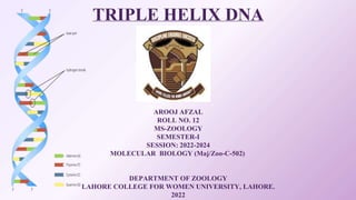
- 1. TRIPLE HELIX DNA BY AROOJ AFZAL ROLL NO. 12 MS-ZOOLOGY SEMESTER-I SESSION: 2022-2024 MOLECULAR BIOLOGY (Maj/Zoo-C-502) DEPARTMENT OF ZOOLOGY LAHORE COLLEGE FOR WOMEN UNIVERSITY, LAHORE. 2022
- 3. 01 Classification of DNA Triple Helices TABLE OF CONTENTS 03 Potential Applications of DNA Triplex Formation in Therapeutics 02 04 Reference Structure
- 4. DISCOVERY
- 5. DISCOVERY • Friedrich Miescher discovered DNA in 1868. • It took more than 70 years to demonstrate that it is the molecule that carries genetic information. • Triple-helical nucleic acids were first described in 1957 by Felsenfeld and Rich.
- 6. STRUCTURE • Multi-stranded DNA • DNA can form multi-stranded helices through either folding of one of the two strands or association of two, three, or four strands of DNA. • A short mixed sequence triplex-forming oligonucleotide (TFO) formed a stable specific triple helical DNA complex • The third strand of DNA in the triplex structure (i.e. the TFO) follows a path through the major groove of the duplex DNA.
- 7. • Hoogsteen hydrogen bonds • Different from those formed in classical Watson-Crick base pairing in duplex DNA. • Because purines contain potential hydrogen bonds with incoming third strand bases, the binding of the third strand is to the purine-rich strand of the DNA duplex Figure 1: Triplex-forming sequences in the human c-MYC gene
- 9. Classification of DNA Triple Helices 1. Intermolecular triplexes 2. Intramolecular triplexes 1. Intermolecular Triplexes • Formed when the triplex-forming strand originates from a second DNA molecule Figure 2: Schematic representation of intermolecular DNA triplex formation
- 10. Role of Intermolecular Triplexes • Potential therapeutic application in inhibiting the expression of genes involved in cancer and other human diseases • For stimulating DNA repair and/or homologous recombination pathways • For targeting disease genes for in-activation • For interfering with DNA replication
- 11. Triplex Formation 1. Parallel 2. Anti-parallel 1. Parallel 2. Anti-Parallel Polypyrimidine third strands (Y) bind to the polypurine strand of the duplex DNA via Hoogsteen hydrogen bonding in a parallel fashion C+:G-C T:A-T triplets The polypurine third strand (R) binds in an antiparallel fashion to the purine strand of the duplex via reverse-Hoogsteen hydrogen bonds G:G-C A:A-T T:A-T C+ represents a protonated cytosine on the N3 position
- 12. Figure 3: Schematic representation of canonical base triplets formed in purine and pyrimidine triplex motifs Dotted lines: Watson-Crick base pairing Broken lines: Hoogsteen base pairing
- 13. 2. Intramolecular Triplexes (H-DNA) • Third strand is provided by one of the strands of the same duplex DNA molecule at a mirror repeat sequence Figure 4: H-DNA (Intramolecular Triplex DNA) Forms of Intramolecular Triplexes: I. H-DNA: When third strand of the triplex is rich in polypyrimidine II. *H-DNA: When third strand of the triplex is rich in polypurine
- 14. Factors Affecting Trplex DNA Length Base Composition Divalent cations Temperature
- 15. Potential Applications of DNA Triplex Formation in Therapeutics
- 16. Potential Applications of DNA Triplex Formation in Therapeutics 1. Targeting Genes as an Approach to Molecular-Targeted Therapeutics: • Ability to target specific genes to modulate their structure and function in the genome • Role in biology, biotechnology and medicine • TFOs represent near-ideal molecules for this purpose because of their ability to bind duplex DNA with high affinity and specificity. TFOs: Triplex-Forming Oligonucleotides Example: Target damage to specific sites in a genome
- 17. 2. Abundance of TFO Binding Sites in Mammalian Genomes : • The gene products involved in many important biological processes such as cell signaling, proliferation, and carcinogenesis have now been identified, providing plausible targets for gene modulation. • Triplex technology represents an approach to regulate these processes by manipulating the structure and/or function of these critical genes • To determine the number of potential TFO binding sites available in mammalian genomes, we designed an algorithm to search the entire human and mouse genomes for such sites, and were surprised to find~2 million in each of these mammalian genomes We found that most annotated genes in both the mouse and human genomes contain at least one unique TFO binding site, and these sites are enriched in the promoter and /or transcribed gene regions.
- 18. • The ability of TFOs to inhibit the expression of a number of genes in a variety of systems. • Mammalian genes that have been targeted by TFOs 3. Modulating Gene Expression via Triplex Formation: Limitations: • TFO stability once in the cells, lack of optimal target site binding affinity and specificity due to intracellular salt concentrations and pH. • Displacement by DNA metabolic activities (e.g. transcription, replication and repair) • Chromatin structure which may present a barrier to target site accessibility.
- 19. 4. Directing Site-Specific DNA Damage: • Another potential strategy for the development of TFOs as “therapeutic agents” is their utility as targeted DNA damaging agents. • This approach differs from TFO-directed transcription inhibition in that TFO-directed DNA damage has been shown to stimulate mutation, recombination, and DNA repair at the targeted sites • Thus, this application has the potential to directly inactivate genes, rather than transiently regulating gene expression Example: Triplex formation can be used to direct site-specific DNA damage and thereby induce DNA repair synthesis locally, independent of replication synthesis
- 20. Thanks!