Downloaded 19 times


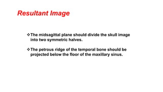


The document discusses the proper positioning for a Waters projection x-ray exam, including tilting the patient's head upward at a 37 degree angle with the canthomeatal line perpendicular to the image receptor. The central x-ray beam should be perpendicular to and centered on the image receptor in the area of the maxillary sinuses. A properly positioned Waters projection will show a symmetric skull image divided in half by the midsagittal plane and the petrous ridge of the temporal bone projected below the floor of the maxillary sinus.


