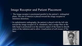
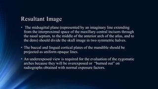
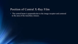
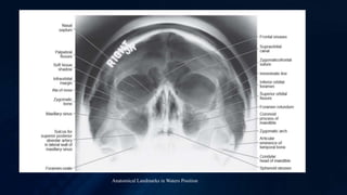
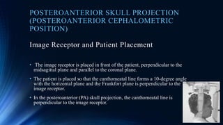
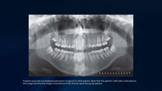
SlideShare a Scribd company logo
1 of 51
NEW HORIZON DENTAL COLLEGE AND RESEARCH INSTITUTE
DEPARTMENT OF ORAL MEDICINE, DIAGNOSIS, ORALAND
MAXILLOFACIAL RADIOLOGY
SEMINAR ON
EXTRAORAL IMAGING TECHNIQUES
SUBMITTED BY: GUIDED BY :
SARBAJIT HALDER DEPARTMENT OF ORAL
ROLL NO. : 15110067 MEDICINE, DIAGNOSIS, ORALAND
BATCH : 2017-2018 MAXILLOFACIAL RADIOLOGY
CONTENTS
ā€¢ Introduction
ā€¢ Technique
ā€¢ Lateral Skull Projection
ā€¢ Submentovertex Projection
ā€¢ Waters projection
ā€¢ Posteroanterior Skull Projection
ā€¢ Reverse-Towne Projection
ā€¢ Mandibular Oblique Lateral Projections
ā€¢ Panaromic Projection
ā€¢ TMJ Imaging
ā€¢ Conclusions
ā€¢ References
INTRODUCTION
ā€¢ In extraoral radiographic examinations both the x-ray source and
image receptor (ļ¬lm or electronic sensor) are placed outside the
patientā€™s mouth.
ā€¢ In most common extraoral radiographic examinations the source and
sensor remain static.
ā€¢ These include the lateral cephalometric projection of the sagittal or
median plane; the submentovertex projection of the transverse or
horizontal plane; the Waters, posteroanterior cephalometric, and
reverse-Towne projections of the coronal or frontal plane; and the
oblique lateral projections of the mandibular body and ramus.
TECHNIQUE
ā€¢ The ļ¬rst step in obtaining a radiograph is the selection of the appropriate projection
for the pertinent diagnostic task.
ā€¢ Extraoral radiographs are produced with conventional dental x-ray machines, certain
models of panoramic machines, or highercapacity medical x-ray units.
ā€¢ Cephalometric and skull views require at least a 20 Ɨ 25 cm (8 Ɨ 10 inch) image
receptor, whereas oblique lateral projections of the mandible can be obtained with a
13 Ɨ 18 cm (5 Ɨ 7 inch) image receptor.
ā€¢ It is critical to correctly and clearly label the right and left sides of the image. This
usually is done by placing a metal marker (an R or an L) on the outside of the
cassette in a corner in which the marker does not obstruct diagnostic information.
ā€¢ The proper exposure parameters depend on the patientā€™s size, anatomy, and head
orientation; image receptor speed; x-ray sourceto-receptor distance; and whether
grids are used.
ā€¢ In cases of known or suspected disease, medium- or high-speed rare-earth
screen-ļ¬ lm combinations provide optimal balance between diagnostic
information and patient exposure.
ā€¢ Proper positioning of the x-ray source, patient, and image receptor requires
patience, attention to detail, and experience.
ā€¢ The main anatomic landmark used in patient positioning during extraoral
radiography is the canthomeatal line.
ā€¢ This line joins the central point of the external auditory canal to the outer
canthus of the eye. The canthomeatal line forms approximately a 10-degree
angle with the Frankfort plane.
ā€¢ This line that connects the superior border of the external auditory canal with
the infraorbital rim.
LATERAL SKULL POSITION ( LATERAL
CEPHALOMETRIC POSITION)
ā€¢ The lateral cephalometric projection is by far the most commonly used in
dentistry.
ā€¢ All cephalometric radiographs, including the lateral view, are made with a
cephalostat that helps maintain a constant relationship among the skull, the ļ¬
lm, and the x-ray beam.
ā€¢ Skeletal, dental, and soft tissue anatomic landmarks delineate lines, planes,
angles, and distances that are used to generate measurements and to classify
patient craniofacial morphologic features.
ā€¢ At the beginning of treatment, these measurements are often compared with
an established standard; during treatment, the measurements are usually
compared with those from previous cephalometric radiographs of the same
patient to monitor growth and development as well as treatment.
Image Receptor and Patient Placement
ā€¢ The image receptor is positioned parallel to the patientā€™s midsagittal
plane. The site of interest is placed toward the image receptor to
minimize distortion.
ā€¢ In cephalometric radiography, the patient is placed with the left side
toward the image receptor (U.S. standards), and a wedge ļ¬lter at the
tube head is positioned over the anterior aspect of the beam to absorb
some of the radiation and to allow visualization of soft tissues of the
face.
Position of Central X-Ray Beam
ā€¢ The central beam is perpendicular to the midsagittal plane of the
patient and the plane of the image receptor and is centered over the
external auditory meatus.
Resultant Image
ā€¢ Exact superimposition of right and left sides is impossible because structures
on the side near the image receptor are magniļ¬ed less than the same
structures on the side far from the image receptor.
ā€¢ Bilateral structures close to the midsagittal plane demonstrate less
discrepancy in size compared with bilateral structures farther away from the
midsagittal plane.
ā€¢ Structures close to the midsagittal plane (e.g., the clinoid processes and
inferior turbinates) should be nearly superimposed.
ā€¢ There are many cephalometric analyses that are based on a variety of
anatomic landmarks. Steiner and Ricketts analyses are two commonly used
analyses that use the skeletal, dental, and soft tissue landmarks.
ā€¢ Precise identiļ¬cation of the various landmarks on the lateral cephalometric
radiograph is necessary to generate accurate cephalometric measurements.
Anatomic Landmarks Identified in lateral cephalometric projection.
Indications
ā€¢ To visualize the relationship between soft & hard tissues of face.
ā€¢ To assess the position of the teeth in relation to each other and hard
tissues and adjacent structures.
ā€¢ To assess the skeletal pattern.
SUBMENTOVERTEX (BASE PROJECTION)
Image Receptor and Patient Placement
ā€¢ The image receptor is positioned parallel to patientā€™s transverse plane
and perpendicular to the midsagittal and coronal planes.
ā€¢ To achieve this, the patientā€™s neck is extended as far backward as
possible, with the canthomeatal line forming a 10-degree angle with
the image receptor.
Position of Central X-ray Film
ā€¢ The central beam is perpendicular to the image receptor, directed from
below the mandible toward the vertex of the skull (hence the name
submentovertex, or SMV), and centered about 2 c m anterior to a line
connecting the right and left condyles.
Resultant Image
ā€¢ The midsagittal plane (represented by an imaginary line extending
from the interproximal space of the maxillary central incisors through
the nasal septum, to the middle of the anterior arch of the atlas, and to
the dens) should divide the skull image in two symmetric halves.
ā€¢ The buccal and lingual cortical plates of the mandible should be
projected as uniform opaque lines.
ā€¢ An underexposed view is required for the evaluation of the zygomatic
arches because they will be overexposed or ā€œburned outā€ on
radiographs obtained with normal exposure factors.
A, Anatomic landmarks identified in the submentovertex projection. B, An underexposed
submentovertex view reveals the zygomatic arches
Indications
ā€¢ Used to evaluate fractures and displaces fractured zygomatic arch.
ā€¢ View base of skull, position of condyles, sphenoid sinuses.
WATERS POSITION (OCCIPETO-MENTON
PROJECTION)
Image Receptor and Patient Placement
ā€¢ The image receptor is placed in front of the patient and perpendicular
to the midsagittal plane.
ā€¢ The patientā€™s head is tilted upward so that the canthomeatal line forms
a 37-degree angle with the image receptor.
ā€¢ If the patient ā€™s mouth is open, the sphenoid sinus will be seen
superimposed over the palate
Position of Central X-Ray Film
ā€¢ The central beam is perpendicular to the image receptor and centered
in the area of the maxillary sinuses.
Resultant Image
ā€¢ The midsagittal plane (represented by an imaginary line extending
from the interproximal space of the maxillary central incisors through
the nasal septum and the middle of the bridge of the nose) should
divide the skull image in two symmetric halves.
ā€¢ The petrous ridge of the temporal bone should be projected below the
ļ¬‚oor of the maxillary sinus.
Anatomical Landmarks in Waters Position
Indications
ā€¢ Evaluation of the maxillary sinus, frontal sinus and ethmoidal sinus.
ā€¢ Evaluation of the orbit and nasal cavity.
ā€¢ Evaluation of zygomaticofrontal suture.
POSTEROANTERIOR SKULL PROJECTION
(POSTEROANTERIOR CEPHALOMETRIC
POSITION)
Image Receptor and Patient Placement
ā€¢ The image receptor is placed in front of the patient, perpendicular to the
midsagittal plane and parallel to the coronal plane.
ā€¢ The patient is placed so that the canthomeatal line forms a 10-degree angle
with the horizontal plane and the Frankfort plane is perpendicular to the
image receptor.
ā€¢ In the posteroanterior (PA) skull projection, the canthomeatal line is
perpendicular to the image receptor.
Position of the Central X-Ray Beam
ā€¢ The central beam is perpendicular to the image receptor, directed from
the posterior to the anterior (hence the name posteroanterior, or PA ),
parallel to patientā€™s midsagittal plane, and is centered at the level of the
bridge of the nose.
Resultant Image
ā€¢ The midsagittal plane (represented by an imaginary line extending
from the interproximal space of the central incisors through the nasal
septum and the middle of the bridge of the nose) should divide the
skull image into two symmetric halves.
ā€¢ The superior border of the petrous ridge should lie in the lower third of
the orbit.
Anatomic landmarks identiļ¬ed in the PA cephalometric projection.
Indications
ā€¢ To assess the position of unerupted canines.
ā€¢ Le Forte I fracture of middle third of face.
ā€¢ Gross displacement of zygomatic buttress.
ā€¢ Displacement of teeth in alveolar fractures.
ā€¢ Deformity of mandible or the maxillofacial area.
REVERSE-TOWNE PROJECTION (OPEN-
MOUTH)
Image Receptor and Patient Placement
ā€¢ The image receptor is placed in front of the patient, perpendicular to the midsagittal and parallel
to the coronal plane.
ā€¢ The patientā€™s head is tilted downward so that the canthomeatal line forms a 25- to 30degree angle
with the image receptor.
ā€¢ To improve the visualization of the condyles, the patientā€™s mouth is opened so that the condylar
heads are located inferior to the articular eminence.
ā€¢ When the clinician requests this image to evaluate the condyles, it is necessary to specify ā€œ open-
mouth, reverse-Towneā€ otherwise a standard Towne view of the occiput may result.
Position of the Central X-Ray Beam
ā€¢ The central beam is perpendicular to the image receptor and parallel to
patientā€™ s midsagittal plane and it is centered at the level of the
condyles.
Resultant Image
ā€¢ The midsagittal plane (represented by an imaginary line extending
from the middle of the foramen magnum and the posterior arch of the
atlas through the middle of the bridge of the nose and the nasal septum)
should divide the skull image into two symmetric halves.
ā€¢ The petrous ridge of the temporal bone should be superimposed at the
inferior part of the occipital bone, and the condylar heads should be
projected inferior to the articular eminence.
Anatomic landmarks identiļ¬ed in the open-mouth reverse-Towne projection
Indications
ā€¢ To observe fractures involving condylar neck.
ā€¢ When the displacement of condyle is suspected.
ā€¢ Developmental anomalies involving the condyle, such as condylar
hyperplasia or hypoplasia.
ā€¢ Good visualization of posterolateral wall of the maxillary antrum.
MANDIBULAR OBLIQUE LATERAL
PROJECTIONS
MANDIBULAR BODY PROJECTION
Image Receptor and Patient Placement
ā€¢ The image receptor is placed against the patientā€™s cheek on the side of interest and
centered in the molar-premolar area.
ā€¢ The lower border of the cassette is parallel and at least 2 cm below the inferior
border of the mandible.
ā€¢ The head is tilted toward the side being examined, and the mandible is protruded.
Position of the Central X-Ray Beam
ā€¢ The central beam is directed toward the molar-premolar region from a point
2 cm below the angle of the opposite side of the mandible.
Resultant Image
ā€¢ A clear image of the teeth, the alveolar ridge, and the body of the mandible
should be obtained.
ā€¢ If significant distortion is present, the head was tilted excessively. If the
contralateral side of the mandible is superimposed over the area of interest,
the head was not tilted suffciently.
Anatomic landmarks identified in the oblique lateral projection of the mandibular body.
MANDIBULAR RAMUS PROJECTION
Image Receptor and Patient Placement
ā€¢ The image receptor is placed over the ramus and far enough posteriorly to
include the condyle.
ā€¢ The lower border of the cassette is parallel and at least 2 cm below the
inferior border of the mandible.
ā€¢ The head is tilted toward the side being examined so that the condyle of the
area of interest and the contralateral angle of the mandible form a
horizontal line.
ā€¢ The mandible is protruded.
Position of the Central X-Ray Beam
ā€¢ The central beam is directed toward the center of the imaged ramus,
from 2 cm below the inferior border of the opposite side of the
mandible at the area of the first molar.
Resultant Image
ā€¢ A clear image of the third molar ā€“ retromolar area, angle of the
mandible, ramus, and condyle head should be obtained.
ā€¢ If significant distortion is present, the head was tilted excessively.
ā€¢ If the contralateral side of the mandible is superimposed over the area
of interest, the head was not tilted sufficiently.
Anatomic landmarks identified in the oblique lateral projection of the mandibular ramus
Indications
ā€¢ When intraoral views are unobtainable in patients having trismus or
severe gagging.
ā€¢ Position of unerupted teeth or impacted third molars.
ā€¢ Fractures of the angle or body of the mandible.
PANAROMIC PROJECTION
Image Receptor and Patient Placement
ā€¢ Dental appliances, earrings, necklaces, hairpins, and any other metallic objects in the head and neck
region should be removed.
ā€¢ It may also be wise to demonstrate the machine to the patient by cycling it while explaining the need
to remain still during the procedure.
ā€¢ This is particularly true for children, who may be anxious. Children should be instructed to look
forward and to not follow the tube head with their eyes.
ā€¢ The anteroposterior position radiograph of the patient is achieved typically by having patients place
the incisal edges of their maxillary and mandibular incisors into a notched positioning device (the bite
block).
ā€¢ Patients should not shift the mandible to either side when making this protrusive movement. The
midsagittal plane must be centered within the image layer of the particular x-ray unit.
ā€¢ Intensifying screens are routinely used in panoramic radiography because they significantly reduce
the amount of radiation required for properly exposing a radiograph.
ā€¢ Fast films combined with high-speed (rare earth) screens are indicated for most examinations.
Resultant Image
ā€¢ It is extremely important to recognize the planes of the patient that
are represented in different parts of the panoramic image.
ā€¢ The panoramic image is actually three images in one: left and right
lateral images posterior to the canines and a PA image anterior to the
canines.
ā€¢ The anterior sextants are also subject to the most dimensional
distortion and to superimposition artifacts from the cervical
vertebrae.
Properly acquired and displayed panoramic image of an adult patient. Note that the patientā€™ s left side is indicated on
the image and that the image is oriented as if the clinician were facing the patient.
Indications
ā€¢ Trauma ofTMJ.
ā€¢ Development anomalies: (hypoplasia, hypertrophy, condyl
malformation).
ā€¢ Inflammatory diseases.
ā€¢ Tumors (osteoplastic or osteolityc).
ā€¢ Degenerative diseases.
TMJ IMAGING
ā€¢ Plain films, usually consisting of a combination of transcranial, transpharyngeal
(Parma), transorbital, and submentovertex (basal) projections allow visualization of
theTMJs in various planes.
ā€¢ Transcranial and transpharyngeal projections provide lateral views.
ā€¢ The transcranial view is taken in the closed and open mouth positions and depicts
the lateral aspect of theTMJ.
ā€¢ Whereas the transpharyngeal projection is taken in the mouth open position only
and depicts the medial aspect of the condyle.
ā€¢ The transorbital projection is taken in the open or protruded position and depicts
the entire medial-lateral aspect of the condyle in the frontal plane and is very useful
in detecting condylar neck fractures.
ā€¢ Transorbital view is rarely being used now due to unwanted radiation exposure to
the eyes.
A B
C D
E
A & B,Transcranial projections providing a profile of the lateral aspect of the condylar head (arrow) in the closed view
and thedegree of translatory movement (arrow) in the open view. C, An example of a transpharyngeal projection
showing a medial profi le of the condyle. D, An example of a transorbital projection showing a frontal view of the
condyle. The lateral pole is indicated with an arrow.This submentovertex projection E, shows the measurement of the
angle of the long axis of thecondylar heads used for tomography.
Indications
ā€¢ To demonstrate the destruction of condyles due to degenerative
changes.
ā€¢ To assess the degree of movement of the joint.
ā€¢ To visualize the head and neck of the condyles.
ā€¢ Developmental anomalies of the condyle neck.
ā€¢ To detect the grossly displaced fracture of the condyle.
Relative usefulness of extraoral radiographic projections to display various anatomic structures.
CONCLUSIONS
ā€¢ Extraoral radiography can provide valuable information for the dental
and craniofacial complex.
ā€¢ After assessing the patientā€™s signs and symptoms, the clinician should
should choose the proper projection that provides the appropriate
diagnostic information for the evaluation of anatomic structures in
question.
ā€¢ Although most extraoral radiographs in dentistry are cephalometric
projections obtained for orthodontic and orthognathic assessment of
asymptomatic patients, anatomic variants that can simulate disease or
affect treatment or even occult pathology can be identified.
REFERENCES
ā€¢ Oral Radiology PRINCIPLES and INTERPRETATION - STUART C.
WHITE and MICHAEL J. PHAROAH, 6th Edition.
ā€¢ Essentials of ORAL & MAXILLOFACIAL RADIOLOGY ā€“ FRENY R
KARJODKAR, 2nd Edition.
THANK YOU

More Related Content

What's hot

EXTRA ORAL RADIOGRAPHY
EXTRA ORAL RADIOGRAPHYEXTRA ORAL RADIOGRAPHY
EXTRA ORAL RADIOGRAPHYAnweshaBiswas13
Ā 
Bitewing radiographry
Bitewing radiographryBitewing radiographry
Bitewing radiographrymaryam jahangiri
Ā 
Periapical radiograph
Periapical radiograph Periapical radiograph
Periapical radiograph UE
Ā 
Extra-oral Radiographic Techniques
Extra-oral Radiographic TechniquesExtra-oral Radiographic Techniques
Extra-oral Radiographic TechniquesArun Panwar
Ā 
Mandible lateral oblique
Mandible lateral obliqueMandible lateral oblique
Mandible lateral obliqueAmila Abeyweera
Ā 
Opg and lateral cephalometric
Opg and lateral cephalometricOpg and lateral cephalometric
Opg and lateral cephalometriczubir shazli
Ā 
TMJ Radiology & Applied Aspect
TMJ Radiology & Applied AspectTMJ Radiology & Applied Aspect
TMJ Radiology & Applied AspectAshish Ranghani
Ā 
Panoramic imaging (1)
Panoramic imaging (1)Panoramic imaging (1)
Panoramic imaging (1)fidesmuriel
Ā 
Radiograph sem
Radiograph semRadiograph sem
Radiograph semHema Latha
Ā 
Chapter 19: Bitewing Technique
Chapter 19: Bitewing TechniqueChapter 19: Bitewing Technique
Chapter 19: Bitewing TechniqueKatieHenkel1
Ā 
Technique of dental radiographic
Technique of dental radiographicTechnique of dental radiographic
Technique of dental radiographicYashawant Yadav
Ā 

What's hot (20)

EXTRA ORAL RADIOGRAPHY
EXTRA ORAL RADIOGRAPHYEXTRA ORAL RADIOGRAPHY
EXTRA ORAL RADIOGRAPHY
Ā 
Orthopantomogram
OrthopantomogramOrthopantomogram
Orthopantomogram
Ā 
Bitewing radiographry
Bitewing radiographryBitewing radiographry
Bitewing radiographry
Ā 
TMJ Imaging
TMJ ImagingTMJ Imaging
TMJ Imaging
Ā 
CBCT
CBCTCBCT
CBCT
Ā 
Periapical radiograph
Periapical radiograph Periapical radiograph
Periapical radiograph
Ā 
Cbct
CbctCbct
Cbct
Ā 
Extra-oral Radiographic Techniques
Extra-oral Radiographic TechniquesExtra-oral Radiographic Techniques
Extra-oral Radiographic Techniques
Ā 
Mandible lateral oblique
Mandible lateral obliqueMandible lateral oblique
Mandible lateral oblique
Ā 
Opg and lateral cephalometric
Opg and lateral cephalometricOpg and lateral cephalometric
Opg and lateral cephalometric
Ā 
Cbct
CbctCbct
Cbct
Ā 
TMJ Radiology & Applied Aspect
TMJ Radiology & Applied AspectTMJ Radiology & Applied Aspect
TMJ Radiology & Applied Aspect
Ā 
Panoramic radiography
Panoramic radiographyPanoramic radiography
Panoramic radiography
Ā 
Panoramic imaging (1)
Panoramic imaging (1)Panoramic imaging (1)
Panoramic imaging (1)
Ā 
Radiograph sem
Radiograph semRadiograph sem
Radiograph sem
Ā 
Stereoscopy in Dentistry
Stereoscopy in DentistryStereoscopy in Dentistry
Stereoscopy in Dentistry
Ā 
Occlusal radiography
Occlusal radiographyOcclusal radiography
Occlusal radiography
Ā 
Orthopantomography
OrthopantomographyOrthopantomography
Orthopantomography
Ā 
Chapter 19: Bitewing Technique
Chapter 19: Bitewing TechniqueChapter 19: Bitewing Technique
Chapter 19: Bitewing Technique
Ā 
Technique of dental radiographic
Technique of dental radiographicTechnique of dental radiographic
Technique of dental radiographic
Ā 

Similar to Extraoral Imaging Techniques

Extraoral radiography
 Extraoral radiography Extraoral radiography
Extraoral radiographyMaedeh Aminian
Ā 
Extra oral radiographic techniques
Extra oral radiographic techniquesExtra oral radiographic techniques
Extra oral radiographic techniquesRuchika Garg
Ā 
OMR PANO.pptx
OMR PANO.pptxOMR PANO.pptx
OMR PANO.pptxsooraj40
Ā 
CEPHALOMETRICS.ppt A Scientific approach to the scrutiny of human craniofacia...
CEPHALOMETRICS.ppt A Scientific approach to the scrutiny of human craniofacia...CEPHALOMETRICS.ppt A Scientific approach to the scrutiny of human craniofacia...
CEPHALOMETRICS.ppt A Scientific approach to the scrutiny of human craniofacia...SadhuAbhijeet
Ā 
Temporomandibular joint imaging part-1
Temporomandibular joint imaging part-1 Temporomandibular joint imaging part-1
Temporomandibular joint imaging part-1 SamruddhiBengal
Ā 
intraoral projection
intraoral projectionintraoral projection
intraoral projectionzohre rafi
Ā 
Seminar extra oral radiograph
Seminar extra oral radiographSeminar extra oral radiograph
Seminar extra oral radiographNegi Singh
Ā 
Radiographic Techniques in Pediatric Dentistry - Part Two.......
Radiographic Techniques in Pediatric Dentistry - Part Two.......Radiographic Techniques in Pediatric Dentistry - Part Two.......
Radiographic Techniques in Pediatric Dentistry - Part Two.......Dr Simran Deepak Vangani
Ā 
8. Cephalometric Radiography.pptx
8. Cephalometric Radiography.pptx8. Cephalometric Radiography.pptx
8. Cephalometric Radiography.pptxManuelKituzi
Ā 
Exraoral and intraoral radiography
Exraoral and intraoral radiographyExraoral and intraoral radiography
Exraoral and intraoral radiographyRahma Mohammed
Ā 
CEPHELOMETRICS IN PEDIATRIC DENTISTRY
CEPHELOMETRICS IN PEDIATRIC DENTISTRYCEPHELOMETRICS IN PEDIATRIC DENTISTRY
CEPHELOMETRICS IN PEDIATRIC DENTISTRYKarishma Sirimulla
Ā 
Panoramic Radiography
Panoramic RadiographyPanoramic Radiography
Panoramic RadiographyDrJamilAlossaimi
Ā 
Cephalometrics analysis
Cephalometrics analysisCephalometrics analysis
Cephalometrics analysisRachael Gupta
Ā 
OPG and Extraoral radiography
OPG and Extraoral radiographyOPG and Extraoral radiography
OPG and Extraoral radiographyNeha Sharma
Ā 
Cephalometrics for orthodontics
Cephalometrics for orthodonticsCephalometrics for orthodontics
Cephalometrics for orthodonticsIndian dental academy
Ā 
Cephalometric-Analysis.pptx
Cephalometric-Analysis.pptxCephalometric-Analysis.pptx
Cephalometric-Analysis.pptxSyed Mohammad
Ā 
Cephalometrics analysis -ih
Cephalometrics  analysis -ihCephalometrics  analysis -ih
Cephalometrics analysis -ihitrat hussain
Ā 

Similar to Extraoral Imaging Techniques (20)

Extraoral radiography
 Extraoral radiography Extraoral radiography
Extraoral radiography
Ā 
Extra oral radiographic techniques
Extra oral radiographic techniquesExtra oral radiographic techniques
Extra oral radiographic techniques
Ā 
Extra
ExtraExtra
Extra
Ā 
OMR PANO.pptx
OMR PANO.pptxOMR PANO.pptx
OMR PANO.pptx
Ā 
CEPHALOMETRICS.ppt A Scientific approach to the scrutiny of human craniofacia...
CEPHALOMETRICS.ppt A Scientific approach to the scrutiny of human craniofacia...CEPHALOMETRICS.ppt A Scientific approach to the scrutiny of human craniofacia...
CEPHALOMETRICS.ppt A Scientific approach to the scrutiny of human craniofacia...
Ā 
Temporomandibular joint imaging part-1
Temporomandibular joint imaging part-1 Temporomandibular joint imaging part-1
Temporomandibular joint imaging part-1
Ā 
intraoral projection
intraoral projectionintraoral projection
intraoral projection
Ā 
Seminar extra oral radiograph
Seminar extra oral radiographSeminar extra oral radiograph
Seminar extra oral radiograph
Ā 
RADIO.pptx
RADIO.pptxRADIO.pptx
RADIO.pptx
Ā 
Radiographic Techniques in Pediatric Dentistry - Part Two.......
Radiographic Techniques in Pediatric Dentistry - Part Two.......Radiographic Techniques in Pediatric Dentistry - Part Two.......
Radiographic Techniques in Pediatric Dentistry - Part Two.......
Ā 
8. Cephalometric Radiography.pptx
8. Cephalometric Radiography.pptx8. Cephalometric Radiography.pptx
8. Cephalometric Radiography.pptx
Ā 
Exraoral and intraoral radiography
Exraoral and intraoral radiographyExraoral and intraoral radiography
Exraoral and intraoral radiography
Ā 
CEPHELOMETRICS IN PEDIATRIC DENTISTRY
CEPHELOMETRICS IN PEDIATRIC DENTISTRYCEPHELOMETRICS IN PEDIATRIC DENTISTRY
CEPHELOMETRICS IN PEDIATRIC DENTISTRY
Ā 
Panoramic Radiography
Panoramic RadiographyPanoramic Radiography
Panoramic Radiography
Ā 
Cephalometrics analysis
Cephalometrics analysisCephalometrics analysis
Cephalometrics analysis
Ā 
Seminar new
Seminar newSeminar new
Seminar new
Ā 
OPG and Extraoral radiography
OPG and Extraoral radiographyOPG and Extraoral radiography
OPG and Extraoral radiography
Ā 
Cephalometrics for orthodontics
Cephalometrics for orthodonticsCephalometrics for orthodontics
Cephalometrics for orthodontics
Ā 
Cephalometric-Analysis.pptx
Cephalometric-Analysis.pptxCephalometric-Analysis.pptx
Cephalometric-Analysis.pptx
Ā 
Cephalometrics analysis -ih
Cephalometrics  analysis -ihCephalometrics  analysis -ih
Cephalometrics analysis -ih
Ā 

Recently uploaded

VIP Call Girls Pune Vrinda 9907093804 Short 1500 Night 6000 Best call girls S...
VIP Call Girls Pune Vrinda 9907093804 Short 1500 Night 6000 Best call girls S...VIP Call Girls Pune Vrinda 9907093804 Short 1500 Night 6000 Best call girls S...
VIP Call Girls Pune Vrinda 9907093804 Short 1500 Night 6000 Best call girls S...Miss joya
Ā 
Call Girls Colaba Mumbai ā¤ļø 9920874524 šŸ‘ˆ Cash on Delivery
Call Girls Colaba Mumbai ā¤ļø 9920874524 šŸ‘ˆ Cash on DeliveryCall Girls Colaba Mumbai ā¤ļø 9920874524 šŸ‘ˆ Cash on Delivery
Call Girls Colaba Mumbai ā¤ļø 9920874524 šŸ‘ˆ Cash on Deliverynehamumbai
Ā 
VIP Service Call Girls Sindhi Colony šŸ“³ 7877925207 For 18+ VIP Call Girl At Th...
VIP Service Call Girls Sindhi Colony šŸ“³ 7877925207 For 18+ VIP Call Girl At Th...VIP Service Call Girls Sindhi Colony šŸ“³ 7877925207 For 18+ VIP Call Girl At Th...
VIP Service Call Girls Sindhi Colony šŸ“³ 7877925207 For 18+ VIP Call Girl At Th...jageshsingh5554
Ā 
Call Girls Service Jaipur Grishma WhatsApp ā¤8445551418 VIP Call Girls Jaipur
Call Girls Service Jaipur Grishma WhatsApp ā¤8445551418 VIP Call Girls JaipurCall Girls Service Jaipur Grishma WhatsApp ā¤8445551418 VIP Call Girls Jaipur
Call Girls Service Jaipur Grishma WhatsApp ā¤8445551418 VIP Call Girls Jaipurparulsinha
Ā 
VIP Call Girls Pune Vani 9907093804 Short 1500 Night 6000 Best call girls Ser...
VIP Call Girls Pune Vani 9907093804 Short 1500 Night 6000 Best call girls Ser...VIP Call Girls Pune Vani 9907093804 Short 1500 Night 6000 Best call girls Ser...
VIP Call Girls Pune Vani 9907093804 Short 1500 Night 6000 Best call girls Ser...Miss joya
Ā 
High Profile Call Girls Coimbatore Saanviā˜Žļø 8250192130 Independent Escort Se...
High Profile Call Girls Coimbatore Saanviā˜Žļø  8250192130 Independent Escort Se...High Profile Call Girls Coimbatore Saanviā˜Žļø  8250192130 Independent Escort Se...
High Profile Call Girls Coimbatore Saanviā˜Žļø 8250192130 Independent Escort Se...narwatsonia7
Ā 
VIP Russian Call Girls in Varanasi Samaira 8250192130 Independent Escort Serv...
VIP Russian Call Girls in Varanasi Samaira 8250192130 Independent Escort Serv...VIP Russian Call Girls in Varanasi Samaira 8250192130 Independent Escort Serv...
VIP Russian Call Girls in Varanasi Samaira 8250192130 Independent Escort Serv...Neha Kaur
Ā 
Low Rate Call Girls Patna Anika 8250192130 Independent Escort Service Patna
Low Rate Call Girls Patna Anika 8250192130 Independent Escort Service PatnaLow Rate Call Girls Patna Anika 8250192130 Independent Escort Service Patna
Low Rate Call Girls Patna Anika 8250192130 Independent Escort Service Patnamakika9823
Ā 
VIP Call Girls Tirunelveli Aaradhya 8250192130 Independent Escort Service Tir...
VIP Call Girls Tirunelveli Aaradhya 8250192130 Independent Escort Service Tir...VIP Call Girls Tirunelveli Aaradhya 8250192130 Independent Escort Service Tir...
VIP Call Girls Tirunelveli Aaradhya 8250192130 Independent Escort Service Tir...narwatsonia7
Ā 
Call Girl Number in Vashi MumbaišŸ“² 9833363713 šŸ’ž Full Night Enjoy
Call Girl Number in Vashi MumbaišŸ“² 9833363713 šŸ’ž Full Night EnjoyCall Girl Number in Vashi MumbaišŸ“² 9833363713 šŸ’ž Full Night Enjoy
Call Girl Number in Vashi MumbaišŸ“² 9833363713 šŸ’ž Full Night Enjoybabeytanya
Ā 
(šŸ‘‘VVIP ISHAAN ) Russian Call Girls Service Navi MumbaišŸ–•9920874524šŸ–•Independent...
(šŸ‘‘VVIP ISHAAN ) Russian Call Girls Service Navi MumbaišŸ–•9920874524šŸ–•Independent...(šŸ‘‘VVIP ISHAAN ) Russian Call Girls Service Navi MumbaišŸ–•9920874524šŸ–•Independent...
(šŸ‘‘VVIP ISHAAN ) Russian Call Girls Service Navi MumbaišŸ–•9920874524šŸ–•Independent...Taniya Sharma
Ā 
Call Girls Service Navi Mumbai Samaira 8617697112 Independent Escort Service ...
Call Girls Service Navi Mumbai Samaira 8617697112 Independent Escort Service ...Call Girls Service Navi Mumbai Samaira 8617697112 Independent Escort Service ...
Call Girls Service Navi Mumbai Samaira 8617697112 Independent Escort Service ...Call girls in Ahmedabad High profile
Ā 
šŸ’ŽVVIP Kolkata Call Girls ParganasšŸ©±7001035870šŸ©±Independent Girl ( Ac Rooms Avai...
šŸ’ŽVVIP Kolkata Call Girls ParganasšŸ©±7001035870šŸ©±Independent Girl ( Ac Rooms Avai...šŸ’ŽVVIP Kolkata Call Girls ParganasšŸ©±7001035870šŸ©±Independent Girl ( Ac Rooms Avai...
šŸ’ŽVVIP Kolkata Call Girls ParganasšŸ©±7001035870šŸ©±Independent Girl ( Ac Rooms Avai...Taniya Sharma
Ā 
Call Girls Service Pune Vaishnavi 9907093804 Short 1500 Night 6000 Best call ...
Call Girls Service Pune Vaishnavi 9907093804 Short 1500 Night 6000 Best call ...Call Girls Service Pune Vaishnavi 9907093804 Short 1500 Night 6000 Best call ...
Call Girls Service Pune Vaishnavi 9907093804 Short 1500 Night 6000 Best call ...Miss joya
Ā 
CALL ON āž„9907093804 šŸ” Call Girls Baramati ( Pune) Girls Service
CALL ON āž„9907093804 šŸ” Call Girls Baramati ( Pune)  Girls ServiceCALL ON āž„9907093804 šŸ” Call Girls Baramati ( Pune)  Girls Service
CALL ON āž„9907093804 šŸ” Call Girls Baramati ( Pune) Girls ServiceMiss joya
Ā 
Artifacts in Nuclear Medicine with Identifying and resolving artifacts.
Artifacts in Nuclear Medicine with Identifying and resolving artifacts.Artifacts in Nuclear Medicine with Identifying and resolving artifacts.
Artifacts in Nuclear Medicine with Identifying and resolving artifacts.MiadAlsulami
Ā 
Russian Escorts Girls Nehru Place ZINATHI šŸ”9711199012 ā˜Ŗ 24/7 Call Girls Delhi
Russian Escorts Girls  Nehru Place ZINATHI šŸ”9711199012 ā˜Ŗ 24/7 Call Girls DelhiRussian Escorts Girls  Nehru Place ZINATHI šŸ”9711199012 ā˜Ŗ 24/7 Call Girls Delhi
Russian Escorts Girls Nehru Place ZINATHI šŸ”9711199012 ā˜Ŗ 24/7 Call Girls DelhiAlinaDevecerski
Ā 
Call Girls Cuttack Just Call 9907093804 Top Class Call Girl Service Available
Call Girls Cuttack Just Call 9907093804 Top Class Call Girl Service AvailableCall Girls Cuttack Just Call 9907093804 Top Class Call Girl Service Available
Call Girls Cuttack Just Call 9907093804 Top Class Call Girl Service AvailableDipal Arora
Ā 
Bangalore Call Girls Nelamangala Number 7001035870 Meetin With Bangalore Esc...
Bangalore Call Girls Nelamangala Number 7001035870  Meetin With Bangalore Esc...Bangalore Call Girls Nelamangala Number 7001035870  Meetin With Bangalore Esc...
Bangalore Call Girls Nelamangala Number 7001035870 Meetin With Bangalore Esc...narwatsonia7
Ā 

Recently uploaded (20)

sauth delhi call girls in Bhajanpura šŸ” 9953056974 šŸ” escort Service
sauth delhi call girls in Bhajanpura šŸ” 9953056974 šŸ” escort Servicesauth delhi call girls in Bhajanpura šŸ” 9953056974 šŸ” escort Service
sauth delhi call girls in Bhajanpura šŸ” 9953056974 šŸ” escort Service
Ā 
VIP Call Girls Pune Vrinda 9907093804 Short 1500 Night 6000 Best call girls S...
VIP Call Girls Pune Vrinda 9907093804 Short 1500 Night 6000 Best call girls S...VIP Call Girls Pune Vrinda 9907093804 Short 1500 Night 6000 Best call girls S...
VIP Call Girls Pune Vrinda 9907093804 Short 1500 Night 6000 Best call girls S...
Ā 
Call Girls Colaba Mumbai ā¤ļø 9920874524 šŸ‘ˆ Cash on Delivery
Call Girls Colaba Mumbai ā¤ļø 9920874524 šŸ‘ˆ Cash on DeliveryCall Girls Colaba Mumbai ā¤ļø 9920874524 šŸ‘ˆ Cash on Delivery
Call Girls Colaba Mumbai ā¤ļø 9920874524 šŸ‘ˆ Cash on Delivery
Ā 
VIP Service Call Girls Sindhi Colony šŸ“³ 7877925207 For 18+ VIP Call Girl At Th...
VIP Service Call Girls Sindhi Colony šŸ“³ 7877925207 For 18+ VIP Call Girl At Th...VIP Service Call Girls Sindhi Colony šŸ“³ 7877925207 For 18+ VIP Call Girl At Th...
VIP Service Call Girls Sindhi Colony šŸ“³ 7877925207 For 18+ VIP Call Girl At Th...
Ā 
Call Girls Service Jaipur Grishma WhatsApp ā¤8445551418 VIP Call Girls Jaipur
Call Girls Service Jaipur Grishma WhatsApp ā¤8445551418 VIP Call Girls JaipurCall Girls Service Jaipur Grishma WhatsApp ā¤8445551418 VIP Call Girls Jaipur
Call Girls Service Jaipur Grishma WhatsApp ā¤8445551418 VIP Call Girls Jaipur
Ā 
VIP Call Girls Pune Vani 9907093804 Short 1500 Night 6000 Best call girls Ser...
VIP Call Girls Pune Vani 9907093804 Short 1500 Night 6000 Best call girls Ser...VIP Call Girls Pune Vani 9907093804 Short 1500 Night 6000 Best call girls Ser...
VIP Call Girls Pune Vani 9907093804 Short 1500 Night 6000 Best call girls Ser...
Ā 
High Profile Call Girls Coimbatore Saanviā˜Žļø 8250192130 Independent Escort Se...
High Profile Call Girls Coimbatore Saanviā˜Žļø  8250192130 Independent Escort Se...High Profile Call Girls Coimbatore Saanviā˜Žļø  8250192130 Independent Escort Se...
High Profile Call Girls Coimbatore Saanviā˜Žļø 8250192130 Independent Escort Se...
Ā 
VIP Russian Call Girls in Varanasi Samaira 8250192130 Independent Escort Serv...
VIP Russian Call Girls in Varanasi Samaira 8250192130 Independent Escort Serv...VIP Russian Call Girls in Varanasi Samaira 8250192130 Independent Escort Serv...
VIP Russian Call Girls in Varanasi Samaira 8250192130 Independent Escort Serv...
Ā 
Low Rate Call Girls Patna Anika 8250192130 Independent Escort Service Patna
Low Rate Call Girls Patna Anika 8250192130 Independent Escort Service PatnaLow Rate Call Girls Patna Anika 8250192130 Independent Escort Service Patna
Low Rate Call Girls Patna Anika 8250192130 Independent Escort Service Patna
Ā 
VIP Call Girls Tirunelveli Aaradhya 8250192130 Independent Escort Service Tir...
VIP Call Girls Tirunelveli Aaradhya 8250192130 Independent Escort Service Tir...VIP Call Girls Tirunelveli Aaradhya 8250192130 Independent Escort Service Tir...
VIP Call Girls Tirunelveli Aaradhya 8250192130 Independent Escort Service Tir...
Ā 
Call Girl Number in Vashi MumbaišŸ“² 9833363713 šŸ’ž Full Night Enjoy
Call Girl Number in Vashi MumbaišŸ“² 9833363713 šŸ’ž Full Night EnjoyCall Girl Number in Vashi MumbaišŸ“² 9833363713 šŸ’ž Full Night Enjoy
Call Girl Number in Vashi MumbaišŸ“² 9833363713 šŸ’ž Full Night Enjoy
Ā 
(šŸ‘‘VVIP ISHAAN ) Russian Call Girls Service Navi MumbaišŸ–•9920874524šŸ–•Independent...
(šŸ‘‘VVIP ISHAAN ) Russian Call Girls Service Navi MumbaišŸ–•9920874524šŸ–•Independent...(šŸ‘‘VVIP ISHAAN ) Russian Call Girls Service Navi MumbaišŸ–•9920874524šŸ–•Independent...
(šŸ‘‘VVIP ISHAAN ) Russian Call Girls Service Navi MumbaišŸ–•9920874524šŸ–•Independent...
Ā 
Call Girls Service Navi Mumbai Samaira 8617697112 Independent Escort Service ...
Call Girls Service Navi Mumbai Samaira 8617697112 Independent Escort Service ...Call Girls Service Navi Mumbai Samaira 8617697112 Independent Escort Service ...
Call Girls Service Navi Mumbai Samaira 8617697112 Independent Escort Service ...
Ā 
šŸ’ŽVVIP Kolkata Call Girls ParganasšŸ©±7001035870šŸ©±Independent Girl ( Ac Rooms Avai...
šŸ’ŽVVIP Kolkata Call Girls ParganasšŸ©±7001035870šŸ©±Independent Girl ( Ac Rooms Avai...šŸ’ŽVVIP Kolkata Call Girls ParganasšŸ©±7001035870šŸ©±Independent Girl ( Ac Rooms Avai...
šŸ’ŽVVIP Kolkata Call Girls ParganasšŸ©±7001035870šŸ©±Independent Girl ( Ac Rooms Avai...
Ā 
Call Girls Service Pune Vaishnavi 9907093804 Short 1500 Night 6000 Best call ...
Call Girls Service Pune Vaishnavi 9907093804 Short 1500 Night 6000 Best call ...Call Girls Service Pune Vaishnavi 9907093804 Short 1500 Night 6000 Best call ...
Call Girls Service Pune Vaishnavi 9907093804 Short 1500 Night 6000 Best call ...
Ā 
CALL ON āž„9907093804 šŸ” Call Girls Baramati ( Pune) Girls Service
CALL ON āž„9907093804 šŸ” Call Girls Baramati ( Pune)  Girls ServiceCALL ON āž„9907093804 šŸ” Call Girls Baramati ( Pune)  Girls Service
CALL ON āž„9907093804 šŸ” Call Girls Baramati ( Pune) Girls Service
Ā 
Artifacts in Nuclear Medicine with Identifying and resolving artifacts.
Artifacts in Nuclear Medicine with Identifying and resolving artifacts.Artifacts in Nuclear Medicine with Identifying and resolving artifacts.
Artifacts in Nuclear Medicine with Identifying and resolving artifacts.
Ā 
Russian Escorts Girls Nehru Place ZINATHI šŸ”9711199012 ā˜Ŗ 24/7 Call Girls Delhi
Russian Escorts Girls  Nehru Place ZINATHI šŸ”9711199012 ā˜Ŗ 24/7 Call Girls DelhiRussian Escorts Girls  Nehru Place ZINATHI šŸ”9711199012 ā˜Ŗ 24/7 Call Girls Delhi
Russian Escorts Girls Nehru Place ZINATHI šŸ”9711199012 ā˜Ŗ 24/7 Call Girls Delhi
Ā 
Call Girls Cuttack Just Call 9907093804 Top Class Call Girl Service Available
Call Girls Cuttack Just Call 9907093804 Top Class Call Girl Service AvailableCall Girls Cuttack Just Call 9907093804 Top Class Call Girl Service Available
Call Girls Cuttack Just Call 9907093804 Top Class Call Girl Service Available
Ā 
Bangalore Call Girls Nelamangala Number 7001035870 Meetin With Bangalore Esc...
Bangalore Call Girls Nelamangala Number 7001035870  Meetin With Bangalore Esc...Bangalore Call Girls Nelamangala Number 7001035870  Meetin With Bangalore Esc...
Bangalore Call Girls Nelamangala Number 7001035870 Meetin With Bangalore Esc...
Ā 

Extraoral Imaging Techniques

  • 1. NEW HORIZON DENTAL COLLEGE AND RESEARCH INSTITUTE DEPARTMENT OF ORAL MEDICINE, DIAGNOSIS, ORALAND MAXILLOFACIAL RADIOLOGY SEMINAR ON EXTRAORAL IMAGING TECHNIQUES SUBMITTED BY: GUIDED BY : SARBAJIT HALDER DEPARTMENT OF ORAL ROLL NO. : 15110067 MEDICINE, DIAGNOSIS, ORALAND BATCH : 2017-2018 MAXILLOFACIAL RADIOLOGY
  • 2. CONTENTS ā€¢ Introduction ā€¢ Technique ā€¢ Lateral Skull Projection ā€¢ Submentovertex Projection ā€¢ Waters projection ā€¢ Posteroanterior Skull Projection ā€¢ Reverse-Towne Projection ā€¢ Mandibular Oblique Lateral Projections ā€¢ Panaromic Projection ā€¢ TMJ Imaging ā€¢ Conclusions ā€¢ References
  • 3. INTRODUCTION ā€¢ In extraoral radiographic examinations both the x-ray source and image receptor (ļ¬lm or electronic sensor) are placed outside the patientā€™s mouth. ā€¢ In most common extraoral radiographic examinations the source and sensor remain static. ā€¢ These include the lateral cephalometric projection of the sagittal or median plane; the submentovertex projection of the transverse or horizontal plane; the Waters, posteroanterior cephalometric, and reverse-Towne projections of the coronal or frontal plane; and the oblique lateral projections of the mandibular body and ramus.
  • 4. TECHNIQUE ā€¢ The ļ¬rst step in obtaining a radiograph is the selection of the appropriate projection for the pertinent diagnostic task. ā€¢ Extraoral radiographs are produced with conventional dental x-ray machines, certain models of panoramic machines, or highercapacity medical x-ray units. ā€¢ Cephalometric and skull views require at least a 20 Ɨ 25 cm (8 Ɨ 10 inch) image receptor, whereas oblique lateral projections of the mandible can be obtained with a 13 Ɨ 18 cm (5 Ɨ 7 inch) image receptor. ā€¢ It is critical to correctly and clearly label the right and left sides of the image. This usually is done by placing a metal marker (an R or an L) on the outside of the cassette in a corner in which the marker does not obstruct diagnostic information. ā€¢ The proper exposure parameters depend on the patientā€™s size, anatomy, and head orientation; image receptor speed; x-ray sourceto-receptor distance; and whether grids are used.
  • 5. ā€¢ In cases of known or suspected disease, medium- or high-speed rare-earth screen-ļ¬ lm combinations provide optimal balance between diagnostic information and patient exposure. ā€¢ Proper positioning of the x-ray source, patient, and image receptor requires patience, attention to detail, and experience. ā€¢ The main anatomic landmark used in patient positioning during extraoral radiography is the canthomeatal line. ā€¢ This line joins the central point of the external auditory canal to the outer canthus of the eye. The canthomeatal line forms approximately a 10-degree angle with the Frankfort plane. ā€¢ This line that connects the superior border of the external auditory canal with the infraorbital rim.
  • 6.
  • 7.
  • 8. LATERAL SKULL POSITION ( LATERAL CEPHALOMETRIC POSITION) ā€¢ The lateral cephalometric projection is by far the most commonly used in dentistry. ā€¢ All cephalometric radiographs, including the lateral view, are made with a cephalostat that helps maintain a constant relationship among the skull, the ļ¬ lm, and the x-ray beam. ā€¢ Skeletal, dental, and soft tissue anatomic landmarks delineate lines, planes, angles, and distances that are used to generate measurements and to classify patient craniofacial morphologic features. ā€¢ At the beginning of treatment, these measurements are often compared with an established standard; during treatment, the measurements are usually compared with those from previous cephalometric radiographs of the same patient to monitor growth and development as well as treatment.
  • 9. Image Receptor and Patient Placement ā€¢ The image receptor is positioned parallel to the patientā€™s midsagittal plane. The site of interest is placed toward the image receptor to minimize distortion. ā€¢ In cephalometric radiography, the patient is placed with the left side toward the image receptor (U.S. standards), and a wedge ļ¬lter at the tube head is positioned over the anterior aspect of the beam to absorb some of the radiation and to allow visualization of soft tissues of the face.
  • 10. Position of Central X-Ray Beam ā€¢ The central beam is perpendicular to the midsagittal plane of the patient and the plane of the image receptor and is centered over the external auditory meatus.
  • 11. Resultant Image ā€¢ Exact superimposition of right and left sides is impossible because structures on the side near the image receptor are magniļ¬ed less than the same structures on the side far from the image receptor. ā€¢ Bilateral structures close to the midsagittal plane demonstrate less discrepancy in size compared with bilateral structures farther away from the midsagittal plane. ā€¢ Structures close to the midsagittal plane (e.g., the clinoid processes and inferior turbinates) should be nearly superimposed. ā€¢ There are many cephalometric analyses that are based on a variety of anatomic landmarks. Steiner and Ricketts analyses are two commonly used analyses that use the skeletal, dental, and soft tissue landmarks. ā€¢ Precise identiļ¬cation of the various landmarks on the lateral cephalometric radiograph is necessary to generate accurate cephalometric measurements.
  • 12. Anatomic Landmarks Identified in lateral cephalometric projection.
  • 13. Indications ā€¢ To visualize the relationship between soft & hard tissues of face. ā€¢ To assess the position of the teeth in relation to each other and hard tissues and adjacent structures. ā€¢ To assess the skeletal pattern.
  • 14. SUBMENTOVERTEX (BASE PROJECTION) Image Receptor and Patient Placement ā€¢ The image receptor is positioned parallel to patientā€™s transverse plane and perpendicular to the midsagittal and coronal planes. ā€¢ To achieve this, the patientā€™s neck is extended as far backward as possible, with the canthomeatal line forming a 10-degree angle with the image receptor.
  • 15. Position of Central X-ray Film ā€¢ The central beam is perpendicular to the image receptor, directed from below the mandible toward the vertex of the skull (hence the name submentovertex, or SMV), and centered about 2 c m anterior to a line connecting the right and left condyles.
  • 16. Resultant Image ā€¢ The midsagittal plane (represented by an imaginary line extending from the interproximal space of the maxillary central incisors through the nasal septum, to the middle of the anterior arch of the atlas, and to the dens) should divide the skull image in two symmetric halves. ā€¢ The buccal and lingual cortical plates of the mandible should be projected as uniform opaque lines. ā€¢ An underexposed view is required for the evaluation of the zygomatic arches because they will be overexposed or ā€œburned outā€ on radiographs obtained with normal exposure factors.
  • 17. A, Anatomic landmarks identified in the submentovertex projection. B, An underexposed submentovertex view reveals the zygomatic arches
  • 18. Indications ā€¢ Used to evaluate fractures and displaces fractured zygomatic arch. ā€¢ View base of skull, position of condyles, sphenoid sinuses.
  • 19. WATERS POSITION (OCCIPETO-MENTON PROJECTION) Image Receptor and Patient Placement ā€¢ The image receptor is placed in front of the patient and perpendicular to the midsagittal plane. ā€¢ The patientā€™s head is tilted upward so that the canthomeatal line forms a 37-degree angle with the image receptor. ā€¢ If the patient ā€™s mouth is open, the sphenoid sinus will be seen superimposed over the palate
  • 20. Position of Central X-Ray Film ā€¢ The central beam is perpendicular to the image receptor and centered in the area of the maxillary sinuses.
  • 21. Resultant Image ā€¢ The midsagittal plane (represented by an imaginary line extending from the interproximal space of the maxillary central incisors through the nasal septum and the middle of the bridge of the nose) should divide the skull image in two symmetric halves. ā€¢ The petrous ridge of the temporal bone should be projected below the ļ¬‚oor of the maxillary sinus.
  • 22. Anatomical Landmarks in Waters Position
  • 23. Indications ā€¢ Evaluation of the maxillary sinus, frontal sinus and ethmoidal sinus. ā€¢ Evaluation of the orbit and nasal cavity. ā€¢ Evaluation of zygomaticofrontal suture.
  • 24. POSTEROANTERIOR SKULL PROJECTION (POSTEROANTERIOR CEPHALOMETRIC POSITION) Image Receptor and Patient Placement ā€¢ The image receptor is placed in front of the patient, perpendicular to the midsagittal plane and parallel to the coronal plane. ā€¢ The patient is placed so that the canthomeatal line forms a 10-degree angle with the horizontal plane and the Frankfort plane is perpendicular to the image receptor. ā€¢ In the posteroanterior (PA) skull projection, the canthomeatal line is perpendicular to the image receptor.
  • 25. Position of the Central X-Ray Beam ā€¢ The central beam is perpendicular to the image receptor, directed from the posterior to the anterior (hence the name posteroanterior, or PA ), parallel to patientā€™s midsagittal plane, and is centered at the level of the bridge of the nose.
  • 26. Resultant Image ā€¢ The midsagittal plane (represented by an imaginary line extending from the interproximal space of the central incisors through the nasal septum and the middle of the bridge of the nose) should divide the skull image into two symmetric halves. ā€¢ The superior border of the petrous ridge should lie in the lower third of the orbit.
  • 27. Anatomic landmarks identiļ¬ed in the PA cephalometric projection.
  • 28. Indications ā€¢ To assess the position of unerupted canines. ā€¢ Le Forte I fracture of middle third of face. ā€¢ Gross displacement of zygomatic buttress. ā€¢ Displacement of teeth in alveolar fractures. ā€¢ Deformity of mandible or the maxillofacial area.
  • 29. REVERSE-TOWNE PROJECTION (OPEN- MOUTH) Image Receptor and Patient Placement ā€¢ The image receptor is placed in front of the patient, perpendicular to the midsagittal and parallel to the coronal plane. ā€¢ The patientā€™s head is tilted downward so that the canthomeatal line forms a 25- to 30degree angle with the image receptor. ā€¢ To improve the visualization of the condyles, the patientā€™s mouth is opened so that the condylar heads are located inferior to the articular eminence. ā€¢ When the clinician requests this image to evaluate the condyles, it is necessary to specify ā€œ open- mouth, reverse-Towneā€ otherwise a standard Towne view of the occiput may result.
  • 30. Position of the Central X-Ray Beam ā€¢ The central beam is perpendicular to the image receptor and parallel to patientā€™ s midsagittal plane and it is centered at the level of the condyles.
  • 31. Resultant Image ā€¢ The midsagittal plane (represented by an imaginary line extending from the middle of the foramen magnum and the posterior arch of the atlas through the middle of the bridge of the nose and the nasal septum) should divide the skull image into two symmetric halves. ā€¢ The petrous ridge of the temporal bone should be superimposed at the inferior part of the occipital bone, and the condylar heads should be projected inferior to the articular eminence.
  • 32. Anatomic landmarks identiļ¬ed in the open-mouth reverse-Towne projection
  • 33. Indications ā€¢ To observe fractures involving condylar neck. ā€¢ When the displacement of condyle is suspected. ā€¢ Developmental anomalies involving the condyle, such as condylar hyperplasia or hypoplasia. ā€¢ Good visualization of posterolateral wall of the maxillary antrum.
  • 34. MANDIBULAR OBLIQUE LATERAL PROJECTIONS MANDIBULAR BODY PROJECTION Image Receptor and Patient Placement ā€¢ The image receptor is placed against the patientā€™s cheek on the side of interest and centered in the molar-premolar area. ā€¢ The lower border of the cassette is parallel and at least 2 cm below the inferior border of the mandible. ā€¢ The head is tilted toward the side being examined, and the mandible is protruded.
  • 35. Position of the Central X-Ray Beam ā€¢ The central beam is directed toward the molar-premolar region from a point 2 cm below the angle of the opposite side of the mandible. Resultant Image ā€¢ A clear image of the teeth, the alveolar ridge, and the body of the mandible should be obtained. ā€¢ If significant distortion is present, the head was tilted excessively. If the contralateral side of the mandible is superimposed over the area of interest, the head was not tilted suffciently.
  • 36. Anatomic landmarks identified in the oblique lateral projection of the mandibular body.
  • 37. MANDIBULAR RAMUS PROJECTION Image Receptor and Patient Placement ā€¢ The image receptor is placed over the ramus and far enough posteriorly to include the condyle. ā€¢ The lower border of the cassette is parallel and at least 2 cm below the inferior border of the mandible. ā€¢ The head is tilted toward the side being examined so that the condyle of the area of interest and the contralateral angle of the mandible form a horizontal line. ā€¢ The mandible is protruded.
  • 38. Position of the Central X-Ray Beam ā€¢ The central beam is directed toward the center of the imaged ramus, from 2 cm below the inferior border of the opposite side of the mandible at the area of the first molar. Resultant Image ā€¢ A clear image of the third molar ā€“ retromolar area, angle of the mandible, ramus, and condyle head should be obtained. ā€¢ If significant distortion is present, the head was tilted excessively. ā€¢ If the contralateral side of the mandible is superimposed over the area of interest, the head was not tilted sufficiently.
  • 39. Anatomic landmarks identified in the oblique lateral projection of the mandibular ramus
  • 40. Indications ā€¢ When intraoral views are unobtainable in patients having trismus or severe gagging. ā€¢ Position of unerupted teeth or impacted third molars. ā€¢ Fractures of the angle or body of the mandible.
  • 41. PANAROMIC PROJECTION Image Receptor and Patient Placement ā€¢ Dental appliances, earrings, necklaces, hairpins, and any other metallic objects in the head and neck region should be removed. ā€¢ It may also be wise to demonstrate the machine to the patient by cycling it while explaining the need to remain still during the procedure. ā€¢ This is particularly true for children, who may be anxious. Children should be instructed to look forward and to not follow the tube head with their eyes. ā€¢ The anteroposterior position radiograph of the patient is achieved typically by having patients place the incisal edges of their maxillary and mandibular incisors into a notched positioning device (the bite block). ā€¢ Patients should not shift the mandible to either side when making this protrusive movement. The midsagittal plane must be centered within the image layer of the particular x-ray unit. ā€¢ Intensifying screens are routinely used in panoramic radiography because they significantly reduce the amount of radiation required for properly exposing a radiograph. ā€¢ Fast films combined with high-speed (rare earth) screens are indicated for most examinations.
  • 42. Resultant Image ā€¢ It is extremely important to recognize the planes of the patient that are represented in different parts of the panoramic image. ā€¢ The panoramic image is actually three images in one: left and right lateral images posterior to the canines and a PA image anterior to the canines. ā€¢ The anterior sextants are also subject to the most dimensional distortion and to superimposition artifacts from the cervical vertebrae.
  • 43. Properly acquired and displayed panoramic image of an adult patient. Note that the patientā€™ s left side is indicated on the image and that the image is oriented as if the clinician were facing the patient.
  • 44. Indications ā€¢ Trauma ofTMJ. ā€¢ Development anomalies: (hypoplasia, hypertrophy, condyl malformation). ā€¢ Inflammatory diseases. ā€¢ Tumors (osteoplastic or osteolityc). ā€¢ Degenerative diseases.
  • 45. TMJ IMAGING ā€¢ Plain films, usually consisting of a combination of transcranial, transpharyngeal (Parma), transorbital, and submentovertex (basal) projections allow visualization of theTMJs in various planes. ā€¢ Transcranial and transpharyngeal projections provide lateral views. ā€¢ The transcranial view is taken in the closed and open mouth positions and depicts the lateral aspect of theTMJ. ā€¢ Whereas the transpharyngeal projection is taken in the mouth open position only and depicts the medial aspect of the condyle. ā€¢ The transorbital projection is taken in the open or protruded position and depicts the entire medial-lateral aspect of the condyle in the frontal plane and is very useful in detecting condylar neck fractures. ā€¢ Transorbital view is rarely being used now due to unwanted radiation exposure to the eyes.
  • 46. A B C D E A & B,Transcranial projections providing a profile of the lateral aspect of the condylar head (arrow) in the closed view and thedegree of translatory movement (arrow) in the open view. C, An example of a transpharyngeal projection showing a medial profi le of the condyle. D, An example of a transorbital projection showing a frontal view of the condyle. The lateral pole is indicated with an arrow.This submentovertex projection E, shows the measurement of the angle of the long axis of thecondylar heads used for tomography.
  • 47. Indications ā€¢ To demonstrate the destruction of condyles due to degenerative changes. ā€¢ To assess the degree of movement of the joint. ā€¢ To visualize the head and neck of the condyles. ā€¢ Developmental anomalies of the condyle neck. ā€¢ To detect the grossly displaced fracture of the condyle.
  • 48. Relative usefulness of extraoral radiographic projections to display various anatomic structures.
  • 49. CONCLUSIONS ā€¢ Extraoral radiography can provide valuable information for the dental and craniofacial complex. ā€¢ After assessing the patientā€™s signs and symptoms, the clinician should should choose the proper projection that provides the appropriate diagnostic information for the evaluation of anatomic structures in question. ā€¢ Although most extraoral radiographs in dentistry are cephalometric projections obtained for orthodontic and orthognathic assessment of asymptomatic patients, anatomic variants that can simulate disease or affect treatment or even occult pathology can be identified.
  • 50. REFERENCES ā€¢ Oral Radiology PRINCIPLES and INTERPRETATION - STUART C. WHITE and MICHAEL J. PHAROAH, 6th Edition. ā€¢ Essentials of ORAL & MAXILLOFACIAL RADIOLOGY ā€“ FRENY R KARJODKAR, 2nd Edition.

Editor's Notes

  1. li