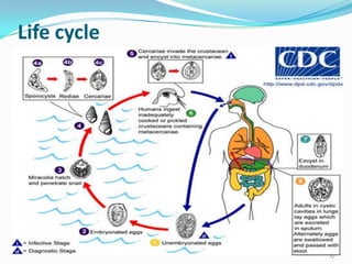
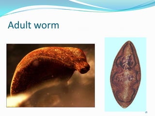
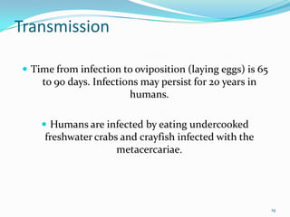
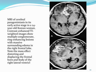
By :group one
Introduction
 Paragonimiasis is a food-borne parasitic infection
 caused by 15 different species of Paragonimus
  trematodes
 cause a sub-acute to chronic inflammatory disease of
  the lung


                                                         2
 P. westermani was discovered in the lungs of a human
  by Ringer in 1879
 and eggs in the sputum were recognized
  independently by Manson and Erwin von Baelz in 1880


                                                         3
 Manson proposed the snail as an intermediate host
 and various Japanese workers detailed the whole life
  cycle in the snail between 1916 and 1922
 The species name P. westermani was named after a
  zookeeper who noted the trematode in a Bengal tiger
  in an Amsterdam Zoo.


                                                         4
Epidemiology

 Humans become infected after eating raw freshwater
  crabs or crayfish that have been encysted with the
  metacerciaria.
 Southeast Asia is more predominately more infected
  because of lifestyles.
 Raw seafood is popular in these countries.
 Crab collectors string raw crabs together and bring
  them miles inland to sell in Taiwan markets.


                                                        5
 These raw crabs are then marinated or pickled in
  vinegar or wine to coagulate the crustacean muscle.
 This process of cooking does not kill the
  metacercariae, consequently infecting the host.
 Smashing rice-eating crabs in rice paddies, splashing
  juices containing metacercariae, can also transmit the
  parasite, or using juices strained from fresh crabs to
  medicinal uses.


                                                           6
 This parasite is easily spread because it is able to infect
  other animals (zoonosis).
 An assortment of mammals and birds can be infected
  and act as paratenic hosts.
 Ingestion of the paratenic host can lead to infection of
  this parasite.
 Paragonimus westermani is distributed in southeast
  Asia and Japan.


                                                                7
 Other species of Paragonimus are common in parts of
  Asia, Africa and South and Central America.
 P. westermani has been increasingly recognized in the
  United States during the past 15 years
 because of the increase of immigrants from endemic
  areas such as Southeast Asia.
 Estimated to infects 22 million people worldwide.


                                                          8
9
10
Life cycle
 The eggs are excreted unembryonated in the sputum,
  or alternately they are swallowed and passed with stool
 In the external environment, the eggs become
  embryonated , and miracidia hatch and seek the first
  intermediate host, a snail, and penetrate its soft tissues


                                                               11
 Miracidia go through several developmental stages
  inside the snail
 sporocysts , rediae , with the latter giving rise to many
  cercariae , which emerge from the snail.
 The cercariae invade the second intermediate host, a
  crustacean such as a crab or crayfish, where they
  encyst and become metacercariae.


                                                              12
 This is the infective stage for the mammalian host
 Human infection with P. westermani occurs by eating
 inadequately cooked or pickled crab or crayfish that
 harbor metacercariae of the parasite


                                                        13
 The metacercariae excyst in the duodenum
 penetrate through the intestinal wall into the
  peritoneal cavity,
 then through the abdominal wall and diaphragm into
  the lungs,
 where they become encapsulated and develop into
  adults (7.5 to 12 mm by 4 to 6 mm).


                                                       14
 The worms can also reach other organs and tissues,
  such as the brain and striated muscles, respectively.
 However, when this takes place completion of the life
  cycles is not achieved, because the eggs laid cannot
  exit these sites.
 Time from infection to oviposition is 65 to 90 days.


                                                          15
 Infections may persist for 20 years in humans. Animals
   such as pigs, dogs, and a variety of feline species can
                 also harbor P. westermani.


                                                             16
Life cycle


             17
Adult worm


             18
Transmission

 Time from infection to oviposition (laying eggs) is 65
    to 90 days. Infections may persist for 20 years in
                        humans.

     Humans are infected by eating undercooked
     freshwater crabs and crayfish infected with the
                     metacercariae.


                                                           19
pathology
 The immature flukes burrow out of the human
  intestine into the peritoneum, where they mature and
  tunnel their way into the lungs.
 Here they cause inflammation, haemorrhage, and
  necrosis of the lung parenchyma.
 Adult flukes (stout, bean-shaped, ~1 cm long) live in
  cavities in proximity to airways.


                                                          20
Clinical features
 Days-weeks after eating infected food, migration of the
    flukes within the peritoneal and pleural cavities causes
    signs of inflammatory and allergic responses
    fever
   rashes
   urticaria
    abdominal and chest pain or discomfort.


                                                               21
 Ova are expelled either in expectorated sputum or in
  the faeces after being swallowed.
 Flukes that miss the lungs produce extrapulmonary
  symptoms (due to cysts, granulomas, and abscesses) in
  muscles, abdominal viscera, brain, genitalia.


                                                         22
o The classic feature of chronic pulmonary disease is
 persistent cough with production of a thick brownish-
  red sputum (due to the presence of ova and flukes).
 Some times hemoptysis
 Physical examination of the chest often reveals little
  and the patients appear quite well.


                                                           23
 Aberrant migration of the flukes may produce signs of
  a cerebral SOL (epilepsy, raised ICP, psychiatric
  syndromes, meningeal irritation) or spinal SOL,
 necrosis of abdominal viscera, transitory subcutaneous
  swellings.
 Extrapulmonary disease may occur in the absence of
  pulmonary signs, but this is uncommon.


                                                          24
 Ectopic lesions from can involve any organ
 including abdominal viscera, the heart, and the
  mediastinum.
 Paragonimus flukes may also invade the liver, spleen,
  intestinal wall, peritoneum, and abdominal lymph
  nodes


                                                          25
In the skin
 P. skrjabini often produces skin nodules, subcutaneous
  abscesses
 a type of creeping eruption known as "trematode larva
  migrans."


                                                       26
In the CNS
 The most clinically recognizable ectopic lesions arise
  from cerebral paragonimiasis,
 which, in highly endemic countries, more commonly
  affects children.
 Many patients with central nervous system disease
  also have pulmonary infections.
 CNS involvement occurs in up to 25% of hospitalized
  patients


                                                           27
 These children present with
 eosinophilic meningoencephalitis,
 seizures
 or signs of space-occupying lesions.


                                         28
Diagnosis:

 presence of ova or adult flukes in the sputum, faeces,
    or effusion
   but these are not present until 2 to 3 months after
    infection.
    serology.
   X-ray the chest cavity and look for worms.
   easily misdiagnosed, because pulmonary infections
    look like tuberculosis, pneumonia, or spirochaetosis


                                                           29
Microscopy
 Sputum examined microscopically may reveal
  Paragonimus eggs released by the flukes in the lungs.
 Keep in mind that the acid-fast stain that is used for
  TB testing of sputum destroys eggs.


                                                           30
 The eggs may also be found by multiple stool exams on
  different days as a result of coughed-up eggs that are
  swallowed.
 The microscopic eggs are yellowish brown, 80-120 µm
  long by 45-70 µm wide, thick-shelled, and with an
  obvious operculum.


                                                           31
32
33
Serology and blood tests
 Serologic tests can be especially useful for early
  infections (prior to maturation of flukes)
 or for ectopic infections where eggs are not passed in
  stool.
 Paragonimus-specific antibody test by micro-ELISA


 Peripheral eosinophilia is common and can be intense,
  especially during the early larval migration stages.


                                                           34
lung biopsy
    can also be used to diagnose this parasite.


                                                  35
Lung biopsy


              36
X-ray the chest
Many patients have a spectrum of abnormalities on chest
  radiographs:
 lobar infiltrates
 coin lesions
 cavities
 calcified nodules,
 hilar enlargement,
 pleural thickening and effusions.


                                                      37
Scattered areas of
patchy pneumonia
with "cotton wool"
opacities and ring
shadows


                     38
There are typical
fluffy, cotton wool
densities in the right
lung base and left
suprahilar area
produced by the
migrating larvae
which have
penetrated the
diaphragm and
pleura in the first
stage

                         39
Lateral chest
radiograph of a 38-
year-old Korean
man with
paragonimiasis
shows a worm
migration tract
identified by the
meandering linear
opacity (arrow)
abutting the major
pleural fissure

                      40
Ring-shaped opacities of contiguous cavities giving the
   characteristic appearance of a bunch of grapes are
    highly suggestive of pulmonary paragonimiasis.


                                                          41
CT and MRI of the brain
  Central nervous system disease may provide similar
   "grapebunch“ findings, characteristically seen in the
 temporal and occipital lobes on computed tomography
                      of the brain


                                                       42
Contrast-enhanced
CT scan shows a
conglomerate of
ring-shaped lesions
in the right parietal
lobe with
surrounding edema.


                        43
MRI of cerebral
paragonimiasis in its
early active stage in a 24-
year-old Korean woman.
Contrast-enhanced TI-
weighted images show
multiple conglomerate,
ring-enhancing lesions
with massive
surrounding edema in
the right frontal lobe.
Note the mass effect
distorting and
displacing the frontal
horn and body of the
right lateral ventricl

                              44
Management:

Praziquantel is the drug of choice:
 adult or pediatric dosage, 25 mg/kg given orally 3
 times per day for 2 consecutive days.
Alternatives:
Triclabendazole (not available in the U.S.), adult or
 pediatric dosage, 10 mg/kg orally once or twice;
 or Bithionol: adult or pediatric dosage, 30-50 mg/kg
 on alternate days for 10-15 doses.


                                                         45
 For cerebral disease, Treat cautiously
 a short course of corticosteroids may be given.
 dexamethasone 4 mg IV q6 h as cover
 neurological deterioration, in some cases producing
  seizures and coma. Beware of raised ICP due to dying
  parasites.


                                                         46
Prevention
 improve health education to decrease consumption of
    undercooked crustaceans
    mass treatment of persons in endemic areas.
   Never eat raw freshwater crabs or crayfish.
   Cook crabs and crayfish for to at least 145°F (~63°C).
   Travelers should be advised to avoid traditional meals
    containing undercooked freshwater crustaceans.


                                                             47
48
49

Paragonimiasis (lung fluke disease)

  • 1.
  • 2.
    Introduction  Paragonimiasis isa food-borne parasitic infection  caused by 15 different species of Paragonimus trematodes  cause a sub-acute to chronic inflammatory disease of the lung 2
  • 3.
     P. westermaniwas discovered in the lungs of a human by Ringer in 1879  and eggs in the sputum were recognized independently by Manson and Erwin von Baelz in 1880 3
  • 4.
     Manson proposedthe snail as an intermediate host  and various Japanese workers detailed the whole life cycle in the snail between 1916 and 1922  The species name P. westermani was named after a zookeeper who noted the trematode in a Bengal tiger in an Amsterdam Zoo. 4
  • 5.
    Epidemiology  Humans becomeinfected after eating raw freshwater crabs or crayfish that have been encysted with the metacerciaria.  Southeast Asia is more predominately more infected because of lifestyles.  Raw seafood is popular in these countries.  Crab collectors string raw crabs together and bring them miles inland to sell in Taiwan markets. 5
  • 6.
     These rawcrabs are then marinated or pickled in vinegar or wine to coagulate the crustacean muscle.  This process of cooking does not kill the metacercariae, consequently infecting the host.  Smashing rice-eating crabs in rice paddies, splashing juices containing metacercariae, can also transmit the parasite, or using juices strained from fresh crabs to medicinal uses. 6
  • 7.
     This parasiteis easily spread because it is able to infect other animals (zoonosis).  An assortment of mammals and birds can be infected and act as paratenic hosts.  Ingestion of the paratenic host can lead to infection of this parasite.  Paragonimus westermani is distributed in southeast Asia and Japan. 7
  • 8.
     Other speciesof Paragonimus are common in parts of Asia, Africa and South and Central America.  P. westermani has been increasingly recognized in the United States during the past 15 years  because of the increase of immigrants from endemic areas such as Southeast Asia.  Estimated to infects 22 million people worldwide. 8
  • 9.
  • 10.
  • 11.
    Life cycle  Theeggs are excreted unembryonated in the sputum, or alternately they are swallowed and passed with stool  In the external environment, the eggs become embryonated , and miracidia hatch and seek the first intermediate host, a snail, and penetrate its soft tissues 11
  • 12.
     Miracidia gothrough several developmental stages inside the snail  sporocysts , rediae , with the latter giving rise to many cercariae , which emerge from the snail.  The cercariae invade the second intermediate host, a crustacean such as a crab or crayfish, where they encyst and become metacercariae. 12
  • 13.
     This isthe infective stage for the mammalian host  Human infection with P. westermani occurs by eating inadequately cooked or pickled crab or crayfish that harbor metacercariae of the parasite 13
  • 14.
     The metacercariaeexcyst in the duodenum  penetrate through the intestinal wall into the peritoneal cavity,  then through the abdominal wall and diaphragm into the lungs,  where they become encapsulated and develop into adults (7.5 to 12 mm by 4 to 6 mm). 14
  • 15.
     The wormscan also reach other organs and tissues, such as the brain and striated muscles, respectively.  However, when this takes place completion of the life cycles is not achieved, because the eggs laid cannot exit these sites.  Time from infection to oviposition is 65 to 90 days. 15
  • 16.
     Infections maypersist for 20 years in humans. Animals such as pigs, dogs, and a variety of feline species can also harbor P. westermani. 16
  • 17.
  • 18.
  • 19.
    Transmission  Time frominfection to oviposition (laying eggs) is 65 to 90 days. Infections may persist for 20 years in humans.  Humans are infected by eating undercooked freshwater crabs and crayfish infected with the metacercariae. 19
  • 20.
    pathology  The immatureflukes burrow out of the human intestine into the peritoneum, where they mature and tunnel their way into the lungs.  Here they cause inflammation, haemorrhage, and necrosis of the lung parenchyma.  Adult flukes (stout, bean-shaped, ~1 cm long) live in cavities in proximity to airways. 20
  • 21.
    Clinical features  Days-weeksafter eating infected food, migration of the flukes within the peritoneal and pleural cavities causes signs of inflammatory and allergic responses  fever  rashes  urticaria  abdominal and chest pain or discomfort. 21
  • 22.
     Ova areexpelled either in expectorated sputum or in the faeces after being swallowed.  Flukes that miss the lungs produce extrapulmonary symptoms (due to cysts, granulomas, and abscesses) in muscles, abdominal viscera, brain, genitalia. 22
  • 23.
    o The classicfeature of chronic pulmonary disease is  persistent cough with production of a thick brownish- red sputum (due to the presence of ova and flukes).  Some times hemoptysis  Physical examination of the chest often reveals little and the patients appear quite well. 23
  • 24.
     Aberrant migrationof the flukes may produce signs of a cerebral SOL (epilepsy, raised ICP, psychiatric syndromes, meningeal irritation) or spinal SOL,  necrosis of abdominal viscera, transitory subcutaneous swellings.  Extrapulmonary disease may occur in the absence of pulmonary signs, but this is uncommon. 24
  • 25.
     Ectopic lesionsfrom can involve any organ  including abdominal viscera, the heart, and the mediastinum.  Paragonimus flukes may also invade the liver, spleen, intestinal wall, peritoneum, and abdominal lymph nodes 25
  • 26.
    In the skin P. skrjabini often produces skin nodules, subcutaneous abscesses  a type of creeping eruption known as "trematode larva migrans." 26
  • 27.
    In the CNS The most clinically recognizable ectopic lesions arise from cerebral paragonimiasis,  which, in highly endemic countries, more commonly affects children.  Many patients with central nervous system disease also have pulmonary infections.  CNS involvement occurs in up to 25% of hospitalized patients 27
  • 28.
     These childrenpresent with  eosinophilic meningoencephalitis,  seizures  or signs of space-occupying lesions. 28
  • 29.
    Diagnosis:  presence ofova or adult flukes in the sputum, faeces, or effusion  but these are not present until 2 to 3 months after infection.  serology.  X-ray the chest cavity and look for worms.  easily misdiagnosed, because pulmonary infections look like tuberculosis, pneumonia, or spirochaetosis 29
  • 30.
    Microscopy  Sputum examinedmicroscopically may reveal Paragonimus eggs released by the flukes in the lungs.  Keep in mind that the acid-fast stain that is used for TB testing of sputum destroys eggs. 30
  • 31.
     The eggsmay also be found by multiple stool exams on different days as a result of coughed-up eggs that are swallowed.  The microscopic eggs are yellowish brown, 80-120 µm long by 45-70 µm wide, thick-shelled, and with an obvious operculum. 31
  • 32.
  • 33.
  • 34.
    Serology and bloodtests  Serologic tests can be especially useful for early infections (prior to maturation of flukes)  or for ectopic infections where eggs are not passed in stool.  Paragonimus-specific antibody test by micro-ELISA  Peripheral eosinophilia is common and can be intense, especially during the early larval migration stages. 34
  • 35.
    lung biopsy can also be used to diagnose this parasite. 35
  • 36.
  • 37.
    X-ray the chest Manypatients have a spectrum of abnormalities on chest radiographs:  lobar infiltrates  coin lesions  cavities  calcified nodules,  hilar enlargement,  pleural thickening and effusions. 37
  • 38.
    Scattered areas of patchypneumonia with "cotton wool" opacities and ring shadows 38
  • 39.
    There are typical fluffy,cotton wool densities in the right lung base and left suprahilar area produced by the migrating larvae which have penetrated the diaphragm and pleura in the first stage 39
  • 40.
    Lateral chest radiograph ofa 38- year-old Korean man with paragonimiasis shows a worm migration tract identified by the meandering linear opacity (arrow) abutting the major pleural fissure 40
  • 41.
    Ring-shaped opacities ofcontiguous cavities giving the characteristic appearance of a bunch of grapes are highly suggestive of pulmonary paragonimiasis. 41
  • 42.
    CT and MRIof the brain Central nervous system disease may provide similar "grapebunch“ findings, characteristically seen in the temporal and occipital lobes on computed tomography of the brain 42
  • 43.
    Contrast-enhanced CT scan showsa conglomerate of ring-shaped lesions in the right parietal lobe with surrounding edema. 43
  • 44.
    MRI of cerebral paragonimiasisin its early active stage in a 24- year-old Korean woman. Contrast-enhanced TI- weighted images show multiple conglomerate, ring-enhancing lesions with massive surrounding edema in the right frontal lobe. Note the mass effect distorting and displacing the frontal horn and body of the right lateral ventricl 44
  • 45.
    Management: Praziquantel is thedrug of choice:  adult or pediatric dosage, 25 mg/kg given orally 3 times per day for 2 consecutive days. Alternatives: Triclabendazole (not available in the U.S.), adult or pediatric dosage, 10 mg/kg orally once or twice;  or Bithionol: adult or pediatric dosage, 30-50 mg/kg on alternate days for 10-15 doses. 45
  • 46.
     For cerebraldisease, Treat cautiously  a short course of corticosteroids may be given.  dexamethasone 4 mg IV q6 h as cover  neurological deterioration, in some cases producing seizures and coma. Beware of raised ICP due to dying parasites. 46
  • 47.
    Prevention  improve healtheducation to decrease consumption of undercooked crustaceans  mass treatment of persons in endemic areas.  Never eat raw freshwater crabs or crayfish.  Cook crabs and crayfish for to at least 145°F (~63°C).  Travelers should be advised to avoid traditional meals containing undercooked freshwater crustaceans. 47
  • 48.
  • 49.