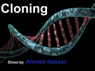
Cloning
•Download as PPTX, PDF•
0 likes•82 views

Cloning involves making genetically identical copies of organisms or genes. Cloning organisms produces clones through asexual reproduction or laboratory techniques. Dolly the sheep was the first mammal cloned in 1997 using nuclei from udder cells implanted into egg cells. Gene cloning involves using restriction enzymes to cut out a target gene, insert it into a bacterial plasmid, and replicate it in bacteria. Common cloning vectors like plasmids and bacteriophages are used to transport cloned sequences between hosts due to characteristics like self-replication and selectable markers. DNA ligase joins DNA strands during cloning, and gel electrophoresis separates cloned DNA fragments by size.

Recommended
More Related Content
What's hot
What's hot (20)
Similar to Cloning
Similar to Cloning (20)
More from GKM
Recently uploaded
Recently uploaded (20)
Cloning
- 2. Cloning • Organisms that are genetically identical are clones • Asexual Reproduction always produces clones • Laboratory Techniques have been developed that have allowed this to happen in Animals
- 3. Cloning in Plants Plant Cloning
- 4. Cloning in Animals • 1950’s first experiments done with Frogs. • Early 1990’s some success found with Mice, Mice were cloned by using nuclei of cells taken from Mice Embryos. • Wilmut et al. Produced “Dolly” in 1997.
- 6. Cloning – Steps of Dolly • Used Genetic Information Taken from Udder of Adult Sheep (donor). • Unfertilized Eggs Collected from sheep and had nuclei removed. • Nuclei from Udder Cells Transplanted into the egg cells. • Resulting Cells were cultured and a few began to divide. • These early embryos implanted into a surrogate mother. One of these developed into a Lamb – Dolly. • DNA Tests confirmed that the Lamb was identical Genetically to the Sheep that had provided the Udder Cells.
- 8. Cloning Genes • Gene cloning: amplifying a specific piece of DNA via a bacteria.
- 9. Gene Cloning Steps 1. The target gene is isolated and cut out using a Restriction Enzyme. 2. The bacterial plasmid is cut open using the same Restriction Enzyme. 3. DNA ligase (Ligase) the target gene into the plasmid. 4. Monitoring (gel electrophoresis). 5. Recombinant plasmid is inserted into the bacterium by transformation.
- 13. Restriction Enzyme A restriction enzyme or restriction endonucleases is an enzyme that cleaves DNA into fragments at or near specific recognition sites within the molecule known as restriction site.
- 15. Type of Restriction Enzymes • Type I: Restriction Endonucleases are single multifunctional enzymes with three different subunits. They required ATP, Mg2+ and S-adenosylmethionine for its activity. Their active cleavage site usually present at least 1000 bp away from the host specificity site. Example: EcoB
- 16. • Type II: restriction endonucleases are most common type. Enzymes Recognize a particular target sequence in a duplex DNA molecule and break the phosphodiester bond within, or near. require no cofactor other than Mg2+ . Example: EcoRI : GAATTC
- 17. • Type III : Restriction enzymes are characterized by their ability to cut DNA in a specific location known and They required ATP , Mg2+ and S- adenosylmethionine for its activity. Example : EcoP1
- 18. ‘5GAATTC3’ ‘3CTTAAG5’ ‘5G AATTC3’ ‘3CTTAA G5’ ‘5CCCGGG3’ ‘3GGGCCC5’ ‘5CCC GGG3’ ‘3GGG CCC5’
- 20. CLONING VECTORS Cloning vectors are DNA molecules that are used to "transport" cloned sequences between biological hosts and the test tube.
- 21. CHARACTERISTICS 1.Self replication, multiple copies. 2.Replication origin site. 3.Selectable marker gene. 4.Small size. 5.Low molecular weight. 6.Easily isolated & purified. 7.Easily isolated into host cell. 8.pass into new cells during cell division. 9.Unique restriction sites to facilitate cloning of insert DNA.
- 22. Type of cloning vector •plasmids •bacteriophageλ , M13 •Cosmids •Artificial chromosomes BAC, YAC, MAC etc.
- 23. What Determines Choice of Vector • Insert size • Vector size • Restriction size • Cloning efficiency Vector Insert size (kb) Plasmid < 10 kb Bacteriophage 3 – 18 kb Cosmids 23 – 45 kb BACs ≤ 300 kb PACs 100 – 300 kb YACs 100 – 3000kb
- 24. Plasmid •Extrachromosomal DNA found in bacteria. •Close circular DNA molecules, supercoiled. •Can replicate independent of chromosome. •Can be transfer to other cells by conjugation. •Can be integrated into the chromosome. •plasmids carry genes like Resistance to Antibiotics. •Number of plasmid per cell - controlled by plasmid itself High copy number > 100 /cell; low copy number < 20 /cell
- 25. pBR322 • pBR322 is a plasmid and was one of the first widely used E.coli cloning vectors. • The p stands for "plasmid," and BR for "Bolivar" and "Rodriguez.“ • 15 copies. • Ampilicin resistance gene • Tetracycline resistance gene • Origin of replication (Ori) • Two selectable markers (Ampilicin,Tetracycline)
- 27. DNA ligase is a specific type of enzyme, a ligase that facilitates the joining of DNA strands together by catalysing the formation of a phosphodiester bond . It plays a role in repairing single-strand breaks in duplex DNA in living organisms, but some forms (such as DNA ligase IV) may specifically repair double-strand breaks. with DNA ligase creating the final phosphodiester bond to fully repair the DNA. DNA ligase
- 31. • Gel electrophoresis is a method for separation and analysis of macromolecules (DNA, RNA and protein) and their fragments, based on their size and charge. • DNA samples are loaded into wells (indentations) at one end of a gel, and an electric current is applied to pull them through the gel. • When a gel is stained with a DNA-binding dye, the DNA fragments can be seen as bands, each representing a group of same-sized DNA fragments. Gel electrophoresis
- 37. Thank you