SlideShare a Scribd company logo
1 of 12
Download to read offline
1
Bioassay of various endocrine hormones, drugs and autocoids
Bioassay of Insulin:
 Standard preparation and unit:
It is pure, dry and crystalline insulin. One unit contains 0.04082 mg. This unit is
specified by Ministry of Health, Government of India and is equivalent to international
unit.
 Preparation of standard solution:
Accurately weigh 20 units of insulin and dissolve it in normal saline. Acidify it with HCl
to pH 2.5. Add 0.5% phenol as preservative. Add 1.4% to 1.8% glycerin. Final volume
should contain 20 units/ml. Store the solution in a cool place and use it within six
months.
 Preparation of test sample solution:
The solution of the test sample is prepared in the same way as the standard solution
described above.
1. Rabbit Method:
 Selection of rabbits: They should be healthy, weighing about 1800-3000 gms. They
should then be maintained on uniform diet but are fasted for 18 hrs before assay.
Water is withdrawn during the experiment.
 Standard and Sample Dilutions: These are freshly prepared by diluting with normal
NaCl solution so as to contain 1 unit/ml. and 2 units/ml.
 Doses: The dose which can produce suitable fall in blood sugar level is calculated for
the standard.
 Principle: The potency of a test sample is estimated by comparing the hypoglycemic
effect of the sample with that of the std. preparation of insulin. Any other suitable
method can also be used.
 Experimental Procedure: Animals are divided into 4 groups of 3 rabbits each. The
rabbits are then put into an animal holder. They should be handled with care to avoid
excitement.
First part of the Test:
 A sample of blood is taken from the marginal ear vein of each rabbit. Presence of
reducing sugar is estimated per 100 ml. of blood by a suitable chemical method. This
concentration is called ‘Initial Blood Sugar Level’.
 The four groups of rabbits are then given sc. injections of insulin as follows:
2
 From each rabbit, a sample of blood is withdrawn up to 5 hrs at the interval of 1 hr.
each. Blood sugar is determined again. This is known as ‘Final Blood Sugar Level’.
Second part of the test (Cross over test):
 The same animals are used for the second part.
 The experiment can be carried out after one week.
 Again they are fasted and initial blood sugar is determined.
 The grouping is reversed, that is to say, those animals which received the standard
are given the test and those which received the test are now given the standard.
 Those animals which received the less dose of the standard are given the higher dose
of the test sample and vice-versa. This test is known as ‘Twin Cross Over Test’.
 Mean percentage decrease in blood sugar of the first and second part is calculated.
2. Mouse Method:
Mice show characteristic convulsions after s.c. inj. of insulin at elevated temperatures.
The percentage convulsions produced by the test and standard preparations are
compared.
Experimental procedure:
Minimum 100 mice weighing between 18-22 gms of the same strain are used. They should
be maintained on constant diet. They should be fasted 18 hrs prior to the experiment.
Standard and sample dilutions: Dilutions are prepared with sterile saline solution, so as
to contain 0.064 units/ml (std. dilution I) and 0.096 units/ml (std. dilution II). Similarly,
test sample solutions are also prepared.
Mice are divided into 4 groups each containing 25 mice and insulin is injected s.c. as
follows:
Mice are put in an air incubator at 33o
C and observed for one and a half hr. An air
incubator with a glass front provided with six shelves is used. The temperature is
thermostatically controlled. Two mice are kept in each of the boxes made up of
perforated sheets of metal.
The mice which convulse or die are taken out of the incubator and observed. These mice
usually convulse severely but failure of the animal to upright itself when placed on its
back, should as well be considered as convulsion. Convulsive mice may be saved by an inj.
of 0.5 ml. of 5% dextrose solution.
Percentage convulsions produced by the test sample are compared with those of the
standard sample. Those animals which survive may be used again for another expt. after
an interval of one week.
3
3. Rat diaphragm method: In this method increase in glycogen content of the muscle or
increase in glucose uptake by muscle in response to insulin is taken as the index of
potency of insulin.
4. Rat epididymal fat-pad method: Here, the ability of insulin to increase CO2 production
by the fat-pad is taken as the parameter for the measurement of potency of the insulin
preparation.
5. Radioimmunoassay: It is the estimation of the concentration of the substance in a unit
quantity of preparation using radiolabelled antigens. A number of drugs are estimated
now days by radioimmunoassy methods because these methods are highly specific and
highly sensitive.
The radioimmunoassay of insulin is based on the ability of human insulin (unlabelled) to
displace beef’s insulin (which may be labelled) from the binding sites (i.e. antibodies).
The method involves the following steps:
I. Bovine insulin is injected into the sheep. After a week the serum containing antibodies
produced against bovine insulin is collected from the blood of the sheep.
II. The serum containing antibodies is exposed to radiolabelled insulin and the bound vs.
free ratio is determined.
III. The mixture of labelled antigen-antibodies is then added in different test-tubes
labelled as standard and test. About 6 concentrations of the standards are taken. They
are then added to different tubes and the bound vs free ratio is again determined using
gamma-counter.
IV. Standard curves are determined and the concentration of test insulin is determined
using this standard curve.
Bioassay of oxytocin:
 About Oxytocin:
 Oxytocin (Oxy- rapid, tocos- labour)
 Synthesized in both sexes, well recognized physiological effects only in women.
 Cyclic Polypeptide hormone - from posterior pituitary gland.
 Pituitary gland consist posterior lobe which produce oxytocin and diuretic hormone.
 Neurosecretory product mainly synthesize in the cell bodies of paraventricular
nuclear of the hypothalamus.
 Role of Oxytocin:
 Stimulate the contraction of the uterine smooth muscle & memory gland.
 Oestrogen, progesterone & prolactin- responsible for production of milk by memory
gland but milk ejection require oxytocin.
 Facilitates the contraction of uterus.
 Oxytocin preparation:
 Animal species have 90 - 110% stated number of units of oxytocin activity.
 It is presented as a solid or solution in a solvent containing an appropriate
antimicrobial preservative such as 0.2% w/v of chlorbutol.
4
 Synthetic: Solid contains NLT 560 units/mg of oxytocin and liquid contain NLT 150
units/ml of oxytocin calculated with reference to the peptide content.
 Actions of Oxytocin:
 Oxytocin (as a neuropeptide) synthesized in hypothalamus stimulates uterine
contractions to expel the infant from uterus.
 Responsible for milk let-down & triggered by the nipple stimulation of suckling called
love & bonding hormone.
 It has a very special effect on mothering It enables the growing sense of love and
attachment to the infant. The more the infant suck, the more oxytocin is produced. In
mothers it increases their attachment to their infant, promoting the feeling of love,
and makes her infant more valuable to her. It also suppresses the fear that would
normally cause her to back off from threat.
 Psychologically, oxytocin promotes a feeling of well-being and tranquillity.
 Principle of bioassay:
Potency of test is determined by comparing its activity with standard preparation of
oxytocin. The activity may characterized as depression of BP, contraction of uterus, milk
ejection pressure, vasopressor effect.
 Standard preparation: Consisting freeze dried synthetic oxytocin peptide with human
albumin citric acid (12.5 units)
1) Depression of the BP in chicken
 Test animals: Healthy cockerel (young male chicken), 1.2 - 2.3 kg,
 Anaesthetized cock-prolonged & constant high B.P
 Expose gluteus primus muscle (thigh) & remove popliteal artery & crural vein.
 Cannulate the popliteal artery & record B.P response.
 Cannulate the crural or brachial vein. Prepare standard solution with saline.
 Inject 0.1 - 0.5ml Inject 2 doses of standard solution into cannulated vein and record
B.P response.
 Dose should cause decrease in B.P (required dose is between 20-100mUnits).
 Interval between 2 doses of injections, 3-10mins depend on rate at which B.P return
normal.
 Dilute the test solution of oxytocin with saline in order to get same response as
standard.
 The ratio between standard & test should be equal.
 If animal rapidly becomes insensitive to repeated injection, another solution must be
used. Measure all responses are calculated result of the assay by standard statistical
method.
2) Contraction of the rat uterus:
 Test animals: Female rat 120 – 200g
 Immediately before assay, select the rats in oestrus or preoestrus phase of estrus
cycle confirmed by vaginal smear.
 Inject 100ug of estradiol benzoate IM into female rat before the assay.
5
 Kill rat & suspend one horn of uterus in organ bath containing PSS solution of
following salts- NaCl, KCl, CaCl2, NaHCO3, Na2HPO4, Mgcl2, and dextrose.
 Maintain the bath at temp at of 32⁰C.
 Oxygenate solution with mix of 95% of O2, 5% of CO2.
 Bath liquid (PSS) required dose between 10-50 units/ml.
 Record contraction of uterine muscle produces by addition of two dose of standard
preparation (Required dose 10 & 50 munits/ml of bath liquid. PSS).
 When maximum contraction has been reached, replaced bath liquid (PSS) by fresh
solution.
 Dose should be added at regular interval [3-5minutes].
 Similarly, record the contraction of uterine muscle produced by test preparation.
 The ratio between two dose of test & two dose of standard should be equal. This
ratio kept constant throughout the assay.
 Measure all response & calculate result of assay by standard statistical method.
3) Milk ejection pressure in Lactating rat:
 Test animals: Select a lactating rat, in the third to twenty-first day after parturition
and weighing about 300 g, separate it from the litter and 30 to 60 minutes later
anaesthetise (for example, by the intraperitoneal injection of a solution of
Pentobarbitone Sodium).
 Tie the rat to an operating table, maintained at 37o, by its hind legs leaving the front
legs free.
 Cannulate the trachea with a short polyethylene tube of internal diameter about 2.5
mm in such a manner so as to ensure a free airway; apply artificial respiration only if
necessary.
 Cannulate an external jugular or femoral vein with a polyethylene tube of internal
diameter about 0.4 mm which is filled with saline solution and closed with a pin.
 Shave the skin surrounding the inguinal and abdominal teats and excise the tip of one
teat, preferably the lower inguinal teat.
 Insert a polyethylene tube of internal diameter about 0.3 mm and external diameter
about 0.6 mm, to a depth sufficient to obtain appropriate measurement of pressure
(3 to 10 mm depth), into the primary teat duct which opens onto the cut surface and
tie firmly in place with a ligature.
 Connect this cannula with a suitable strain gauge transducer (such as that used for
recording arterial blood pressure in the rat) and fill the whole system with a 3.8% w/v
solution of sodium citrate or saline solution containing 50 Units of heparin sodium per
ml to prevent clotting of milk.
 After cannulation, inject a small volume (0.05 to 0.2 ml) of this solution into the teat
duct through the transducer to clear the milk from the tip of the cannula. (This
procedure may be repeated during the assay should obstruction arise from milk
ejected into the cannula).
6
 Clamp the strain gauge so that a slight tension is applied to the teat and its natural
alignment is preserved and connect the gauge to a potentiometric recorder adjusted
to give full-scale deflection for an increase in milk-ejection pressure of about 5.3 kPa.
 Inject all solutions through the venous cannula using a 1-ml syringe graduated in 0.01
ml and wash them in with 0.2 ml of saline solution.
 Prepare a solution of the Standard Preparation and a solution of the preparation
being examined in saline solution so that the volume to be injected is between 0.1 ml
and 0.4 ml.
 Choose two doses of the Standard Preparation such that the increase in milk-ejection
pressure is about 1.35 kPa for the lower dose and about 2.7 kPa for the higher dose.
As an initial approximation, a lower dose of between 0.1 and 0.4 milliUnit and an
upper dose of 1.5 to 2 times this amount may be tried.
 Choose two doses of the preparation being examined with the same inter-dose ratio,
matching the effects of the doses of the Standard Preparation as closely as possible.
 Inject the four doses (two doses of the Standard Preparation and two doses of the
preparation being examined) at intervals of 3 to 5 minutes.
 The two doses of Standard Preparation and the two doses of the preparation being
examined should be given according to a randomised block or a Latin square design
and at least four responses to each should be recorded.
 Measure all the responses and calculate the result of the assay by standard statistical
methods.
Bioassay of Vasopressin:
 Principle:
The vasopressor activity is estimated by comparing the activity of the preparation being
examined with that of the Standard Preparation of arginine vasopressin under the
conditions of a suitable method of assay.
 Standard Preparation:
The Standard Preparation is the Ist
International Standard for Arginine vasopressin,
established in 1978, consisting of freeze-dried synthetic arginine vasopressin peptide
acetate with human albumin and citric acid (supplied in ampoules containing 8.20 Units),
or another suitable preparation the potency of which has been determined in relation to
that of the International Standard.
 Suggested Method:
 Inject slowly into the tail vein of a male albino rat weighing about 300 g a solution of a
suitable alpha -adrenoceptor blocking agent, for example 10 ml per kg of body weight
of a solution prepared by dissolving 5 mg of phenoxybenzamine hydrochloride in 0.1
ml of ethanol (95%), adding 0.05 ml of 1M hydrochloride acid and diluting to 5 ml with
saline solution.
 After 18 hours, anaesthetise the rat with an anaesthetic that will maintain a
prolonged and uniform blood pressure. After 45 to 60 minutes, tie the rat on its back
to the operating table by its hind legs.
7
 Cannulate the trachea with a short polyethylene tube of external diameter about 2.5
mm and dissect a carotid artery ready for cannulation.
 Then cannulate the femoral vein close to the inguinal ligament.
 Retract the abdominal muscles to expose the inguinal ligament.
 Retract the superficial pudendal vein to one side and dissect the femoral vein
towards the inguinal ligament from the corresponding artery.
 When dissecting, a deep branch reaching the femoral vein must be found and tied off
to prevent bleeding during cannulation.
 Tie a short polyethylene cannula of external diameter about 1 mm into the femoral
vein by two ligatures and join by a short piece of flexible tubing to a 1-ml burette with
an attached thistle funnel containing saline solution at about 37o.
 Firmly fix a wet absorbent cotton swab to the thigh so as to cover the incision and
cannula.
 At this stage, inject through the venous cannula 200 Units of heparin dissolved in
saline solution per 100 g of body weight.
 Then tie in a carotid cannula of external diameter about 1 mm and connect by a
column of saline solution containing heparin with a suitable pressure measuring
device such as a mercury manometer of internal diameter about 2 to 3 mm.
 The central and peripheral nervous system including both vagus and associated
sympathetic nerves is left intact. No artificial respiration is necessary. Taking care that
no air is injected, inject all solutions through the venous cannula by means of a 1-ml
syringe graduated in 0.01 ml and wash in with 0.2 ml of saline solution from the
burette.
 Dilute the extract of the standard preparation and the preparation being examined
with saline solution so that the volume to be injected is between 0.1 ml and 0.5 ml.
 Choose two doses of the standard preparation such that the elevation of the blood
pressure is about 4 kPa for the lower dose and about 7 kPa but always submaximal
for the higher dose, the ratio of low to high dose being determined by the response
and usually being 3 to 5.
 As an initial approximation doses of 3 and 5 milliUnits may be tried.
 Choose two doses of the preparation being examined with the same inter-dose ratio,
matching the effects of the dose of the standard preparation as closely as possible.
 Inject doses at intervals of 10 to 15 minutes.
 The two doses of the standard preparation and the two doses of the preparation
being examined should be given in a randomised block or a Latin square design and
four to five responses to each should be recorded.
 Measure all the responses and calculate the result of the assay by standard statistical
methods.
Bioassay of d-Tubocurarine:
1. Rabbit Head-drop Method:
 Principle:
8
d-Tubocurarine hydrochloride is injected into the marginal vein of a rabbit’s ear till the
rabbit’s neck muscles are relaxed such that the animal cannot hold its head up. The total
amount of test sample required to produce the endpoint is compared with the total
amount of the standard sample required to produce similar endpoint.
 Selection of Rabbits:
Rabbits weighing 2 kg are used. Animals should be free from disease, obtained from a
healthy colony and should be accustomed with the experimental procedure.
 Experimental Procedure:
 Each rabbit is placed in a holder with its head protruding outside. The head should be
freely movable. Minimum 8 rabbits are used. They are divided into two groups each
containing 4 rabbits. First group will receive standard sample and the second group
will receive the sample under test. D-Tubocurarine solution is injected at a constant
speed by infusion apparatus through the marginal vein.
 Injection should be given at a rate of 0.4 ml/min and should take about 10 min.
Infusion is continued till the rabbit will not be in a position to hold its head erect or
there will be no response by focussing light on the eyes and the neck gets elongated
and toneless.
 Suitable dose of d-tubocurarine is 0.012% w/v in saline. Rabbits recover immediately
from the effect of curarization. During the expt. there is a possibility or respiratory
embarrassment which is treated by injecting neostigmine methyl sulphate (0.05 mg.)
and atropine sulphate immediately through the marginal ear vein.
 Cross-over test is carried out to minimise biological error due to animal variation.
Those rabbits which received the standard sample on the first day will be given test
sample on the second day of expt. and vice versa. Mean dose which produces head
drop of the test sample is compared with the mean dose of standard preparation.
2) Frog’s Rectus Abdominis muscle Preparation:
 A frog is pithed and laid on its back on a cork covered board to which it is pinned. The
skin covering the abdomen is cut away and the rectus abdominis muscle of one side is
dissected from the pelvic girdle to its insertion in the cartilage of the pectoral girdle.
 The muscle is then pinned to the cork by four pins to keep its normal length while a
thread is sewn through each end.
 It is then mounted in the organ bath containing frog’s Ringer solution which contains
NaCl, 6.5gm; KCl, 0.29 gm; CaCl2, 0.24 gm; NaHCO3, 0.4 gm; glucose, 1.5 gm and
distilled water 2000 ml . Oxygenation is carried out to keep the tissue alive. The
muscle is stabilized for 30-45 min. in order to get critical quantitative response. The
responses are recorded using isotonic frontal writing lever with 1 G. tension.
 Two similar contractions with the same concentration of acetylcholine are obtained.
Three doses of the standard sample and one intermediate dose of the test sample are
selected and the reduction in height of contraction induced by acetylcholine is noted
down.
9
 Acetylcholine contraction is recorded on slow moving drum for 90 sec. d-
Tubocurarine is allowed to act for 30 sec. The percentage reduction at each dose
levels is calculated and log dose response curve of the standard drug is plotted. A
linear response will be obtained. The potency of test sample is calculated from the
standard curve.
Bioassay of digitalis:
 Principle:
Potency of the test sample is compared with that of the standard preparation by
determining the action on the cardiac muscle. Any other equivalent method, which gives
results similar to those obtained by this method as also valid.
 Standard Preparation and Units:
The standard preparation is a mixture of dried and powdered digitalis leaves (1 unit = 76
mg.)
 Preparation of Extracts:
Exact amount of the powder is extracted with dehydrated alcohol in a continuous
extraction apparatus for six hours. The final extract should contain 10 ml. (5 ml. alcohol +
5 ml. water) per 10 g. of digitalis powder. It should be stored in between 5 ⁰C and –5 ⁰C.
Pigeon Method:
Minimum 6 pigeons are used for testing each sample. They should be free from gross
evidence of disease or emaciation. The weight of the heaviest pigeon should not exceed
twice the weight of the lightest pigeon. Food is withheld 16-28 hours before the
experiment.
Pigeons are divided on the basis of their sex, weight and breed, into two groups. They
are anaesthetized with anaesthetic ether. One side of the wing is dissected and the alar
vein is cannulated by means of a venous cannula. Dilutions are made with normal saline.
Average lethal dose of each sample is determined; results are tabulated and calculated as
per guinea pig method.
The lethal dose per kg of body weight is determined for each pigeon. The potency of the
test sample is determined by dividing the mean lethal dose of standard by the mean
lethal dose of the test sample.
In pigeons, stoppage of heart is associated with a characteristic vomiting response called
‘emesis. The milk from the crop sac of pigeons is being ejected out. This may be taken as
the end point response of digitalis.
Bioassay of histamine:
1. Contractions of isolated guinea pig ileum
Bioassay of histamine on isolated guinea pig ileum can be determined by graded
response bioassays like matching, interpolation, bracketing and multiple point bioassays.
 Matching Method:
In this method a constant dose of the test is bracketed by varying doses of standard till
the exact match is obtained between test dose and the standard dose.
10
Initially, two responses of the standard are taken. The doses are adjusted such that one is
giving response of approximately 20% and other 70% of the maximum. The response of
unknown which lies between two responses of standard dose is taken. The panel is
repeated by increasing or decreasing the dose s of standard till all three equal responses
are obtained. The dose of test sample is kept constant. At the end, a response of the
double dose of the standard and test which match each other are taken. These should
give equal responses. Concentration of the test sample can be determined as follows:
This method has following limitations:
1. It occupies a larger area of the drum as far as tracings are concerned.
2. The match is purely subjective, so chances of error are there and one cannot determine
them.
3. It does not give any idea of dose-response relationship.
However, this method is particularly useful if the sensitivity of the preparation is not
stable. Bioassay of histamine, on guinea pig ileum is preferably carried out by matching
method is shown below-
 Graphical/ interpolation method:
This method is based on the assumption of the dose-response relationship. Log-dose-
response curve is plotted and the dose of standard producing the same response as
produced by the test sample is directly read from the graph. In simpler design, 5-6
responses of the graded doses of the standard are taken and then two equiactive
responses of the test sample are taken. The height of contraction is measured and
plotted against the log-dose. The dose of standard producing the same response as
produced by the test is read directly from the graph and the concentration of test sample
is determined by the same formula as mentioned before.
11
The characteristic of log-dose response curve is that it is linear in the middle (20-80%).
Thus, the comparison should be done within this range only. In other words, the
response of test sample must lie within this range.
Advantage of this method is that, it is a simple method and chances of errors are less if
the sensitivity of the preparation is not changed.
 Multiple point bioassays:
These include 3 point, 4 point and 6 point methods. In these 4 methods, the responses
are repeated several times and the mean of each is taken. Thus, chances of error are
minimized in these methods. In 3 point assay method 2 doses of the standard and one
dose of the test are used. In 4 point method 2 doses of standard and 2 doses of the test
are used. In 6 point method 3 doses of standard and 3 doses of the test are used.
Similarly one can design 8 point method also. The sequence of responses is followed as
per the Latin square method of randomization in order to avoid any bias.
The mean responses are calculated and plotted against log-dose and amount of standard
producing the same response as produced by the test is determined graphically as well
as mathematically:
Where, n1 = Lower Standard dose, n2 = Higher Standard dose, t = Test dose, S1 =
Response of n1, S2 = Response of n2, T = Response of test (t), and Cs = Concentration of
standard.
12
Similarly, in 4 point method, amount of standard producing the same response as
produced by the test can be determined by graphical method. It is determined
mathematically as follows:
Where, t1 = lower dose of test, t2 = higher dose of test, T1 = response of t1, T2 = response of t2.
Histamine receptors in ileum:
 Histamine receptors in ileum H1 receptors
 Receptor type: G-protein-coupled receptor
 Agonist: Histamine
 Mechanism of contraction:
++ G-protein→ ++ phospholipase C→ splitting of PIP2 into- 1) DAG that increases the
opening of calcium channels, and 2) IP3 which increases calcium mobilization from
sarcoplasmic stores.DAG & IP3 lead to increase in the intracellular concentration of
calcium and smooth muscles contraction.
2) Fall in BP in anaesthetised and atropinized cat or dog:
Cat or dog is anaesthetised with chloralose or barbiturate and prepared for recording of
BP. Sensitivity is determined by injecting standard solution of 0.05, 0.1, 0.15 µg of
histamine base per kg body weight for 5min interval.
A fixed dose of standard producing a fall in BP about 20 mm Hg is injected with changing
doses of test at regular intervals and matching assay is done.
 Mechanism of BP fall:
Histamine binds with histamine H1 receptor of endothelium causes release of EDRF
(endothelium derived relaxing factor). This EDRF diffuses out and reaches the smooth
muscle of the arteriole and causes generation of cyclic-GMP→ causing reduction of Ca2+
in smooth muscle relaxation of the smooth muscle→ arteriolar dilatation. Another
possibility is that combination of H1 with the Histamine causes release of PGI2 which
causes vasodilatation.
Bioassay of 5-hydroxytryptamine (5HT/ serotonin):
1) Contraction of isolated atropinized rat uterus,
2) Contraction of isolated terminal colon of rat,
3) Contraction of isolated fundus strip of rat stomach, and
4) Constriction of blood vessels of perfused rabbit ear.

More Related Content

What's hot

Preclinical screening methods of Sedative and hypnotics by syed
Preclinical screening methods of Sedative and hypnotics by syedPreclinical screening methods of Sedative and hypnotics by syed
Preclinical screening methods of Sedative and hypnotics by syedMubasheer syed
 
Pharmacological screening of analgesic activity
Pharmacological screening of analgesic activityPharmacological screening of analgesic activity
Pharmacological screening of analgesic activityBiswash Sapkota
 
Screening methods of immunomodulators
Screening methods of immunomodulatorsScreening methods of immunomodulators
Screening methods of immunomodulatorsAanchal46
 
Screening of antihypertensive agents
Screening of antihypertensive agentsScreening of antihypertensive agents
Screening of antihypertensive agentsKanthlal SK
 
screening method of cardiovascular activity
screening method of cardiovascular activityscreening method of cardiovascular activity
screening method of cardiovascular activityArpitSuralkar
 
Recording of Rat ECG, BP and heart rate
Recording of Rat ECG, BP and heart rateRecording of Rat ECG, BP and heart rate
Recording of Rat ECG, BP and heart rateShivanshu Bajaj
 
Preclinical screening of anti fertility agents
Preclinical screening of anti fertility agentsPreclinical screening of anti fertility agents
Preclinical screening of anti fertility agentsNaveen K L
 
Oecd principles of good laboratory practices (glp)
Oecd principles of good laboratory practices (glp)Oecd principles of good laboratory practices (glp)
Oecd principles of good laboratory practices (glp)Sriram Mamidi
 
Anti-diarrhoeal screening models and methods
Anti-diarrhoeal screening models and methodsAnti-diarrhoeal screening models and methods
Anti-diarrhoeal screening models and methodsPervej Alom
 
Preclinical Screening of Antihypertensive Agents | In-Vitro & In-Vivo Models
Preclinical Screening of Antihypertensive Agents | In-Vitro & In-Vivo ModelsPreclinical Screening of Antihypertensive Agents | In-Vitro & In-Vivo Models
Preclinical Screening of Antihypertensive Agents | In-Vitro & In-Vivo ModelsChetan Prakash
 
Antiulcer screening models
Antiulcer screening modelsAntiulcer screening models
Antiulcer screening modelsDRx Priya Shukla
 
Drug screening methods for antiarrhythmic agents
Drug screening methods for antiarrhythmic agentsDrug screening methods for antiarrhythmic agents
Drug screening methods for antiarrhythmic agentsDr. Abhishek Vyas
 
Screening methof of Local anaesthesia
Screening methof of Local anaesthesiaScreening methof of Local anaesthesia
Screening methof of Local anaesthesiaGaurabi Chakrabortty
 
Screening models on aphrodisiac agents
Screening models on aphrodisiac agentsScreening models on aphrodisiac agents
Screening models on aphrodisiac agentsJaineel Dharod
 
Pharmacological screening of Anti-psychotic agents
Pharmacological screening of Anti-psychotic agentsPharmacological screening of Anti-psychotic agents
Pharmacological screening of Anti-psychotic agentsAbin Joy
 
Preclinical screening of antiallergics
Preclinical screening of antiallergicsPreclinical screening of antiallergics
Preclinical screening of antiallergicsPrajitha p
 
Screening Methods of Anti Anxiety Agents
Screening Methods of Anti Anxiety AgentsScreening Methods of Anti Anxiety Agents
Screening Methods of Anti Anxiety AgentsAnupam dubey
 
Anesthesia and euthanasia of experimental animal by vivek and naveen
Anesthesia and euthanasia of experimental animal by vivek and naveenAnesthesia and euthanasia of experimental animal by vivek and naveen
Anesthesia and euthanasia of experimental animal by vivek and naveenAnimatedWorld
 

What's hot (20)

Preclinical screening methods of Sedative and hypnotics by syed
Preclinical screening methods of Sedative and hypnotics by syedPreclinical screening methods of Sedative and hypnotics by syed
Preclinical screening methods of Sedative and hypnotics by syed
 
Pharmacological screening of analgesic activity
Pharmacological screening of analgesic activityPharmacological screening of analgesic activity
Pharmacological screening of analgesic activity
 
Screening methods of immunomodulators
Screening methods of immunomodulatorsScreening methods of immunomodulators
Screening methods of immunomodulators
 
Screening of anti hypertensives2003
Screening of anti hypertensives2003Screening of anti hypertensives2003
Screening of anti hypertensives2003
 
Screening of antihypertensive agents
Screening of antihypertensive agentsScreening of antihypertensive agents
Screening of antihypertensive agents
 
screening method of cardiovascular activity
screening method of cardiovascular activityscreening method of cardiovascular activity
screening method of cardiovascular activity
 
BIOASSAY OF TETANUS ANTITOXIN
BIOASSAY OF TETANUS ANTITOXINBIOASSAY OF TETANUS ANTITOXIN
BIOASSAY OF TETANUS ANTITOXIN
 
Recording of Rat ECG, BP and heart rate
Recording of Rat ECG, BP and heart rateRecording of Rat ECG, BP and heart rate
Recording of Rat ECG, BP and heart rate
 
Preclinical screening of anti fertility agents
Preclinical screening of anti fertility agentsPreclinical screening of anti fertility agents
Preclinical screening of anti fertility agents
 
Oecd principles of good laboratory practices (glp)
Oecd principles of good laboratory practices (glp)Oecd principles of good laboratory practices (glp)
Oecd principles of good laboratory practices (glp)
 
Anti-diarrhoeal screening models and methods
Anti-diarrhoeal screening models and methodsAnti-diarrhoeal screening models and methods
Anti-diarrhoeal screening models and methods
 
Preclinical Screening of Antihypertensive Agents | In-Vitro & In-Vivo Models
Preclinical Screening of Antihypertensive Agents | In-Vitro & In-Vivo ModelsPreclinical Screening of Antihypertensive Agents | In-Vitro & In-Vivo Models
Preclinical Screening of Antihypertensive Agents | In-Vitro & In-Vivo Models
 
Antiulcer screening models
Antiulcer screening modelsAntiulcer screening models
Antiulcer screening models
 
Drug screening methods for antiarrhythmic agents
Drug screening methods for antiarrhythmic agentsDrug screening methods for antiarrhythmic agents
Drug screening methods for antiarrhythmic agents
 
Screening methof of Local anaesthesia
Screening methof of Local anaesthesiaScreening methof of Local anaesthesia
Screening methof of Local anaesthesia
 
Screening models on aphrodisiac agents
Screening models on aphrodisiac agentsScreening models on aphrodisiac agents
Screening models on aphrodisiac agents
 
Pharmacological screening of Anti-psychotic agents
Pharmacological screening of Anti-psychotic agentsPharmacological screening of Anti-psychotic agents
Pharmacological screening of Anti-psychotic agents
 
Preclinical screening of antiallergics
Preclinical screening of antiallergicsPreclinical screening of antiallergics
Preclinical screening of antiallergics
 
Screening Methods of Anti Anxiety Agents
Screening Methods of Anti Anxiety AgentsScreening Methods of Anti Anxiety Agents
Screening Methods of Anti Anxiety Agents
 
Anesthesia and euthanasia of experimental animal by vivek and naveen
Anesthesia and euthanasia of experimental animal by vivek and naveenAnesthesia and euthanasia of experimental animal by vivek and naveen
Anesthesia and euthanasia of experimental animal by vivek and naveen
 

Similar to b2. Bioassay of various endocrine hormones, drugs and autocoids.pdf

Bioassay of insulin & Bioassay of Vasopressin
Bioassay of insulin & Bioassay of VasopressinBioassay of insulin & Bioassay of Vasopressin
Bioassay of insulin & Bioassay of VasopressinHeena Parveen
 
Bioassy of insulin according to Indian pharmacopoeia
Bioassy of insulin according to Indian pharmacopoeiaBioassy of insulin according to Indian pharmacopoeia
Bioassy of insulin according to Indian pharmacopoeiaSONALPANDE5
 
Bioassay of insulin
Bioassay of insulinBioassay of insulin
Bioassay of insulinKomal Sathe
 
Bioassay vasopressin digitalis ACTH
Bioassay vasopressin digitalis ACTHBioassay vasopressin digitalis ACTH
Bioassay vasopressin digitalis ACTHmuthulakshmi623285
 
SCREENING OF immuno.pptx
SCREENING OF immuno.pptxSCREENING OF immuno.pptx
SCREENING OF immuno.pptxSourinDe2
 
Screening models for immunomodulatory agents
Screening models for immunomodulatory agentsScreening models for immunomodulatory agents
Screening models for immunomodulatory agentsChandragiri Siva Sai
 
Chapter-1; Bioassay (Part-3).pptx
Chapter-1; Bioassay (Part-3).pptxChapter-1; Bioassay (Part-3).pptx
Chapter-1; Bioassay (Part-3).pptxAbdulWahid922079
 
Immunodulators Screening methods
Immunodulators Screening methodsImmunodulators Screening methods
Immunodulators Screening methodsANANYAPANDEY71
 
Expt. 8 Hypoglycemic effect of insulin in rabbit
Expt. 8 Hypoglycemic effect of insulin in rabbitExpt. 8 Hypoglycemic effect of insulin in rabbit
Expt. 8 Hypoglycemic effect of insulin in rabbitVISHALJADHAV100
 
Invivo screening methods for anti inflammatory agents
Invivo screening methods for anti inflammatory  agentsInvivo screening methods for anti inflammatory  agents
Invivo screening methods for anti inflammatory agentsSravani Ganti
 
Antiallergic activity by mast cell stabilization assay
Antiallergic activity by mast cell stabilization assayAntiallergic activity by mast cell stabilization assay
Antiallergic activity by mast cell stabilization assayHimikaRathi
 
Respiratory and reproduction pharmacology
Respiratory and reproduction pharmacology Respiratory and reproduction pharmacology
Respiratory and reproduction pharmacology ManjuJhakhar
 
INVESTIGATION OF ANTI DIABETIC ACTIVITY OF STEM OF MURRAYA KOENIGII
INVESTIGATION OF ANTI DIABETIC ACTIVITY OF STEM OF MURRAYA KOENIGIIINVESTIGATION OF ANTI DIABETIC ACTIVITY OF STEM OF MURRAYA KOENIGII
INVESTIGATION OF ANTI DIABETIC ACTIVITY OF STEM OF MURRAYA KOENIGIIpharmaindexing
 
Expt 9- To evaluate the effect of insulin in rabbits at different intervals
Expt 9- To evaluate the effect of insulin in rabbits at different intervalsExpt 9- To evaluate the effect of insulin in rabbits at different intervals
Expt 9- To evaluate the effect of insulin in rabbits at different intervalsMirzaAnwarBaig1
 
Biological assay of oxytocin
Biological assay of oxytocinBiological assay of oxytocin
Biological assay of oxytocinKUNDLAJAYALAKSHMI
 
Antifertility screening
Antifertility screening Antifertility screening
Antifertility screening Prerna Singh
 

Similar to b2. Bioassay of various endocrine hormones, drugs and autocoids.pdf (20)

Bioassay of insulin & Bioassay of Vasopressin
Bioassay of insulin & Bioassay of VasopressinBioassay of insulin & Bioassay of Vasopressin
Bioassay of insulin & Bioassay of Vasopressin
 
Bio assays of insulin
Bio assays of insulinBio assays of insulin
Bio assays of insulin
 
Bioassay of insulin- Lokesh.pptx
Bioassay of insulin- Lokesh.pptxBioassay of insulin- Lokesh.pptx
Bioassay of insulin- Lokesh.pptx
 
Bioassy of insulin according to Indian pharmacopoeia
Bioassy of insulin according to Indian pharmacopoeiaBioassy of insulin according to Indian pharmacopoeia
Bioassy of insulin according to Indian pharmacopoeia
 
Bioassay of insulin
Bioassay of insulinBioassay of insulin
Bioassay of insulin
 
Bioassay vasopressin digitalis ACTH
Bioassay vasopressin digitalis ACTHBioassay vasopressin digitalis ACTH
Bioassay vasopressin digitalis ACTH
 
SCREENING OF immuno.pptx
SCREENING OF immuno.pptxSCREENING OF immuno.pptx
SCREENING OF immuno.pptx
 
Screening models for immunomodulatory agents
Screening models for immunomodulatory agentsScreening models for immunomodulatory agents
Screening models for immunomodulatory agents
 
Chapter-1; Bioassay (Part-3).pptx
Chapter-1; Bioassay (Part-3).pptxChapter-1; Bioassay (Part-3).pptx
Chapter-1; Bioassay (Part-3).pptx
 
Immunodulators Screening methods
Immunodulators Screening methodsImmunodulators Screening methods
Immunodulators Screening methods
 
Expt. 8 Hypoglycemic effect of insulin in rabbit
Expt. 8 Hypoglycemic effect of insulin in rabbitExpt. 8 Hypoglycemic effect of insulin in rabbit
Expt. 8 Hypoglycemic effect of insulin in rabbit
 
Invivo screening methods for anti inflammatory agents
Invivo screening methods for anti inflammatory  agentsInvivo screening methods for anti inflammatory  agents
Invivo screening methods for anti inflammatory agents
 
Antiallergic activity by mast cell stabilization assay
Antiallergic activity by mast cell stabilization assayAntiallergic activity by mast cell stabilization assay
Antiallergic activity by mast cell stabilization assay
 
Respiratory and reproduction pharmacology
Respiratory and reproduction pharmacology Respiratory and reproduction pharmacology
Respiratory and reproduction pharmacology
 
INVESTIGATION OF ANTI DIABETIC ACTIVITY OF STEM OF MURRAYA KOENIGII
INVESTIGATION OF ANTI DIABETIC ACTIVITY OF STEM OF MURRAYA KOENIGIIINVESTIGATION OF ANTI DIABETIC ACTIVITY OF STEM OF MURRAYA KOENIGII
INVESTIGATION OF ANTI DIABETIC ACTIVITY OF STEM OF MURRAYA KOENIGII
 
Expt 9- To evaluate the effect of insulin in rabbits at different intervals
Expt 9- To evaluate the effect of insulin in rabbits at different intervalsExpt 9- To evaluate the effect of insulin in rabbits at different intervals
Expt 9- To evaluate the effect of insulin in rabbits at different intervals
 
Bioassay of oxytocin
Bioassay of oxytocinBioassay of oxytocin
Bioassay of oxytocin
 
Biological assay of oxytocin
Biological assay of oxytocinBiological assay of oxytocin
Biological assay of oxytocin
 
pha2.pdfo
pha2.pdfopha2.pdfo
pha2.pdfo
 
Antifertility screening
Antifertility screening Antifertility screening
Antifertility screening
 

More from VISHALJADHAV100

Acute and Chronic Renal Failure.........
Acute and Chronic Renal Failure.........Acute and Chronic Renal Failure.........
Acute and Chronic Renal Failure.........VISHALJADHAV100
 
Chronic Obstructive Pulmonary Disease...
Chronic Obstructive Pulmonary Disease...Chronic Obstructive Pulmonary Disease...
Chronic Obstructive Pulmonary Disease...VISHALJADHAV100
 
Pathophysiology of Bronchial Asthma.....
Pathophysiology of Bronchial Asthma.....Pathophysiology of Bronchial Asthma.....
Pathophysiology of Bronchial Asthma.....VISHALJADHAV100
 
Cardiovascular Pathophysiology- Coronary Atherosclerosis and Arteriosclerosis
Cardiovascular Pathophysiology- Coronary Atherosclerosis and ArteriosclerosisCardiovascular Pathophysiology- Coronary Atherosclerosis and Arteriosclerosis
Cardiovascular Pathophysiology- Coronary Atherosclerosis and ArteriosclerosisVISHALJADHAV100
 
Cardiovascular Pathophysiology- Coronary Artery Disease (CAD)/Ischaemic Heart...
Cardiovascular Pathophysiology- Coronary Artery Disease (CAD)/Ischaemic Heart...Cardiovascular Pathophysiology- Coronary Artery Disease (CAD)/Ischaemic Heart...
Cardiovascular Pathophysiology- Coronary Artery Disease (CAD)/Ischaemic Heart...VISHALJADHAV100
 
Cardiovascular Pathophysiology- Congestive Cardiac Failure (CCF)/Congestive H...
Cardiovascular Pathophysiology- Congestive Cardiac Failure (CCF)/Congestive H...Cardiovascular Pathophysiology- Congestive Cardiac Failure (CCF)/Congestive H...
Cardiovascular Pathophysiology- Congestive Cardiac Failure (CCF)/Congestive H...VISHALJADHAV100
 
Cardiovascular Pathophysiology- Hypertension
Cardiovascular Pathophysiology- HypertensionCardiovascular Pathophysiology- Hypertension
Cardiovascular Pathophysiology- HypertensionVISHALJADHAV100
 
b1. Bioassay of ACTH.pdf
b1. Bioassay of ACTH.pdfb1. Bioassay of ACTH.pdf
b1. Bioassay of ACTH.pdfVISHALJADHAV100
 
g. Antirheumatic drugs.pdf
g. Antirheumatic drugs.pdfg. Antirheumatic drugs.pdf
g. Antirheumatic drugs.pdfVISHALJADHAV100
 
d3. Substance P (SP).pdf
d3. Substance P (SP).pdfd3. Substance P (SP).pdf
d3. Substance P (SP).pdfVISHALJADHAV100
 
c. Prostaglandins, Thromboxanes and Leukotrienes.pdf
c. Prostaglandins, Thromboxanes and Leukotrienes.pdfc. Prostaglandins, Thromboxanes and Leukotrienes.pdf
c. Prostaglandins, Thromboxanes and Leukotrienes.pdfVISHALJADHAV100
 
b5. 5HT- Antagonists.pdf
b5. 5HT- Antagonists.pdfb5. 5HT- Antagonists.pdf
b5. 5HT- Antagonists.pdfVISHALJADHAV100
 

More from VISHALJADHAV100 (20)

Acute and Chronic Renal Failure.........
Acute and Chronic Renal Failure.........Acute and Chronic Renal Failure.........
Acute and Chronic Renal Failure.........
 
Chronic Obstructive Pulmonary Disease...
Chronic Obstructive Pulmonary Disease...Chronic Obstructive Pulmonary Disease...
Chronic Obstructive Pulmonary Disease...
 
Pathophysiology of Bronchial Asthma.....
Pathophysiology of Bronchial Asthma.....Pathophysiology of Bronchial Asthma.....
Pathophysiology of Bronchial Asthma.....
 
Cardiovascular Pathophysiology- Coronary Atherosclerosis and Arteriosclerosis
Cardiovascular Pathophysiology- Coronary Atherosclerosis and ArteriosclerosisCardiovascular Pathophysiology- Coronary Atherosclerosis and Arteriosclerosis
Cardiovascular Pathophysiology- Coronary Atherosclerosis and Arteriosclerosis
 
Cardiovascular Pathophysiology- Coronary Artery Disease (CAD)/Ischaemic Heart...
Cardiovascular Pathophysiology- Coronary Artery Disease (CAD)/Ischaemic Heart...Cardiovascular Pathophysiology- Coronary Artery Disease (CAD)/Ischaemic Heart...
Cardiovascular Pathophysiology- Coronary Artery Disease (CAD)/Ischaemic Heart...
 
Cardiovascular Pathophysiology- Congestive Cardiac Failure (CCF)/Congestive H...
Cardiovascular Pathophysiology- Congestive Cardiac Failure (CCF)/Congestive H...Cardiovascular Pathophysiology- Congestive Cardiac Failure (CCF)/Congestive H...
Cardiovascular Pathophysiology- Congestive Cardiac Failure (CCF)/Congestive H...
 
Cardiovascular Pathophysiology- Hypertension
Cardiovascular Pathophysiology- HypertensionCardiovascular Pathophysiology- Hypertension
Cardiovascular Pathophysiology- Hypertension
 
b1. Bioassay of ACTH.pdf
b1. Bioassay of ACTH.pdfb1. Bioassay of ACTH.pdf
b1. Bioassay of ACTH.pdf
 
d1. Angiotensin.pdf
d1. Angiotensin.pdfd1. Angiotensin.pdf
d1. Angiotensin.pdf
 
g. Antirheumatic drugs.pdf
g. Antirheumatic drugs.pdfg. Antirheumatic drugs.pdf
g. Antirheumatic drugs.pdf
 
e. NSADIs.pdf
e. NSADIs.pdfe. NSADIs.pdf
e. NSADIs.pdf
 
d3. Substance P (SP).pdf
d3. Substance P (SP).pdfd3. Substance P (SP).pdf
d3. Substance P (SP).pdf
 
d2. Plasma Kinins.pdf
d2. Plasma Kinins.pdfd2. Plasma Kinins.pdf
d2. Plasma Kinins.pdf
 
f. Antigout Drugs.pdf
f. Antigout Drugs.pdff. Antigout Drugs.pdf
f. Antigout Drugs.pdf
 
c. Prostaglandins, Thromboxanes and Leukotrienes.pdf
c. Prostaglandins, Thromboxanes and Leukotrienes.pdfc. Prostaglandins, Thromboxanes and Leukotrienes.pdf
c. Prostaglandins, Thromboxanes and Leukotrienes.pdf
 
b3. H2-Antagonists.pdf
b3. H2-Antagonists.pdfb3. H2-Antagonists.pdf
b3. H2-Antagonists.pdf
 
b1. Histamine.pdf
b1. Histamine.pdfb1. Histamine.pdf
b1. Histamine.pdf
 
b5. 5HT- Antagonists.pdf
b5. 5HT- Antagonists.pdfb5. 5HT- Antagonists.pdf
b5. 5HT- Antagonists.pdf
 
b4. Serotonin.pdf
b4. Serotonin.pdfb4. Serotonin.pdf
b4. Serotonin.pdf
 
b2. H1-Antagonists.pdf
b2. H1-Antagonists.pdfb2. H1-Antagonists.pdf
b2. H1-Antagonists.pdf
 

Recently uploaded

Simple, Complex, and Compound Sentences Exercises.pdf
Simple, Complex, and Compound Sentences Exercises.pdfSimple, Complex, and Compound Sentences Exercises.pdf
Simple, Complex, and Compound Sentences Exercises.pdfstareducators107
 
Transparency, Recognition and the role of eSealing - Ildiko Mazar and Koen No...
Transparency, Recognition and the role of eSealing - Ildiko Mazar and Koen No...Transparency, Recognition and the role of eSealing - Ildiko Mazar and Koen No...
Transparency, Recognition and the role of eSealing - Ildiko Mazar and Koen No...EADTU
 
On_Translating_a_Tamil_Poem_by_A_K_Ramanujan.pptx
On_Translating_a_Tamil_Poem_by_A_K_Ramanujan.pptxOn_Translating_a_Tamil_Poem_by_A_K_Ramanujan.pptx
On_Translating_a_Tamil_Poem_by_A_K_Ramanujan.pptxPooja Bhuva
 
PANDITA RAMABAI- Indian political thought GENDER.pptx
PANDITA RAMABAI- Indian political thought GENDER.pptxPANDITA RAMABAI- Indian political thought GENDER.pptx
PANDITA RAMABAI- Indian political thought GENDER.pptxakanksha16arora
 
UGC NET Paper 1 Unit 7 DATA INTERPRETATION.pdf
UGC NET Paper 1 Unit 7 DATA INTERPRETATION.pdfUGC NET Paper 1 Unit 7 DATA INTERPRETATION.pdf
UGC NET Paper 1 Unit 7 DATA INTERPRETATION.pdfNirmal Dwivedi
 
Unit 3 Emotional Intelligence and Spiritual Intelligence.pdf
Unit 3 Emotional Intelligence and Spiritual Intelligence.pdfUnit 3 Emotional Intelligence and Spiritual Intelligence.pdf
Unit 3 Emotional Intelligence and Spiritual Intelligence.pdfDr Vijay Vishwakarma
 
HMCS Vancouver Pre-Deployment Brief - May 2024 (Web Version).pptx
HMCS Vancouver Pre-Deployment Brief - May 2024 (Web Version).pptxHMCS Vancouver Pre-Deployment Brief - May 2024 (Web Version).pptx
HMCS Vancouver Pre-Deployment Brief - May 2024 (Web Version).pptxmarlenawright1
 
Towards a code of practice for AI in AT.pptx
Towards a code of practice for AI in AT.pptxTowards a code of practice for AI in AT.pptx
Towards a code of practice for AI in AT.pptxJisc
 
21st_Century_Skills_Framework_Final_Presentation_2.pptx
21st_Century_Skills_Framework_Final_Presentation_2.pptx21st_Century_Skills_Framework_Final_Presentation_2.pptx
21st_Century_Skills_Framework_Final_Presentation_2.pptxJoelynRubio1
 
Wellbeing inclusion and digital dystopias.pptx
Wellbeing inclusion and digital dystopias.pptxWellbeing inclusion and digital dystopias.pptx
Wellbeing inclusion and digital dystopias.pptxJisc
 
FICTIONAL SALESMAN/SALESMAN SNSW 2024.pdf
FICTIONAL SALESMAN/SALESMAN SNSW 2024.pdfFICTIONAL SALESMAN/SALESMAN SNSW 2024.pdf
FICTIONAL SALESMAN/SALESMAN SNSW 2024.pdfPondicherry University
 
Jamworks pilot and AI at Jisc (20/03/2024)
Jamworks pilot and AI at Jisc (20/03/2024)Jamworks pilot and AI at Jisc (20/03/2024)
Jamworks pilot and AI at Jisc (20/03/2024)Jisc
 
COMMUNICATING NEGATIVE NEWS - APPROACHES .pptx
COMMUNICATING NEGATIVE NEWS - APPROACHES .pptxCOMMUNICATING NEGATIVE NEWS - APPROACHES .pptx
COMMUNICATING NEGATIVE NEWS - APPROACHES .pptxannathomasp01
 
How to Manage Global Discount in Odoo 17 POS
How to Manage Global Discount in Odoo 17 POSHow to Manage Global Discount in Odoo 17 POS
How to Manage Global Discount in Odoo 17 POSCeline George
 
Model Attribute _rec_name in the Odoo 17
Model Attribute _rec_name in the Odoo 17Model Attribute _rec_name in the Odoo 17
Model Attribute _rec_name in the Odoo 17Celine George
 
Interdisciplinary_Insights_Data_Collection_Methods.pptx
Interdisciplinary_Insights_Data_Collection_Methods.pptxInterdisciplinary_Insights_Data_Collection_Methods.pptx
Interdisciplinary_Insights_Data_Collection_Methods.pptxPooja Bhuva
 
Tatlong Kwento ni Lola basyang-1.pdf arts
Tatlong Kwento ni Lola basyang-1.pdf artsTatlong Kwento ni Lola basyang-1.pdf arts
Tatlong Kwento ni Lola basyang-1.pdf artsNbelano25
 
Understanding Accommodations and Modifications
Understanding  Accommodations and ModificationsUnderstanding  Accommodations and Modifications
Understanding Accommodations and ModificationsMJDuyan
 

Recently uploaded (20)

VAMOS CUIDAR DO NOSSO PLANETA! .
VAMOS CUIDAR DO NOSSO PLANETA!                    .VAMOS CUIDAR DO NOSSO PLANETA!                    .
VAMOS CUIDAR DO NOSSO PLANETA! .
 
Simple, Complex, and Compound Sentences Exercises.pdf
Simple, Complex, and Compound Sentences Exercises.pdfSimple, Complex, and Compound Sentences Exercises.pdf
Simple, Complex, and Compound Sentences Exercises.pdf
 
Our Environment Class 10 Science Notes pdf
Our Environment Class 10 Science Notes pdfOur Environment Class 10 Science Notes pdf
Our Environment Class 10 Science Notes pdf
 
Transparency, Recognition and the role of eSealing - Ildiko Mazar and Koen No...
Transparency, Recognition and the role of eSealing - Ildiko Mazar and Koen No...Transparency, Recognition and the role of eSealing - Ildiko Mazar and Koen No...
Transparency, Recognition and the role of eSealing - Ildiko Mazar and Koen No...
 
On_Translating_a_Tamil_Poem_by_A_K_Ramanujan.pptx
On_Translating_a_Tamil_Poem_by_A_K_Ramanujan.pptxOn_Translating_a_Tamil_Poem_by_A_K_Ramanujan.pptx
On_Translating_a_Tamil_Poem_by_A_K_Ramanujan.pptx
 
PANDITA RAMABAI- Indian political thought GENDER.pptx
PANDITA RAMABAI- Indian political thought GENDER.pptxPANDITA RAMABAI- Indian political thought GENDER.pptx
PANDITA RAMABAI- Indian political thought GENDER.pptx
 
UGC NET Paper 1 Unit 7 DATA INTERPRETATION.pdf
UGC NET Paper 1 Unit 7 DATA INTERPRETATION.pdfUGC NET Paper 1 Unit 7 DATA INTERPRETATION.pdf
UGC NET Paper 1 Unit 7 DATA INTERPRETATION.pdf
 
Unit 3 Emotional Intelligence and Spiritual Intelligence.pdf
Unit 3 Emotional Intelligence and Spiritual Intelligence.pdfUnit 3 Emotional Intelligence and Spiritual Intelligence.pdf
Unit 3 Emotional Intelligence and Spiritual Intelligence.pdf
 
HMCS Vancouver Pre-Deployment Brief - May 2024 (Web Version).pptx
HMCS Vancouver Pre-Deployment Brief - May 2024 (Web Version).pptxHMCS Vancouver Pre-Deployment Brief - May 2024 (Web Version).pptx
HMCS Vancouver Pre-Deployment Brief - May 2024 (Web Version).pptx
 
Towards a code of practice for AI in AT.pptx
Towards a code of practice for AI in AT.pptxTowards a code of practice for AI in AT.pptx
Towards a code of practice for AI in AT.pptx
 
21st_Century_Skills_Framework_Final_Presentation_2.pptx
21st_Century_Skills_Framework_Final_Presentation_2.pptx21st_Century_Skills_Framework_Final_Presentation_2.pptx
21st_Century_Skills_Framework_Final_Presentation_2.pptx
 
Wellbeing inclusion and digital dystopias.pptx
Wellbeing inclusion and digital dystopias.pptxWellbeing inclusion and digital dystopias.pptx
Wellbeing inclusion and digital dystopias.pptx
 
FICTIONAL SALESMAN/SALESMAN SNSW 2024.pdf
FICTIONAL SALESMAN/SALESMAN SNSW 2024.pdfFICTIONAL SALESMAN/SALESMAN SNSW 2024.pdf
FICTIONAL SALESMAN/SALESMAN SNSW 2024.pdf
 
Jamworks pilot and AI at Jisc (20/03/2024)
Jamworks pilot and AI at Jisc (20/03/2024)Jamworks pilot and AI at Jisc (20/03/2024)
Jamworks pilot and AI at Jisc (20/03/2024)
 
COMMUNICATING NEGATIVE NEWS - APPROACHES .pptx
COMMUNICATING NEGATIVE NEWS - APPROACHES .pptxCOMMUNICATING NEGATIVE NEWS - APPROACHES .pptx
COMMUNICATING NEGATIVE NEWS - APPROACHES .pptx
 
How to Manage Global Discount in Odoo 17 POS
How to Manage Global Discount in Odoo 17 POSHow to Manage Global Discount in Odoo 17 POS
How to Manage Global Discount in Odoo 17 POS
 
Model Attribute _rec_name in the Odoo 17
Model Attribute _rec_name in the Odoo 17Model Attribute _rec_name in the Odoo 17
Model Attribute _rec_name in the Odoo 17
 
Interdisciplinary_Insights_Data_Collection_Methods.pptx
Interdisciplinary_Insights_Data_Collection_Methods.pptxInterdisciplinary_Insights_Data_Collection_Methods.pptx
Interdisciplinary_Insights_Data_Collection_Methods.pptx
 
Tatlong Kwento ni Lola basyang-1.pdf arts
Tatlong Kwento ni Lola basyang-1.pdf artsTatlong Kwento ni Lola basyang-1.pdf arts
Tatlong Kwento ni Lola basyang-1.pdf arts
 
Understanding Accommodations and Modifications
Understanding  Accommodations and ModificationsUnderstanding  Accommodations and Modifications
Understanding Accommodations and Modifications
 

b2. Bioassay of various endocrine hormones, drugs and autocoids.pdf

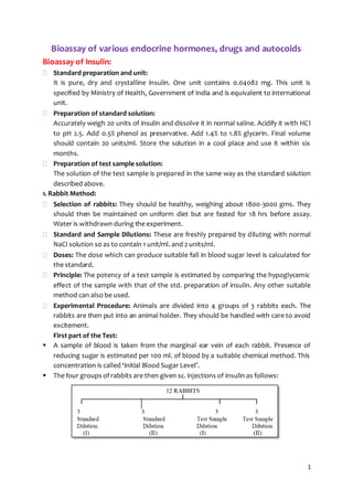
  • 1. 1 Bioassay of various endocrine hormones, drugs and autocoids Bioassay of Insulin:  Standard preparation and unit: It is pure, dry and crystalline insulin. One unit contains 0.04082 mg. This unit is specified by Ministry of Health, Government of India and is equivalent to international unit.  Preparation of standard solution: Accurately weigh 20 units of insulin and dissolve it in normal saline. Acidify it with HCl to pH 2.5. Add 0.5% phenol as preservative. Add 1.4% to 1.8% glycerin. Final volume should contain 20 units/ml. Store the solution in a cool place and use it within six months.  Preparation of test sample solution: The solution of the test sample is prepared in the same way as the standard solution described above. 1. Rabbit Method:  Selection of rabbits: They should be healthy, weighing about 1800-3000 gms. They should then be maintained on uniform diet but are fasted for 18 hrs before assay. Water is withdrawn during the experiment.  Standard and Sample Dilutions: These are freshly prepared by diluting with normal NaCl solution so as to contain 1 unit/ml. and 2 units/ml.  Doses: The dose which can produce suitable fall in blood sugar level is calculated for the standard.  Principle: The potency of a test sample is estimated by comparing the hypoglycemic effect of the sample with that of the std. preparation of insulin. Any other suitable method can also be used.  Experimental Procedure: Animals are divided into 4 groups of 3 rabbits each. The rabbits are then put into an animal holder. They should be handled with care to avoid excitement. First part of the Test:  A sample of blood is taken from the marginal ear vein of each rabbit. Presence of reducing sugar is estimated per 100 ml. of blood by a suitable chemical method. This concentration is called ‘Initial Blood Sugar Level’.  The four groups of rabbits are then given sc. injections of insulin as follows:
  • 2. 2  From each rabbit, a sample of blood is withdrawn up to 5 hrs at the interval of 1 hr. each. Blood sugar is determined again. This is known as ‘Final Blood Sugar Level’. Second part of the test (Cross over test):  The same animals are used for the second part.  The experiment can be carried out after one week.  Again they are fasted and initial blood sugar is determined.  The grouping is reversed, that is to say, those animals which received the standard are given the test and those which received the test are now given the standard.  Those animals which received the less dose of the standard are given the higher dose of the test sample and vice-versa. This test is known as ‘Twin Cross Over Test’.  Mean percentage decrease in blood sugar of the first and second part is calculated. 2. Mouse Method: Mice show characteristic convulsions after s.c. inj. of insulin at elevated temperatures. The percentage convulsions produced by the test and standard preparations are compared. Experimental procedure: Minimum 100 mice weighing between 18-22 gms of the same strain are used. They should be maintained on constant diet. They should be fasted 18 hrs prior to the experiment. Standard and sample dilutions: Dilutions are prepared with sterile saline solution, so as to contain 0.064 units/ml (std. dilution I) and 0.096 units/ml (std. dilution II). Similarly, test sample solutions are also prepared. Mice are divided into 4 groups each containing 25 mice and insulin is injected s.c. as follows: Mice are put in an air incubator at 33o C and observed for one and a half hr. An air incubator with a glass front provided with six shelves is used. The temperature is thermostatically controlled. Two mice are kept in each of the boxes made up of perforated sheets of metal. The mice which convulse or die are taken out of the incubator and observed. These mice usually convulse severely but failure of the animal to upright itself when placed on its back, should as well be considered as convulsion. Convulsive mice may be saved by an inj. of 0.5 ml. of 5% dextrose solution. Percentage convulsions produced by the test sample are compared with those of the standard sample. Those animals which survive may be used again for another expt. after an interval of one week.
  • 3. 3 3. Rat diaphragm method: In this method increase in glycogen content of the muscle or increase in glucose uptake by muscle in response to insulin is taken as the index of potency of insulin. 4. Rat epididymal fat-pad method: Here, the ability of insulin to increase CO2 production by the fat-pad is taken as the parameter for the measurement of potency of the insulin preparation. 5. Radioimmunoassay: It is the estimation of the concentration of the substance in a unit quantity of preparation using radiolabelled antigens. A number of drugs are estimated now days by radioimmunoassy methods because these methods are highly specific and highly sensitive. The radioimmunoassay of insulin is based on the ability of human insulin (unlabelled) to displace beef’s insulin (which may be labelled) from the binding sites (i.e. antibodies). The method involves the following steps: I. Bovine insulin is injected into the sheep. After a week the serum containing antibodies produced against bovine insulin is collected from the blood of the sheep. II. The serum containing antibodies is exposed to radiolabelled insulin and the bound vs. free ratio is determined. III. The mixture of labelled antigen-antibodies is then added in different test-tubes labelled as standard and test. About 6 concentrations of the standards are taken. They are then added to different tubes and the bound vs free ratio is again determined using gamma-counter. IV. Standard curves are determined and the concentration of test insulin is determined using this standard curve. Bioassay of oxytocin:  About Oxytocin:  Oxytocin (Oxy- rapid, tocos- labour)  Synthesized in both sexes, well recognized physiological effects only in women.  Cyclic Polypeptide hormone - from posterior pituitary gland.  Pituitary gland consist posterior lobe which produce oxytocin and diuretic hormone.  Neurosecretory product mainly synthesize in the cell bodies of paraventricular nuclear of the hypothalamus.  Role of Oxytocin:  Stimulate the contraction of the uterine smooth muscle & memory gland.  Oestrogen, progesterone & prolactin- responsible for production of milk by memory gland but milk ejection require oxytocin.  Facilitates the contraction of uterus.  Oxytocin preparation:  Animal species have 90 - 110% stated number of units of oxytocin activity.  It is presented as a solid or solution in a solvent containing an appropriate antimicrobial preservative such as 0.2% w/v of chlorbutol.
  • 4. 4  Synthetic: Solid contains NLT 560 units/mg of oxytocin and liquid contain NLT 150 units/ml of oxytocin calculated with reference to the peptide content.  Actions of Oxytocin:  Oxytocin (as a neuropeptide) synthesized in hypothalamus stimulates uterine contractions to expel the infant from uterus.  Responsible for milk let-down & triggered by the nipple stimulation of suckling called love & bonding hormone.  It has a very special effect on mothering It enables the growing sense of love and attachment to the infant. The more the infant suck, the more oxytocin is produced. In mothers it increases their attachment to their infant, promoting the feeling of love, and makes her infant more valuable to her. It also suppresses the fear that would normally cause her to back off from threat.  Psychologically, oxytocin promotes a feeling of well-being and tranquillity.  Principle of bioassay: Potency of test is determined by comparing its activity with standard preparation of oxytocin. The activity may characterized as depression of BP, contraction of uterus, milk ejection pressure, vasopressor effect.  Standard preparation: Consisting freeze dried synthetic oxytocin peptide with human albumin citric acid (12.5 units) 1) Depression of the BP in chicken  Test animals: Healthy cockerel (young male chicken), 1.2 - 2.3 kg,  Anaesthetized cock-prolonged & constant high B.P  Expose gluteus primus muscle (thigh) & remove popliteal artery & crural vein.  Cannulate the popliteal artery & record B.P response.  Cannulate the crural or brachial vein. Prepare standard solution with saline.  Inject 0.1 - 0.5ml Inject 2 doses of standard solution into cannulated vein and record B.P response.  Dose should cause decrease in B.P (required dose is between 20-100mUnits).  Interval between 2 doses of injections, 3-10mins depend on rate at which B.P return normal.  Dilute the test solution of oxytocin with saline in order to get same response as standard.  The ratio between standard & test should be equal.  If animal rapidly becomes insensitive to repeated injection, another solution must be used. Measure all responses are calculated result of the assay by standard statistical method. 2) Contraction of the rat uterus:  Test animals: Female rat 120 – 200g  Immediately before assay, select the rats in oestrus or preoestrus phase of estrus cycle confirmed by vaginal smear.  Inject 100ug of estradiol benzoate IM into female rat before the assay.
  • 5. 5  Kill rat & suspend one horn of uterus in organ bath containing PSS solution of following salts- NaCl, KCl, CaCl2, NaHCO3, Na2HPO4, Mgcl2, and dextrose.  Maintain the bath at temp at of 32⁰C.  Oxygenate solution with mix of 95% of O2, 5% of CO2.  Bath liquid (PSS) required dose between 10-50 units/ml.  Record contraction of uterine muscle produces by addition of two dose of standard preparation (Required dose 10 & 50 munits/ml of bath liquid. PSS).  When maximum contraction has been reached, replaced bath liquid (PSS) by fresh solution.  Dose should be added at regular interval [3-5minutes].  Similarly, record the contraction of uterine muscle produced by test preparation.  The ratio between two dose of test & two dose of standard should be equal. This ratio kept constant throughout the assay.  Measure all response & calculate result of assay by standard statistical method. 3) Milk ejection pressure in Lactating rat:  Test animals: Select a lactating rat, in the third to twenty-first day after parturition and weighing about 300 g, separate it from the litter and 30 to 60 minutes later anaesthetise (for example, by the intraperitoneal injection of a solution of Pentobarbitone Sodium).  Tie the rat to an operating table, maintained at 37o, by its hind legs leaving the front legs free.  Cannulate the trachea with a short polyethylene tube of internal diameter about 2.5 mm in such a manner so as to ensure a free airway; apply artificial respiration only if necessary.  Cannulate an external jugular or femoral vein with a polyethylene tube of internal diameter about 0.4 mm which is filled with saline solution and closed with a pin.  Shave the skin surrounding the inguinal and abdominal teats and excise the tip of one teat, preferably the lower inguinal teat.  Insert a polyethylene tube of internal diameter about 0.3 mm and external diameter about 0.6 mm, to a depth sufficient to obtain appropriate measurement of pressure (3 to 10 mm depth), into the primary teat duct which opens onto the cut surface and tie firmly in place with a ligature.  Connect this cannula with a suitable strain gauge transducer (such as that used for recording arterial blood pressure in the rat) and fill the whole system with a 3.8% w/v solution of sodium citrate or saline solution containing 50 Units of heparin sodium per ml to prevent clotting of milk.  After cannulation, inject a small volume (0.05 to 0.2 ml) of this solution into the teat duct through the transducer to clear the milk from the tip of the cannula. (This procedure may be repeated during the assay should obstruction arise from milk ejected into the cannula).
  • 6. 6  Clamp the strain gauge so that a slight tension is applied to the teat and its natural alignment is preserved and connect the gauge to a potentiometric recorder adjusted to give full-scale deflection for an increase in milk-ejection pressure of about 5.3 kPa.  Inject all solutions through the venous cannula using a 1-ml syringe graduated in 0.01 ml and wash them in with 0.2 ml of saline solution.  Prepare a solution of the Standard Preparation and a solution of the preparation being examined in saline solution so that the volume to be injected is between 0.1 ml and 0.4 ml.  Choose two doses of the Standard Preparation such that the increase in milk-ejection pressure is about 1.35 kPa for the lower dose and about 2.7 kPa for the higher dose. As an initial approximation, a lower dose of between 0.1 and 0.4 milliUnit and an upper dose of 1.5 to 2 times this amount may be tried.  Choose two doses of the preparation being examined with the same inter-dose ratio, matching the effects of the doses of the Standard Preparation as closely as possible.  Inject the four doses (two doses of the Standard Preparation and two doses of the preparation being examined) at intervals of 3 to 5 minutes.  The two doses of Standard Preparation and the two doses of the preparation being examined should be given according to a randomised block or a Latin square design and at least four responses to each should be recorded.  Measure all the responses and calculate the result of the assay by standard statistical methods. Bioassay of Vasopressin:  Principle: The vasopressor activity is estimated by comparing the activity of the preparation being examined with that of the Standard Preparation of arginine vasopressin under the conditions of a suitable method of assay.  Standard Preparation: The Standard Preparation is the Ist International Standard for Arginine vasopressin, established in 1978, consisting of freeze-dried synthetic arginine vasopressin peptide acetate with human albumin and citric acid (supplied in ampoules containing 8.20 Units), or another suitable preparation the potency of which has been determined in relation to that of the International Standard.  Suggested Method:  Inject slowly into the tail vein of a male albino rat weighing about 300 g a solution of a suitable alpha -adrenoceptor blocking agent, for example 10 ml per kg of body weight of a solution prepared by dissolving 5 mg of phenoxybenzamine hydrochloride in 0.1 ml of ethanol (95%), adding 0.05 ml of 1M hydrochloride acid and diluting to 5 ml with saline solution.  After 18 hours, anaesthetise the rat with an anaesthetic that will maintain a prolonged and uniform blood pressure. After 45 to 60 minutes, tie the rat on its back to the operating table by its hind legs.
  • 7. 7  Cannulate the trachea with a short polyethylene tube of external diameter about 2.5 mm and dissect a carotid artery ready for cannulation.  Then cannulate the femoral vein close to the inguinal ligament.  Retract the abdominal muscles to expose the inguinal ligament.  Retract the superficial pudendal vein to one side and dissect the femoral vein towards the inguinal ligament from the corresponding artery.  When dissecting, a deep branch reaching the femoral vein must be found and tied off to prevent bleeding during cannulation.  Tie a short polyethylene cannula of external diameter about 1 mm into the femoral vein by two ligatures and join by a short piece of flexible tubing to a 1-ml burette with an attached thistle funnel containing saline solution at about 37o.  Firmly fix a wet absorbent cotton swab to the thigh so as to cover the incision and cannula.  At this stage, inject through the venous cannula 200 Units of heparin dissolved in saline solution per 100 g of body weight.  Then tie in a carotid cannula of external diameter about 1 mm and connect by a column of saline solution containing heparin with a suitable pressure measuring device such as a mercury manometer of internal diameter about 2 to 3 mm.  The central and peripheral nervous system including both vagus and associated sympathetic nerves is left intact. No artificial respiration is necessary. Taking care that no air is injected, inject all solutions through the venous cannula by means of a 1-ml syringe graduated in 0.01 ml and wash in with 0.2 ml of saline solution from the burette.  Dilute the extract of the standard preparation and the preparation being examined with saline solution so that the volume to be injected is between 0.1 ml and 0.5 ml.  Choose two doses of the standard preparation such that the elevation of the blood pressure is about 4 kPa for the lower dose and about 7 kPa but always submaximal for the higher dose, the ratio of low to high dose being determined by the response and usually being 3 to 5.  As an initial approximation doses of 3 and 5 milliUnits may be tried.  Choose two doses of the preparation being examined with the same inter-dose ratio, matching the effects of the dose of the standard preparation as closely as possible.  Inject doses at intervals of 10 to 15 minutes.  The two doses of the standard preparation and the two doses of the preparation being examined should be given in a randomised block or a Latin square design and four to five responses to each should be recorded.  Measure all the responses and calculate the result of the assay by standard statistical methods. Bioassay of d-Tubocurarine: 1. Rabbit Head-drop Method:  Principle:
  • 8. 8 d-Tubocurarine hydrochloride is injected into the marginal vein of a rabbit’s ear till the rabbit’s neck muscles are relaxed such that the animal cannot hold its head up. The total amount of test sample required to produce the endpoint is compared with the total amount of the standard sample required to produce similar endpoint.  Selection of Rabbits: Rabbits weighing 2 kg are used. Animals should be free from disease, obtained from a healthy colony and should be accustomed with the experimental procedure.  Experimental Procedure:  Each rabbit is placed in a holder with its head protruding outside. The head should be freely movable. Minimum 8 rabbits are used. They are divided into two groups each containing 4 rabbits. First group will receive standard sample and the second group will receive the sample under test. D-Tubocurarine solution is injected at a constant speed by infusion apparatus through the marginal vein.  Injection should be given at a rate of 0.4 ml/min and should take about 10 min. Infusion is continued till the rabbit will not be in a position to hold its head erect or there will be no response by focussing light on the eyes and the neck gets elongated and toneless.  Suitable dose of d-tubocurarine is 0.012% w/v in saline. Rabbits recover immediately from the effect of curarization. During the expt. there is a possibility or respiratory embarrassment which is treated by injecting neostigmine methyl sulphate (0.05 mg.) and atropine sulphate immediately through the marginal ear vein.  Cross-over test is carried out to minimise biological error due to animal variation. Those rabbits which received the standard sample on the first day will be given test sample on the second day of expt. and vice versa. Mean dose which produces head drop of the test sample is compared with the mean dose of standard preparation. 2) Frog’s Rectus Abdominis muscle Preparation:  A frog is pithed and laid on its back on a cork covered board to which it is pinned. The skin covering the abdomen is cut away and the rectus abdominis muscle of one side is dissected from the pelvic girdle to its insertion in the cartilage of the pectoral girdle.  The muscle is then pinned to the cork by four pins to keep its normal length while a thread is sewn through each end.  It is then mounted in the organ bath containing frog’s Ringer solution which contains NaCl, 6.5gm; KCl, 0.29 gm; CaCl2, 0.24 gm; NaHCO3, 0.4 gm; glucose, 1.5 gm and distilled water 2000 ml . Oxygenation is carried out to keep the tissue alive. The muscle is stabilized for 30-45 min. in order to get critical quantitative response. The responses are recorded using isotonic frontal writing lever with 1 G. tension.  Two similar contractions with the same concentration of acetylcholine are obtained. Three doses of the standard sample and one intermediate dose of the test sample are selected and the reduction in height of contraction induced by acetylcholine is noted down.
  • 9. 9  Acetylcholine contraction is recorded on slow moving drum for 90 sec. d- Tubocurarine is allowed to act for 30 sec. The percentage reduction at each dose levels is calculated and log dose response curve of the standard drug is plotted. A linear response will be obtained. The potency of test sample is calculated from the standard curve. Bioassay of digitalis:  Principle: Potency of the test sample is compared with that of the standard preparation by determining the action on the cardiac muscle. Any other equivalent method, which gives results similar to those obtained by this method as also valid.  Standard Preparation and Units: The standard preparation is a mixture of dried and powdered digitalis leaves (1 unit = 76 mg.)  Preparation of Extracts: Exact amount of the powder is extracted with dehydrated alcohol in a continuous extraction apparatus for six hours. The final extract should contain 10 ml. (5 ml. alcohol + 5 ml. water) per 10 g. of digitalis powder. It should be stored in between 5 ⁰C and –5 ⁰C. Pigeon Method: Minimum 6 pigeons are used for testing each sample. They should be free from gross evidence of disease or emaciation. The weight of the heaviest pigeon should not exceed twice the weight of the lightest pigeon. Food is withheld 16-28 hours before the experiment. Pigeons are divided on the basis of their sex, weight and breed, into two groups. They are anaesthetized with anaesthetic ether. One side of the wing is dissected and the alar vein is cannulated by means of a venous cannula. Dilutions are made with normal saline. Average lethal dose of each sample is determined; results are tabulated and calculated as per guinea pig method. The lethal dose per kg of body weight is determined for each pigeon. The potency of the test sample is determined by dividing the mean lethal dose of standard by the mean lethal dose of the test sample. In pigeons, stoppage of heart is associated with a characteristic vomiting response called ‘emesis. The milk from the crop sac of pigeons is being ejected out. This may be taken as the end point response of digitalis. Bioassay of histamine: 1. Contractions of isolated guinea pig ileum Bioassay of histamine on isolated guinea pig ileum can be determined by graded response bioassays like matching, interpolation, bracketing and multiple point bioassays.  Matching Method: In this method a constant dose of the test is bracketed by varying doses of standard till the exact match is obtained between test dose and the standard dose.
  • 10. 10 Initially, two responses of the standard are taken. The doses are adjusted such that one is giving response of approximately 20% and other 70% of the maximum. The response of unknown which lies between two responses of standard dose is taken. The panel is repeated by increasing or decreasing the dose s of standard till all three equal responses are obtained. The dose of test sample is kept constant. At the end, a response of the double dose of the standard and test which match each other are taken. These should give equal responses. Concentration of the test sample can be determined as follows: This method has following limitations: 1. It occupies a larger area of the drum as far as tracings are concerned. 2. The match is purely subjective, so chances of error are there and one cannot determine them. 3. It does not give any idea of dose-response relationship. However, this method is particularly useful if the sensitivity of the preparation is not stable. Bioassay of histamine, on guinea pig ileum is preferably carried out by matching method is shown below-  Graphical/ interpolation method: This method is based on the assumption of the dose-response relationship. Log-dose- response curve is plotted and the dose of standard producing the same response as produced by the test sample is directly read from the graph. In simpler design, 5-6 responses of the graded doses of the standard are taken and then two equiactive responses of the test sample are taken. The height of contraction is measured and plotted against the log-dose. The dose of standard producing the same response as produced by the test is read directly from the graph and the concentration of test sample is determined by the same formula as mentioned before.
  • 11. 11 The characteristic of log-dose response curve is that it is linear in the middle (20-80%). Thus, the comparison should be done within this range only. In other words, the response of test sample must lie within this range. Advantage of this method is that, it is a simple method and chances of errors are less if the sensitivity of the preparation is not changed.  Multiple point bioassays: These include 3 point, 4 point and 6 point methods. In these 4 methods, the responses are repeated several times and the mean of each is taken. Thus, chances of error are minimized in these methods. In 3 point assay method 2 doses of the standard and one dose of the test are used. In 4 point method 2 doses of standard and 2 doses of the test are used. In 6 point method 3 doses of standard and 3 doses of the test are used. Similarly one can design 8 point method also. The sequence of responses is followed as per the Latin square method of randomization in order to avoid any bias. The mean responses are calculated and plotted against log-dose and amount of standard producing the same response as produced by the test is determined graphically as well as mathematically: Where, n1 = Lower Standard dose, n2 = Higher Standard dose, t = Test dose, S1 = Response of n1, S2 = Response of n2, T = Response of test (t), and Cs = Concentration of standard.
  • 12. 12 Similarly, in 4 point method, amount of standard producing the same response as produced by the test can be determined by graphical method. It is determined mathematically as follows: Where, t1 = lower dose of test, t2 = higher dose of test, T1 = response of t1, T2 = response of t2. Histamine receptors in ileum:  Histamine receptors in ileum H1 receptors  Receptor type: G-protein-coupled receptor  Agonist: Histamine  Mechanism of contraction: ++ G-protein→ ++ phospholipase C→ splitting of PIP2 into- 1) DAG that increases the opening of calcium channels, and 2) IP3 which increases calcium mobilization from sarcoplasmic stores.DAG & IP3 lead to increase in the intracellular concentration of calcium and smooth muscles contraction. 2) Fall in BP in anaesthetised and atropinized cat or dog: Cat or dog is anaesthetised with chloralose or barbiturate and prepared for recording of BP. Sensitivity is determined by injecting standard solution of 0.05, 0.1, 0.15 µg of histamine base per kg body weight for 5min interval. A fixed dose of standard producing a fall in BP about 20 mm Hg is injected with changing doses of test at regular intervals and matching assay is done.  Mechanism of BP fall: Histamine binds with histamine H1 receptor of endothelium causes release of EDRF (endothelium derived relaxing factor). This EDRF diffuses out and reaches the smooth muscle of the arteriole and causes generation of cyclic-GMP→ causing reduction of Ca2+ in smooth muscle relaxation of the smooth muscle→ arteriolar dilatation. Another possibility is that combination of H1 with the Histamine causes release of PGI2 which causes vasodilatation. Bioassay of 5-hydroxytryptamine (5HT/ serotonin): 1) Contraction of isolated atropinized rat uterus, 2) Contraction of isolated terminal colon of rat, 3) Contraction of isolated fundus strip of rat stomach, and 4) Constriction of blood vessels of perfused rabbit ear.