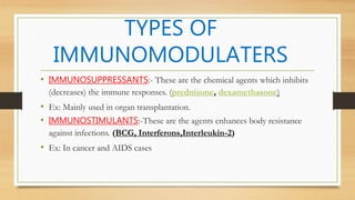
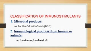
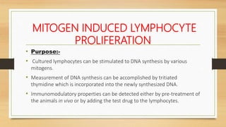
Screening models for immunomodulatory agents:- Introduction for immunostimulants and immunosuppressant, Models for immunomodulatory agents, Screening for immunostimulants, screening for immunosuppressant
Screening methods of immunomodulators by shivam diwakerShivam Diwaker
Immune Modulators are the substances or drugs or chemical compounds that are used for the modification in the Immune system such as stimulate and suppress.
Screening methods of immunomodulators by shivam diwakerShivam Diwaker
Immune Modulators are the substances or drugs or chemical compounds that are used for the modification in the Immune system such as stimulate and suppress.
Assignment on Preclinical Screening of ImmunomodulatorsDeepak Kumar
Assignment on Preclinical screening of new substances for the pharmacological activity using in vivo, in vitro, and other possible animal alternative models
Introduction to Screening Models of Anti-Atherosclerosis
Atherosclerosis, Screening models, In vitro models, In vivo models
Presented by
SHAIK FIRDOUS BANU
Department of Pharmacology
Extrapolation of in vitro data to preclinical and.pptxARSHIKHANAM4
Extrapolation of in vitro data to preclinical.
the topic is included in m.pharmacy 1st sem syllabus. which is essential for the study and that include the details about how you deal with the preclinical data that will help to decide the NOEAL and LOEAL, the humane dose of the drug can be calculated and further formation is also done.
Screening models For immunosuppressants ..pptxRahulTipare
Explore the dynamic world of screening models for immunosuppressants in our comprehensive academic seminar. Delve into cutting-edge methodologies and advancements aimed at enhancing efficacy and safety profiles. Uncover the latest innovations driving the development of immunosuppressive therapies, vital for transplant success and autoimmune disorder management. Join us on this enlightening journey through the evolving landscape of immunosuppressant screening models.
(May 29th, 2024) Advancements in Intravital Microscopy- Insights for Preclini...Scintica Instrumentation
Intravital microscopy (IVM) is a powerful tool utilized to study cellular behavior over time and space in vivo. Much of our understanding of cell biology has been accomplished using various in vitro and ex vivo methods; however, these studies do not necessarily reflect the natural dynamics of biological processes. Unlike traditional cell culture or fixed tissue imaging, IVM allows for the ultra-fast high-resolution imaging of cellular processes over time and space and were studied in its natural environment. Real-time visualization of biological processes in the context of an intact organism helps maintain physiological relevance and provide insights into the progression of disease, response to treatments or developmental processes.
In this webinar we give an overview of advanced applications of the IVM system in preclinical research. IVIM technology is a provider of all-in-one intravital microscopy systems and solutions optimized for in vivo imaging of live animal models at sub-micron resolution. The system’s unique features and user-friendly software enables researchers to probe fast dynamic biological processes such as immune cell tracking, cell-cell interaction as well as vascularization and tumor metastasis with exceptional detail. This webinar will also give an overview of IVM being utilized in drug development, offering a view into the intricate interaction between drugs/nanoparticles and tissues in vivo and allows for the evaluation of therapeutic intervention in a variety of tissues and organs. This interdisciplinary collaboration continues to drive the advancements of novel therapeutic strategies.
Observation of Io’s Resurfacing via Plume Deposition Using Ground-based Adapt...Sérgio Sacani
Since volcanic activity was first discovered on Io from Voyager images in 1979, changes
on Io’s surface have been monitored from both spacecraft and ground-based telescopes.
Here, we present the highest spatial resolution images of Io ever obtained from a groundbased telescope. These images, acquired by the SHARK-VIS instrument on the Large
Binocular Telescope, show evidence of a major resurfacing event on Io’s trailing hemisphere. When compared to the most recent spacecraft images, the SHARK-VIS images
show that a plume deposit from a powerful eruption at Pillan Patera has covered part
of the long-lived Pele plume deposit. Although this type of resurfacing event may be common on Io, few have been detected due to the rarity of spacecraft visits and the previously low spatial resolution available from Earth-based telescopes. The SHARK-VIS instrument ushers in a new era of high resolution imaging of Io’s surface using adaptive
optics at visible wavelengths.
Introduction:
RNA interference (RNAi) or Post-Transcriptional Gene Silencing (PTGS) is an important biological process for modulating eukaryotic gene expression.
It is highly conserved process of posttranscriptional gene silencing by which double stranded RNA (dsRNA) causes sequence-specific degradation of mRNA sequences.
dsRNA-induced gene silencing (RNAi) is reported in a wide range of eukaryotes ranging from worms, insects, mammals and plants.
This process mediates resistance to both endogenous parasitic and exogenous pathogenic nucleic acids, and regulates the expression of protein-coding genes.
What are small ncRNAs?
micro RNA (miRNA)
short interfering RNA (siRNA)
Properties of small non-coding RNA:
Involved in silencing mRNA transcripts.
Called “small” because they are usually only about 21-24 nucleotides long.
Synthesized by first cutting up longer precursor sequences (like the 61nt one that Lee discovered).
Silence an mRNA by base pairing with some sequence on the mRNA.
Discovery of siRNA?
The first small RNA:
In 1993 Rosalind Lee (Victor Ambros lab) was studying a non- coding gene in C. elegans, lin-4, that was involved in silencing of another gene, lin-14, at the appropriate time in the
development of the worm C. elegans.
Two small transcripts of lin-4 (22nt and 61nt) were found to be complementary to a sequence in the 3' UTR of lin-14.
Because lin-4 encoded no protein, she deduced that it must be these transcripts that are causing the silencing by RNA-RNA interactions.
Types of RNAi ( non coding RNA)
MiRNA
Length (23-25 nt)
Trans acting
Binds with target MRNA in mismatch
Translation inhibition
Si RNA
Length 21 nt.
Cis acting
Bind with target Mrna in perfect complementary sequence
Piwi-RNA
Length ; 25 to 36 nt.
Expressed in Germ Cells
Regulates trnasposomes activity
MECHANISM OF RNAI:
First the double-stranded RNA teams up with a protein complex named Dicer, which cuts the long RNA into short pieces.
Then another protein complex called RISC (RNA-induced silencing complex) discards one of the two RNA strands.
The RISC-docked, single-stranded RNA then pairs with the homologous mRNA and destroys it.
THE RISC COMPLEX:
RISC is large(>500kD) RNA multi- protein Binding complex which triggers MRNA degradation in response to MRNA
Unwinding of double stranded Si RNA by ATP independent Helicase
Active component of RISC is Ago proteins( ENDONUCLEASE) which cleave target MRNA.
DICER: endonuclease (RNase Family III)
Argonaute: Central Component of the RNA-Induced Silencing Complex (RISC)
One strand of the dsRNA produced by Dicer is retained in the RISC complex in association with Argonaute
ARGONAUTE PROTEIN :
1.PAZ(PIWI/Argonaute/ Zwille)- Recognition of target MRNA
2.PIWI (p-element induced wimpy Testis)- breaks Phosphodiester bond of mRNA.)RNAse H activity.
MiRNA:
The Double-stranded RNAs are naturally produced in eukaryotic cells during development, and they have a key role in regulating gene expression .
The increased availability of biomedical data, particularly in the public domain, offers the opportunity to better understand human health and to develop effective therapeutics for a wide range of unmet medical needs. However, data scientists remain stymied by the fact that data remain hard to find and to productively reuse because data and their metadata i) are wholly inaccessible, ii) are in non-standard or incompatible representations, iii) do not conform to community standards, and iv) have unclear or highly restricted terms and conditions that preclude legitimate reuse. These limitations require a rethink on data can be made machine and AI-ready - the key motivation behind the FAIR Guiding Principles. Concurrently, while recent efforts have explored the use of deep learning to fuse disparate data into predictive models for a wide range of biomedical applications, these models often fail even when the correct answer is already known, and fail to explain individual predictions in terms that data scientists can appreciate. These limitations suggest that new methods to produce practical artificial intelligence are still needed.
In this talk, I will discuss our work in (1) building an integrative knowledge infrastructure to prepare FAIR and "AI-ready" data and services along with (2) neurosymbolic AI methods to improve the quality of predictions and to generate plausible explanations. Attention is given to standards, platforms, and methods to wrangle knowledge into simple, but effective semantic and latent representations, and to make these available into standards-compliant and discoverable interfaces that can be used in model building, validation, and explanation. Our work, and those of others in the field, creates a baseline for building trustworthy and easy to deploy AI models in biomedicine.
Bio
Dr. Michel Dumontier is the Distinguished Professor of Data Science at Maastricht University, founder and executive director of the Institute of Data Science, and co-founder of the FAIR (Findable, Accessible, Interoperable and Reusable) data principles. His research explores socio-technological approaches for responsible discovery science, which includes collaborative multi-modal knowledge graphs, privacy-preserving distributed data mining, and AI methods for drug discovery and personalized medicine. His work is supported through the Dutch National Research Agenda, the Netherlands Organisation for Scientific Research, Horizon Europe, the European Open Science Cloud, the US National Institutes of Health, and a Marie-Curie Innovative Training Network. He is the editor-in-chief for the journal Data Science and is internationally recognized for his contributions in bioinformatics, biomedical informatics, and semantic technologies including ontologies and linked data.
Slide 1: Title Slide
Extrachromosomal Inheritance
Slide 2: Introduction to Extrachromosomal Inheritance
Definition: Extrachromosomal inheritance refers to the transmission of genetic material that is not found within the nucleus.
Key Components: Involves genes located in mitochondria, chloroplasts, and plasmids.
Slide 3: Mitochondrial Inheritance
Mitochondria: Organelles responsible for energy production.
Mitochondrial DNA (mtDNA): Circular DNA molecule found in mitochondria.
Inheritance Pattern: Maternally inherited, meaning it is passed from mothers to all their offspring.
Diseases: Examples include Leber’s hereditary optic neuropathy (LHON) and mitochondrial myopathy.
Slide 4: Chloroplast Inheritance
Chloroplasts: Organelles responsible for photosynthesis in plants.
Chloroplast DNA (cpDNA): Circular DNA molecule found in chloroplasts.
Inheritance Pattern: Often maternally inherited in most plants, but can vary in some species.
Examples: Variegation in plants, where leaf color patterns are determined by chloroplast DNA.
Slide 5: Plasmid Inheritance
Plasmids: Small, circular DNA molecules found in bacteria and some eukaryotes.
Features: Can carry antibiotic resistance genes and can be transferred between cells through processes like conjugation.
Significance: Important in biotechnology for gene cloning and genetic engineering.
Slide 6: Mechanisms of Extrachromosomal Inheritance
Non-Mendelian Patterns: Do not follow Mendel’s laws of inheritance.
Cytoplasmic Segregation: During cell division, organelles like mitochondria and chloroplasts are randomly distributed to daughter cells.
Heteroplasmy: Presence of more than one type of organellar genome within a cell, leading to variation in expression.
Slide 7: Examples of Extrachromosomal Inheritance
Four O’clock Plant (Mirabilis jalapa): Shows variegated leaves due to different cpDNA in leaf cells.
Petite Mutants in Yeast: Result from mutations in mitochondrial DNA affecting respiration.
Slide 8: Importance of Extrachromosomal Inheritance
Evolution: Provides insight into the evolution of eukaryotic cells.
Medicine: Understanding mitochondrial inheritance helps in diagnosing and treating mitochondrial diseases.
Agriculture: Chloroplast inheritance can be used in plant breeding and genetic modification.
Slide 9: Recent Research and Advances
Gene Editing: Techniques like CRISPR-Cas9 are being used to edit mitochondrial and chloroplast DNA.
Therapies: Development of mitochondrial replacement therapy (MRT) for preventing mitochondrial diseases.
Slide 10: Conclusion
Summary: Extrachromosomal inheritance involves the transmission of genetic material outside the nucleus and plays a crucial role in genetics, medicine, and biotechnology.
Future Directions: Continued research and technological advancements hold promise for new treatments and applications.
Slide 11: Questions and Discussion
Invite Audience: Open the floor for any questions or further discussion on the topic.
THE IMPORTANCE OF MARTIAN ATMOSPHERE SAMPLE RETURN.Sérgio Sacani
The return of a sample of near-surface atmosphere from Mars would facilitate answers to several first-order science questions surrounding the formation and evolution of the planet. One of the important aspects of terrestrial planet formation in general is the role that primary atmospheres played in influencing the chemistry and structure of the planets and their antecedents. Studies of the martian atmosphere can be used to investigate the role of a primary atmosphere in its history. Atmosphere samples would also inform our understanding of the near-surface chemistry of the planet, and ultimately the prospects for life. High-precision isotopic analyses of constituent gases are needed to address these questions, requiring that the analyses are made on returned samples rather than in situ.
2. IMMUNOMODULATORS
These are the chemical agents which modifies the
Immune responses or the functioning of the
Immune system.
Either by inducing or inhibition of immune
responses.
3. TYPES OF
IMMUNOMODULATERS
• IMMUNOSUPPRESSANTS:- These are the chemical agents which inhibits
(decreases) the immune responses. (prednisone, dexamethasone)
• Ex: Mainly used in organ transplantation.
• IMMUNOSTIMULANTS:-These are the agents enhances body resistance
against infections. (BCG, Interferons,Interleukin-2)
• Ex: In cancer and AIDS cases
4. CLASSIFICATION OF IMMUNOSTIMULANTS
1. Microbial products:
ex: Bacillus Calmette-Guerin(BCG)
2. Immunological products from human or
animals:
ex: Interferons,Interleukin-2
6. SCREENING MODELS FOR
IMMUNOMODULATORS
• IN VITRO METHODS:-
• Inhibition of Histamine Release from mast Cells.
• Mitogen induced lymphocyte proliferation.
• IN VIVO METHODS:-
• Delayed Type Hypersensitivity.
9. INHIBITION OF HISTAMINE RELEASE
FROM MAST CELLS
• PURPOSE
• Hypersensitivity reactions can be elicited by various factors:
• Immunologically induced,
• Non- Immunologically induced,
• Mediation through immune responses.
10. Mediators are responsible for hypersensitivity reactions are released from mast cells.
An important performed mediators of allergic reactions found in these cells of histamine.
Mediators are responsible
for hypersensitivity
reactions are released
from mast cells.
An important performed
mediators of allergic
reactions found in these
cells of histamine.
11. Procedure:-
Preparation of mast cell suspension:-
Wistar rats are decapitated and exsanguinated
50ml of HBSS(Hanks balanced salt solution)
injected into peritoneal cavity
Fluid containing peritoneal cells collect in a
centrifuge tube
and centrifuged at 2000rpm
12. The cells are Resuspended in HBSS.
Finally Brought to concentration of 105mast cells/100ul
13. TEST COMPOUND ADMISTRATION AND
INDUCTION OF HISTAMINE RELEASE
1ml of test drug and mast cells suspension incubated at 37degres
for 15min.
The cells are made up of volume 3ml with HBSS equal to specific
allergen is added.
The suspension incubated at 37degres for 30 min followed by
centrifugation at 2500rpm
14. DETERMINATION OF HISTAMINE RELEASE
• The total sample is transferred to an autosampler
vial.
• Then histamine concentration is determined by a
fluorescence detector
15. EVALUATION
• Percentage histamine release can be Expressed by the
following formulae:-
Sample His.relese. – spontaneous his.rel.
100per His.relese. – spontaneous his.rel.
* 100
16. MITOGEN INDUCED LYMPHOCYTE
PROLIFERATION
• Purpose:-
• Cultured lymphocytes can be stimulated to DNA synthesis by various
mitogens.
• Measurement of DNA synthesis can be accomplished by tritiated
thymidine which is incorporated into the newly synthesized DNA.
• Immunomodulatory properties can be detected either by pre-treatment of
the animals in vivo or by adding the test drug to the lymphocytes.
17. PROCEDURE
• For this procedure Mice or rats are used.
• Animals receive the test compound once a day for 5 days.
• After 5 days sacrificed, spleens are removed and a single cell suspension of
5×10⁶ cells/ml prepared.
• Mitogens are titrated and 0.1ml of the suspensions is added.
• Plates are incubated at 37degrees in 5% Carbon Dioxide in air for 48-60 hrs and
for another 8hrs after addition of 3H-thymidine per well.
18. Cells are harvested on glass fibres filters and drying the
degree of radioactivity.
19. EVALUATION
• Stimulation index = proliferation ratio according to
positive control either with or without mean
spleen weight.
21. DELAYED TYPE
HYPERSENSITIVITY
• PURPOSE:-
• Delayed type hypersensitivity is a reaction of cell mediated
immunity and becomes visible only after 16-24hours.
• The same methods as for testing immediate type
hypersensitivity can be used.
22. PROCEDURE
• Rats are sensitized by Intra muscular administration of 0.5 ml
ovalbumin suspension 7days prior to the start of the
experiment.
• They are challenged by injection of 0.1ml of 0.04% solution of
highly purified ovalbumin in the left hind paw.
• Footpad thickness is measured immediately and 24 hours after
ovalbumin administration.
23. ANTI-ANAPHYLACTIC ACTIVITY
(Schultz-Dale reaction)
• Purpose:-
• Guinea pig are sensitized against egg albumin.
• Challenge after 3 weeks causes in isolated organs release of mediators.
• e.g. Histamine, which induce contraction in isolated ileum.
24. PROCEDURE
• Guinea pigs of either sex (300-350gm) are sensitized with alum ppt egg
albumin.
• Alum egg albumin is prepared by dissolving egg albumin (1mg/ml) in six
percent aluminium hydroxide gel, suspended in saline.
25. • The mixture is stirred and kept at room temperature.
• Each animal receives at the same time injections of 0.125 ml of this
mixture in each foot pad and 0.5ml
• After 4weeks the animals are killed and ileum is dissected out.
26. • Take 2-3cm ileum and mounted in organ bath containing Tyrode solution
at 37degrees.
• Leave for resting time for 15min and tested for contractility of ileum by 10-
4gm/ml BaCl2 solution.
• To one organ bath standard and to other vials the test compounds are
added.
27. • One organ bath is serves as control.
• After 3min ovalbumin in a final concentrations of 2×10⁻⁶g/ml is added.
• The contractions are recorded with strain gauges by a polygraph.
28. EVALUATION
• The results are expressed as presence or absence of blocking
activity (percentage inhibition).
• If anti-anaphylactic activity is observed, ED50 values using
different doses are calculated.
29. ACUTE SYSTEMIC ANAPHYLAXIS IN
RATS• PURPOSE:-
• Rats are immunized with ovalbumin.
• After 11days the animals are challenged by intravenous injection of
ovalbumin.
• The shock symptoms can by inhibited by corticosteroids and I.V disodium
cromoglycate.
30. PROCEDURE
• Female Sprague-Dawley rats (120gm) are immunized by i.m Injection of
10mg/kg highly purified ovalbumin.
• 1ml of Bordetella pertussis suspension is injected intraperitoneally.
• IgE antibodies are induced and attached surface of mast cells and
basophilic granulocytes.
31. • 11days later by i.v injection of 25 mg/kg highly purified ovalbumin to
animals. This results in formation of antigen-antibody-complexes.
• On the surface of mast cells and basophilic granulocytes in blood with
immediate release of various mediators such as histamine, serotonin,
prostaglandins.
32. EVALUATION
• The shock symptoms are scored and mortality counted.
• Results after treatment are compared with untreated controls.
• Pre-treatment with corticosteroids or disodium cromoglycate can inhibit
death and ameliorate shock symptoms.
34. PROCEDURE
• On initial day, all groups were sensitized with 0.1 mL of SRBC containing 1×10*8 cells, i.p.
• Group I served as control and was administered 1% Gum acacia suspension in saline.
• Group II received 100 mg/kg bd.wt. of MESI(Methanolic Extract of Sphaeranthus Indicus)
p.o. respectively (1 to 7 days).
• Group III received 200 mg/kg bd.wt. of MESI p.o. respectively (1 to 7 days).
• Group IV received 400 mg/kg bd.wt. of MESI p.o. respectively (1 to 7 days).
• Group V received 50 mg/kg bd.wt. of standard, Levamisole, p.o. respectively (1 to 7 days)
35. • On 7th day before challenge, blood was withdrawn from retro-orbital plexus of
each animal.
• Blood was centrifuged, and serum was separated. Serial two fold dilutions were
made and 50 µL of serum was added to 1st well of 96-well micro titer plate
containing 50 µL normal saline.
• To this 1% SRBC (50 µL) dissolved in normal saline was mixed.
36. From 1st well 50 µl of diluted serum was added to 2nd well containing 50 µl normal
saline and 50µl 1% SRBC. Such dilutions were done till 24th well.
• Plates were incubated at 37ºC for 1h.
• Highest dilution that has shown visible agglutination was considered as
hemagglutination(HA) antibody .
37. REFERENCE
• Hans Gerhard Vogel. Drug discovery and evaluation:
pharmacological assays, vol 2 and 3rd edition,
Page.no: 792-799.
• Harsh Mohan, Text book pathophysiology, 6th
edition, J.P bros., New Delhi, 2010, Page. no:115-120.
38. • IOSR Journal of Pharmacy and Biological Sciences (IOSR-
JPBS) e-ISSN: 2278-3008, p-ISSN:2319-7676.
• Volume 10, Issue 6 Ver. II (Nov - Dec. 2015), PP 91-99
Page Screening of immunomodulatory activity of
Sphaeranthus indicus Linn.