Cardiac Cycle
•Download as PPT, PDF•
2 likes•165 views

This is for undergraduate students from Zoology or physiology.

Recommended
More Related Content
What's hot
What's hot (20)
Similar to Cardiac Cycle
Similar to Cardiac Cycle (20)
Recently uploaded
Recently uploaded (20)
Cardiac Cycle
- 1. CARDIAC CYCLE Dr Sutapa Datta Assistant Professor of Zoology dattasutapa@gmail.com
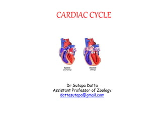
- 2. CARDIAC CYCLE Definition: The cardiac events that occur from the beginning of one heartbeat to the beginning of the next are called the cardiac cycle. oSystole and diastole of atria and ventricles alternately contract and relax During atrial systole, ventricles are relaxed During ventricle systole, atria are relaxed oForces blood from higher Pressure to lower Pressure dattasutapa@gmail.com
- 3. 1.Atrial systole(0.11s-0.53s) 2. Isovolumetric ventricular contraction (0.05s) 3. Rapid ventricular Ejection Phase (0.22-0.27s) 4. Slow ventricular Ejection Phase (0.22-0.27s) 5.Isovolumetric ventricular relaxation(0.08s) 6. Rapid Passive Ventricular filling Phase (0.11s) 7. Slow Passive Ventricular filling Phase (0.19s) There are seven phases of the cardiac cycle (and duration of phases): (Systole- when the ventricles are contracting) (Diastole- when the ventricles are relaxing) dattasutapa@gmail.com
- 4. 1.Atrial systole(0.11s-0.53s) 2. Isovolumetric ventricular contraction (0.05s) 3. Rapid ventricular Ejection Phase (0.22-0.27s) 4. Slow ventricular Ejection Phase (0.22-0.27s) 5.Isovolumetric ventricular relaxation(0.08s) 6. Rapid Passive Ventricular filling Phase (0.11s) 7. Slow Passive Ventricular filling Phase (0.19s) dattasutapa@gmail.com Phases of the cardiac cycle
- 5. 1. Atrial Contraction dattasutapa@gmail.com LEFT ATRIUM Before atrial contraction 80% of Ventral filling done 20% by atrial contraction (required during exercise) Atria contract- intraatrial Pressure increases (a wave)- blood moves to LV - Pressure transferred to LV LV Before atria contracts- Pressure in LV is 0 – LV in relaxed cond- start of atria contraction slight increase in Pressure LV AORTA Aortic valves remain closed Pressure in aorta is more than LV
- 6. 2. Isovolumetric contraction dattasutapa@gmail.com LEFT ATRIUM •As Pressure in LV increases more than LA •Mitral valve closes- a slight increase (jerk) in Pressure in LA (c wave) Blood from lungs still coming- Pressure started to build up slightly- start of v wave LV •LV starts contraction- intraventricular Pressure build up- more than LA •Mitral valve closed- S1 (tricuspid in RA also closed, both closure at the same time) •Pressure not that enough to open aortic valve •Contracts as closed chamber- blood vol and Pressure remain same- isovolumic, isometric; isovolumetric contraction (no blood leaving, no blood coming) • Pressure same as aorta AORTA Pressure still more than LV Aortic valve closed
- 7. 3. Rapid Ventricular Ejection Phase dattasutapa@gmail.com •LV •Pressure reaches to 81 mm of Hg more than aorta – aortic valves open- termination of IVC •Next phase RVEP •Still contraction •Pressure reaches to 120 mm of Hg •Blood rushes to aorta •AORTA •Aorta elastic, dilates- huge ventricular inflow •Aorta and LV having same Pressure •Act as a same chamber RVEP cannot last long •LA •Receiving blood from lungs •reservoir
- 8. 4. Slow Ventricular Ejection Phase dattasutapa@gmail.com •LV •Pressure more than LA- mitral valve still closed •Still contracting •Aortic valve still opened •Pressure starts falling- bcoz blood flow to aorta •Ejection rate slow- SEP / SVEP •Still Pressure more than aorta- blood flow, but slow •AORTA • Pressure decreases as that of LV •LA •Still getting blood from lungs
- 9. 5. Isovolumetric relaxation dattasutapa@gmail.com LV 1. Pressure falls 2. Aortic blood tries to backflow- closure of aortic valve- S2 (both aortic and pulmonary valves,in RV, closed with sound, but aortic closure is slightly earlier) 3. Pressure less than aorta but still more than LA 4. Relaxes as closed chamber – start of IVR •AORTA •Pressure more- valves closed •LA •Accumulates blood from lungs •Pressure Less than LV •Mitral valve closed •Due course Pressure LA more than LV
- 10. 6. Rapid Passive Ventricular Filling dattasutapa@gmail.com •LV •After slow ejection, left over blood- ESV •Pressure less than LA •LA •Accumulated blood from LA moves rapidly to LV- mitral valve opens •Without atrial contraction •This is RPVF •AORTA •Pressure more than LV
- 11. 7. Slow Passive Ventricular Filling (Diastasis) dattasutapa@gmail.com •LA •Mitral valve opened- •atrium not a reservoir- a passage •directly carry blood passively to LV ,slowly • Pressure difference less •This is SPVF •LV •Receives blood from LA •Pressure less than LA •AORTA •Pressure still more than LV •Aortic valves closed
- 12. Atrial depolarization Ventricular depolarization Ventricular repolarization Atrial systole begins and forces blood to into ventricles Ventricular contraction pushes AV valves closed Semilunar valves open and blood is ejected Semilunar valves close and blood flows into aorta Chambers relax and blood fills ventricles passively Atrial systole Ventricular systole Ventricular diasystole 1st phase 2nd phase Early Late PHASES OF CARDIAC CYCLE dattasutapa@gmail.com
- 13. THANK YOU dattasutapa@gmail.com Reference reading: 1. https://www.cvphysiology.com/Cardiac%20Function/CF024 2. https://www.researchgate.net/figure/Cardiac-cycle-top-diagram-depicting- cardiac-signals-ECG-Pressureessures-volumes-and_fig6_281383702 3. https://sites.google.com/site/stethoscope2011/physiology 4. https://www.wikiwand.com/en/Wiggers_diagram 5. https://www.wikiwand.com/en/Wiggers_diagram 6. Review of Medical Physiology: Ganong 7. https://www.youtube.com/watch?v=jVvQaqFOJpY&t=2823s