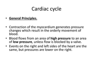
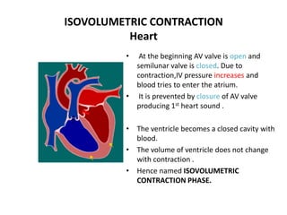
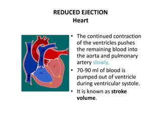
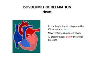
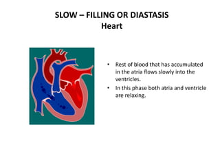
Cardiac Cycle
Blood Flow Through Heart
Definitions
• Systole = period of ventricular contraction.
• Diastole = period of ventricular relaxation.
• NOTE: Normally diastole is longer than
systole.
Cardiac cycle
• General Principles.
• Contraction of the myocardium generates pressure
changes which result in the orderly movement of
blood.
• Blood flows from an area of high pressure to an area
of low pressure, unless flow is blocked by a valve.
• Events on the right and left sides of the heart are the
same, but pressures are lower on the right.
CARDIAC CYCLE
• The normal duration of the cardiac cycle is 0.8
second.
• Each beat of the heart consists of systole and
diastole of atria and ventricles.
• Systole is the contraction of the heart during which
blood is ejected out from the heart
• Diastole is the relaxation of the heart during which
the chambers of the heart are filled with blood
• Ventricular Systole:
1. Isovolumetric contraction phase - 0.05 sec
2. Rapid Ejection phase - 0.10 sec
3. Reduced Ejection phase - 0.15 sec
Ventricular diastole
1. Proto diastolic phase - 0.04 sec
2. Isovolumetric Relaxation phase - 0.06 sec
3. First Rapid filling phase - 0.10 sec
4. Slow Filling - 0.20 sec
5. Last Rapid Filling Phase - 0.10 sec
FACTS TO REMEMBER
• SYSTOLE IS CONTRACTION
-THE RISE IN PRESSURE IN THE CONTRACTING
CHAMBER
-BLOOD BEING EJECTED BY THE CONTRACTING
CHAMBER
• DIASTOLE IS RELAXATION
- A FALL IN PRESSURE OF THE RELAXING
CHAMBER
- FILLING OF THE RELAXING CHAMBER
AV valves*
Semilunar
valves†
Status of ventricles and atria
1.Late diastole open closed
• whole heart is relaxed
• ventricles are expanding and filling (passive filling,
~80% of volume)
2. Atrial systole open closed
• atria contract and pump blood
• additional 10–40% filling of ventricles due to active
contraction of atria
3.Isovolumic
ventricular
contraction
closed closed
• ventricular myocytes begin to contract
• ventricle volume unchanged
4.Ventricular
ejection
closed open
• ventricles fully contract
• pump blood to rest of body
5.Isovolumic
ventricular
relaxation
closed closed
• ventricles relax
• ventricle volume unchanged
• atria expand and are filling
* AV (atrioventricular) valves:
1) mitral valve – between the left atrium and the left ventricle
2) tricuspid valve – between the right atrium and the right ventricle
† Semilunar valves:
1) aortic valve – between the left ventricle and the aorta
ATRIAL SYSTOLE - Heart
• Prior to atrial systole, blood has
been flowing passively from the
atrium into the ventricle through the
open AV valve.
• Contraction of atria propels some
additional blood into the ventricles.
Atrial contraction is complete before
the ventricle begins to contract.
ISOVOLUMETRIC CONTRACTION
Heart
• At the beginning AV valve is open and
semilunar valve is closed. Due to
contraction,IV pressure increases and
blood tries to enter the atrium.
• It is prevented by closure of AV valve
producing 1st heart sound .
• The ventricle becomes a closed cavity with
blood.
• The volume of ventricle does not change
with contraction .
• Hence named ISOVOLUMETRIC
CONTRACTION PHASE.
RAPID EJECTION
Heart
• Due to the increased
IV pressure than
aorta and pulmonary
artery semilunar
valves open.
• This forces blood to
eject rapidly.
REDUCED EJECTION
Heart
• The continued contraction
of the ventricles pushes
the remaining blood into
the aorta and pulmonary
artery slowly.
• 70-90 ml of blood is
pumped out of ventricle
during ventricular systole.
• It is known as stroke
volume.
PROTODIASTOLIC PHASE
• In this, ventricles begin to
relax.The AV valve is closed
and semilunar valve is
open.
• As the IV pressure
decreases below the
pressure in aorta and
pulmonary artery, blood
tries to come back.
• It is prevented by closure
of the semilunar valve
producing 2nd heart sound
ISOVOLUMETRIC RELAXATION
Heart
• At the beginning of this phase the
AV valves are closed.
• Now ventricle is a closed cavity.
• IV pressure goes below the atrial
pressure
FIRST RAPID FILLING
Heart
• The reduced IV pressure causes
opening of AV valves.
• Once the AV valves open,
blood that has accumulated in
the atria flows rapidly into the
ventricles.
• Turbulence to the flow of blood
produces 3rd heart sound .
SLOW – FILLING OR DIASTASIS
Heart
• Rest of blood that has accumulated
in the atria flows slowly into the
ventricles.
• In this phase both atria and ventricle
are relaxing.
LAST RAPID- FILLING
HEART
• It coincides with atrial
contraction.
• The atrial systole forces
blood from atria to
ventricle.
• Turbulence due to rapid
rush of blood produces
4th heart sound .
• At the end , ventricle
contain 130 ml of blood.
• It is the end – diastolic
ventricular volume.

Cardiac Cycle 5.pptx

  • 1.
  • 2.
  • 3.
    Definitions • Systole =period of ventricular contraction. • Diastole = period of ventricular relaxation. • NOTE: Normally diastole is longer than systole.
  • 4.
    Cardiac cycle • GeneralPrinciples. • Contraction of the myocardium generates pressure changes which result in the orderly movement of blood. • Blood flows from an area of high pressure to an area of low pressure, unless flow is blocked by a valve. • Events on the right and left sides of the heart are the same, but pressures are lower on the right.
  • 5.
    CARDIAC CYCLE • Thenormal duration of the cardiac cycle is 0.8 second. • Each beat of the heart consists of systole and diastole of atria and ventricles. • Systole is the contraction of the heart during which blood is ejected out from the heart • Diastole is the relaxation of the heart during which the chambers of the heart are filled with blood
  • 6.
    • Ventricular Systole: 1.Isovolumetric contraction phase - 0.05 sec 2. Rapid Ejection phase - 0.10 sec 3. Reduced Ejection phase - 0.15 sec
  • 7.
    Ventricular diastole 1. Protodiastolic phase - 0.04 sec 2. Isovolumetric Relaxation phase - 0.06 sec 3. First Rapid filling phase - 0.10 sec 4. Slow Filling - 0.20 sec 5. Last Rapid Filling Phase - 0.10 sec
  • 8.
    FACTS TO REMEMBER •SYSTOLE IS CONTRACTION -THE RISE IN PRESSURE IN THE CONTRACTING CHAMBER -BLOOD BEING EJECTED BY THE CONTRACTING CHAMBER • DIASTOLE IS RELAXATION - A FALL IN PRESSURE OF THE RELAXING CHAMBER - FILLING OF THE RELAXING CHAMBER
  • 9.
    AV valves* Semilunar valves† Status ofventricles and atria 1.Late diastole open closed • whole heart is relaxed • ventricles are expanding and filling (passive filling, ~80% of volume) 2. Atrial systole open closed • atria contract and pump blood • additional 10–40% filling of ventricles due to active contraction of atria 3.Isovolumic ventricular contraction closed closed • ventricular myocytes begin to contract • ventricle volume unchanged 4.Ventricular ejection closed open • ventricles fully contract • pump blood to rest of body 5.Isovolumic ventricular relaxation closed closed • ventricles relax • ventricle volume unchanged • atria expand and are filling * AV (atrioventricular) valves: 1) mitral valve – between the left atrium and the left ventricle 2) tricuspid valve – between the right atrium and the right ventricle † Semilunar valves: 1) aortic valve – between the left ventricle and the aorta
  • 10.
    ATRIAL SYSTOLE -Heart • Prior to atrial systole, blood has been flowing passively from the atrium into the ventricle through the open AV valve. • Contraction of atria propels some additional blood into the ventricles. Atrial contraction is complete before the ventricle begins to contract.
  • 11.
    ISOVOLUMETRIC CONTRACTION Heart • Atthe beginning AV valve is open and semilunar valve is closed. Due to contraction,IV pressure increases and blood tries to enter the atrium. • It is prevented by closure of AV valve producing 1st heart sound . • The ventricle becomes a closed cavity with blood. • The volume of ventricle does not change with contraction . • Hence named ISOVOLUMETRIC CONTRACTION PHASE.
  • 12.
    RAPID EJECTION Heart • Dueto the increased IV pressure than aorta and pulmonary artery semilunar valves open. • This forces blood to eject rapidly.
  • 13.
    REDUCED EJECTION Heart • Thecontinued contraction of the ventricles pushes the remaining blood into the aorta and pulmonary artery slowly. • 70-90 ml of blood is pumped out of ventricle during ventricular systole. • It is known as stroke volume.
  • 14.
    PROTODIASTOLIC PHASE • Inthis, ventricles begin to relax.The AV valve is closed and semilunar valve is open. • As the IV pressure decreases below the pressure in aorta and pulmonary artery, blood tries to come back. • It is prevented by closure of the semilunar valve producing 2nd heart sound
  • 15.
    ISOVOLUMETRIC RELAXATION Heart • Atthe beginning of this phase the AV valves are closed. • Now ventricle is a closed cavity. • IV pressure goes below the atrial pressure
  • 16.
    FIRST RAPID FILLING Heart •The reduced IV pressure causes opening of AV valves. • Once the AV valves open, blood that has accumulated in the atria flows rapidly into the ventricles. • Turbulence to the flow of blood produces 3rd heart sound .
  • 17.
    SLOW – FILLINGOR DIASTASIS Heart • Rest of blood that has accumulated in the atria flows slowly into the ventricles. • In this phase both atria and ventricle are relaxing.
  • 18.
    LAST RAPID- FILLING HEART •It coincides with atrial contraction. • The atrial systole forces blood from atria to ventricle. • Turbulence due to rapid rush of blood produces 4th heart sound . • At the end , ventricle contain 130 ml of blood. • It is the end – diastolic ventricular volume.