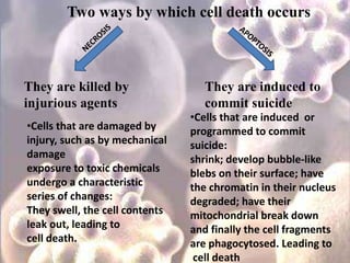
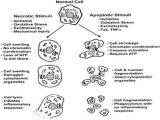
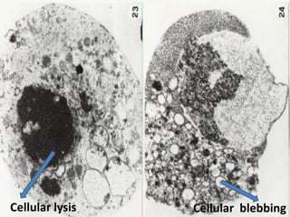
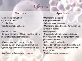
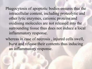
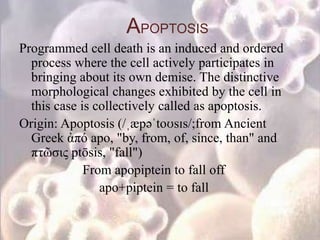
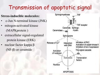
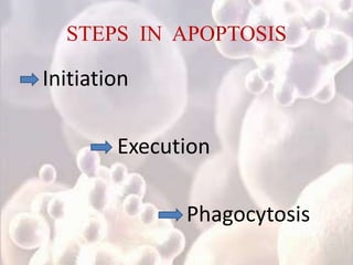
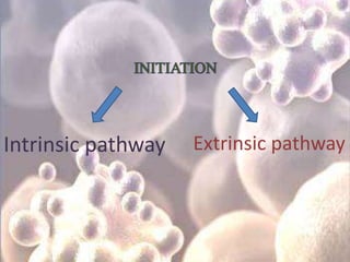
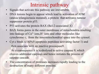
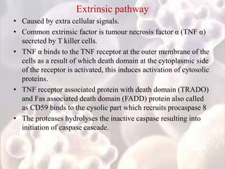
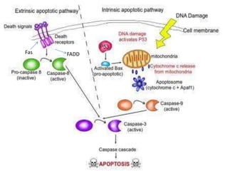
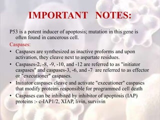
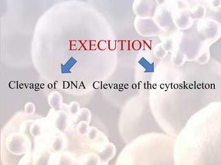
Apoptosis
Every cell in our body
is immortal,
it has a time to
live and die
Two ways by which cell death occurs
They are killed by
injurious agents
They are induced to
commit suicide
•Cells that are damaged by
injury, such as by mechanical
damage
exposure to toxic chemicals
undergo a characteristic
series of changes:
They swell, the cell contents
leak out, leading to
cell death.
•Cells that are induced or
programmed to commit
suicide:
shrink; develop bubble-like
blebs on their surface; have
the chromatin in their nucleus
degraded; have their
mitochondrial break down
and finally the cell fragments
are phagocytosed. Leading to
cell death
Cellular lysis Cellular blebbing
Difference
Necrosis Apoptosis
•Membrane disruption
•Cytoplasm swells
•Cellular lysis
•Vesicle formation absent
•Passive process
•Random digestion of DNA resulting into a
smear after gel electrophoresis
•Affects group of contiguous cells
•Caused by non physiological stimuli like
hypoxia, hyperthermia, viral infection etc
•Significant inflammatory response
•Membrane blebbing
•Cytoplasm shrinks
•Cellular fragmentation
•Membrane bound vesicle formation i.e.
apoptotic bodies
•Active process
•Non random length fragmentation of
DNA resulting into ladder pattern after
gel electrophoresis
•Affects individual cells
•Caused by physiological stimuli like lack
of growth factor, changes in hormonal
environment
•No inflammatory response
Cell Death Without Inflammation???
Phagocytosis of apoptotic bodies ensures that the
intracellular content, including proteolytic and
other lytic enzymes, cationic proteins and
oxidising molecules are not released into the
surrounding tissue thus does not induce a local
inflammatory response.
whereas in case of necrosis , injured cells swell,
burst and release their contents thus inducing
an inflammatory response.
APOPTOSIS
Programmed cell death is an induced and ordered
process where the cell actively participates in
bringing about its own demise. The distinctive
morphological changes exhibited by the cell in
this case is collectively called as apoptosis.
Origin: Apoptosis (/ˌæpəˈtoʊsɪs/;from Ancient
Greek ἀπό apo, "by, from, of, since, than" and
πτῶσις ptōsis, "fall")
From apopiptein to fall off
apo+piptein = to fall
Why should a cell commit suicide?
First Reason
Programmed cell death is needed for proper
development:
Examples :
• Resorption of the tadpole tail at the time of its metamorphosis
into a frog.
• Formation of the fingers and toes of the fetus
• Sloughing off of the inner lining of the uterus (the
endometrium) at the start of menstruation
• Formation of the proper connections (synapses) between
neurons in the brain
• Elimination of T cells that might otherwise mount an
autoimmune attack on the body
• During the pupal stage of insects most of the cells of the larva
die by apoptosis thus providing the nutrients for the
development of the structures of the adult.
Second reason
Programmed cell death is needed to destroy cells that
represent a threat to the integrity
of the organism
Examples:
• Cytotoxic T lymphocytes (CTLs) kill virus-infected cells is by
inducing apoptosis
• The effector cells of immune system must be removed to
prevent autoimmunity
• Removal of cells with damaged DNA for proper embryonic
development
• Radiation and chemicals used in cancer therapy induce
apoptosis in some types of cancer cells.
Transmission of apoptotic signal
Stress-inducible molecules:
• c-Jun N-terminal kinase (JNK)
• mitogen-activated kinase
(MAPKprotein )
• extracellular signal-regulated
protein kinase (ERK)
• nuclear factor kappa β
(NF-β) or ceramide
Mechanism of apoptosis
STEPS IN APOPTOSIS
Initiation
Execution
Phagocytosis
Intrinsic pathway Extrinsic pathway
Intrinsic pathway
• Signals that activate this pathway are irreversible.
• DNA lesions begin to appear which lead to activation of ATM
(ataxia telangiectasia mutated), a protein that activates tumour
suppressor protein p53.
• P53 activates the protein BAX (Bcl-2-associated X)
• BAX forms pores in the outer membrane of mitochondria resulting
into leakage of Ca2+ ions,H+ ions and other molecules like
cytochrome c, from the innermitochondrial space into the cytosol.
• Cyt c binds to APAF(apoptosis peptidase activating factor 1) and
then associate with an inactive procaspase9.
As a consequence it is hydrolysed to active caspase 9, which
cleaves additional caspases and hence initiates the process of
caspase cascade.
• The concentration of proteases increases rapidly leading to the
destruction of many different proteins.
Extrinsic pathway
• Caused by extra cellular signals.
• Common extrinsic factor is tumour necrosis factor α (TNF α)
secreted by T killer cells.
• TNF α binds to the TNF receptor at the outer membrane of the
cells as a result of which death domain at the cytoplasmic side
of the receptor is activated, this induces activation of cytosolic
proteins.
• TNF receptor associated protein with death domain (TRADO)
and Fas associated death domain (FADD) protein also called
as CD59 binds to the cysolic part which recruits procaspase 8
• The proteases hydrolyses the inactive caspase resulting into
initiation of caspase cascade.
Fas FADD
IMPORTANT NOTES:
P53 is a potent inducer of apoptosis; mutation in this gene is
often found in cancerous cell.
Caspases:
• Caspases are synthesized as inactive proforms and upon
activation, they cleave next to aspartate residues.
• Caspases-2, -8, -9, -10, and -12 are referred to as "initiator
caspases" and caspases-3, -6, and -7 are referred to as effector
or "executioner" caspases.
• Initiator caspases cleave and activate "executioner" caspases
that modify proteins responsible for programmed cell death
• Caspases can be inhibited by inhibitor of apoptosis (IAP)
proteins :- c-IAP1/2, XIAP, livin, survivin
EXECUTION
Clevage of DNA Clevage of the cytoskeleton
Clevage of DNA
• After the activation of caspase cascade the caspase 3 cleaves
the inactive inhibitor complexed to DNA
• Thus, DNAase cleaves DNA at clevage sites located at regular
intervals of 180bp.
Clevage of the cytoskeleton
• Caspase cleaves proteins of cytoskeleton as a result cell looses
its structure.
• Caspase cleaves proteins which covert cells to apoptotic blebs.
• polynucleosomal DNA fragmentation is
mediated by cleavage of ICAD (inhibitor of
caspase-activated DNase), the inhibitor of the
endonuclease CAD (caspase-activated DNase)
that cleaves DNA into the characteristic
oligomeric fragments
• proteolysis of several cytoskeletal proteins
such as actin or fodrin leads to loss of overall
cell shape, whereas degradation of lamin
results in nuclear shrinking
Phagocytosis
• Execution leads to modification of outer
membranes of the apoptotic blebs.
• Therefore phagocyts like macrophages
recognise them.
• The blebs fuse with lysosomes.
• Lysosomal enzymes finally metabolise the
blebs and their components leading to cellular
“SUICIDE”.
Why the rate of apoptosis be constant to
maintain homeostasis?
When apoptosis does not work correctly, cells that
should be eliminated may persist and become
immortal,
for example, in cancer and leukemia.
When apoptosis works overly well, it kills too many
cells and results into great tissue damage.
For example in strokes and neurodegenerative
disorders such as Alzheimer's, Huntington's, and
Parkinson's diseases.
Defects in the apoptotic machinery is associated
with autoimmune diseases
For example systemic lupus erythematosus and
rheumatoid arthritis
Role of apoptosis in cancer treatment
Current cancer therapies, for example,
chemotherapy, irradiation, immunotherapy or
suicide gene therapy is based on activation of
apoptosis signal transduction pathways in
cancer cells such as the intrinsic and/or
extrinsic pathway
THANK YOU 

Apoptosis

  • 1.
  • 2.
    Every cell inour body is immortal, it has a time to live and die
  • 3.
    Two ways bywhich cell death occurs They are killed by injurious agents They are induced to commit suicide •Cells that are damaged by injury, such as by mechanical damage exposure to toxic chemicals undergo a characteristic series of changes: They swell, the cell contents leak out, leading to cell death. •Cells that are induced or programmed to commit suicide: shrink; develop bubble-like blebs on their surface; have the chromatin in their nucleus degraded; have their mitochondrial break down and finally the cell fragments are phagocytosed. Leading to cell death
  • 5.
  • 6.
    Difference Necrosis Apoptosis •Membrane disruption •Cytoplasmswells •Cellular lysis •Vesicle formation absent •Passive process •Random digestion of DNA resulting into a smear after gel electrophoresis •Affects group of contiguous cells •Caused by non physiological stimuli like hypoxia, hyperthermia, viral infection etc •Significant inflammatory response •Membrane blebbing •Cytoplasm shrinks •Cellular fragmentation •Membrane bound vesicle formation i.e. apoptotic bodies •Active process •Non random length fragmentation of DNA resulting into ladder pattern after gel electrophoresis •Affects individual cells •Caused by physiological stimuli like lack of growth factor, changes in hormonal environment •No inflammatory response
  • 7.
    Cell Death WithoutInflammation???
  • 8.
    Phagocytosis of apoptoticbodies ensures that the intracellular content, including proteolytic and other lytic enzymes, cationic proteins and oxidising molecules are not released into the surrounding tissue thus does not induce a local inflammatory response. whereas in case of necrosis , injured cells swell, burst and release their contents thus inducing an inflammatory response.
  • 9.
    APOPTOSIS Programmed cell deathis an induced and ordered process where the cell actively participates in bringing about its own demise. The distinctive morphological changes exhibited by the cell in this case is collectively called as apoptosis. Origin: Apoptosis (/ˌæpəˈtoʊsɪs/;from Ancient Greek ἀπό apo, "by, from, of, since, than" and πτῶσις ptōsis, "fall") From apopiptein to fall off apo+piptein = to fall
  • 10.
    Why should acell commit suicide?
  • 11.
    First Reason Programmed celldeath is needed for proper development: Examples : • Resorption of the tadpole tail at the time of its metamorphosis into a frog. • Formation of the fingers and toes of the fetus • Sloughing off of the inner lining of the uterus (the endometrium) at the start of menstruation • Formation of the proper connections (synapses) between neurons in the brain • Elimination of T cells that might otherwise mount an autoimmune attack on the body • During the pupal stage of insects most of the cells of the larva die by apoptosis thus providing the nutrients for the development of the structures of the adult.
  • 12.
    Second reason Programmed celldeath is needed to destroy cells that represent a threat to the integrity of the organism Examples: • Cytotoxic T lymphocytes (CTLs) kill virus-infected cells is by inducing apoptosis • The effector cells of immune system must be removed to prevent autoimmunity • Removal of cells with damaged DNA for proper embryonic development • Radiation and chemicals used in cancer therapy induce apoptosis in some types of cancer cells.
  • 13.
    Transmission of apoptoticsignal Stress-inducible molecules: • c-Jun N-terminal kinase (JNK) • mitogen-activated kinase (MAPKprotein ) • extracellular signal-regulated protein kinase (ERK) • nuclear factor kappa β (NF-β) or ceramide
  • 14.
  • 15.
  • 16.
  • 17.
    Intrinsic pathway • Signalsthat activate this pathway are irreversible. • DNA lesions begin to appear which lead to activation of ATM (ataxia telangiectasia mutated), a protein that activates tumour suppressor protein p53. • P53 activates the protein BAX (Bcl-2-associated X) • BAX forms pores in the outer membrane of mitochondria resulting into leakage of Ca2+ ions,H+ ions and other molecules like cytochrome c, from the innermitochondrial space into the cytosol. • Cyt c binds to APAF(apoptosis peptidase activating factor 1) and then associate with an inactive procaspase9. As a consequence it is hydrolysed to active caspase 9, which cleaves additional caspases and hence initiates the process of caspase cascade. • The concentration of proteases increases rapidly leading to the destruction of many different proteins.
  • 18.
    Extrinsic pathway • Causedby extra cellular signals. • Common extrinsic factor is tumour necrosis factor α (TNF α) secreted by T killer cells. • TNF α binds to the TNF receptor at the outer membrane of the cells as a result of which death domain at the cytoplasmic side of the receptor is activated, this induces activation of cytosolic proteins. • TNF receptor associated protein with death domain (TRADO) and Fas associated death domain (FADD) protein also called as CD59 binds to the cysolic part which recruits procaspase 8 • The proteases hydrolyses the inactive caspase resulting into initiation of caspase cascade.
  • 19.
  • 20.
    IMPORTANT NOTES: P53 isa potent inducer of apoptosis; mutation in this gene is often found in cancerous cell. Caspases: • Caspases are synthesized as inactive proforms and upon activation, they cleave next to aspartate residues. • Caspases-2, -8, -9, -10, and -12 are referred to as "initiator caspases" and caspases-3, -6, and -7 are referred to as effector or "executioner" caspases. • Initiator caspases cleave and activate "executioner" caspases that modify proteins responsible for programmed cell death • Caspases can be inhibited by inhibitor of apoptosis (IAP) proteins :- c-IAP1/2, XIAP, livin, survivin
  • 21.
    EXECUTION Clevage of DNAClevage of the cytoskeleton
  • 22.
    Clevage of DNA •After the activation of caspase cascade the caspase 3 cleaves the inactive inhibitor complexed to DNA • Thus, DNAase cleaves DNA at clevage sites located at regular intervals of 180bp. Clevage of the cytoskeleton • Caspase cleaves proteins of cytoskeleton as a result cell looses its structure. • Caspase cleaves proteins which covert cells to apoptotic blebs.
  • 23.
    • polynucleosomal DNAfragmentation is mediated by cleavage of ICAD (inhibitor of caspase-activated DNase), the inhibitor of the endonuclease CAD (caspase-activated DNase) that cleaves DNA into the characteristic oligomeric fragments • proteolysis of several cytoskeletal proteins such as actin or fodrin leads to loss of overall cell shape, whereas degradation of lamin results in nuclear shrinking
  • 24.
    Phagocytosis • Execution leadsto modification of outer membranes of the apoptotic blebs. • Therefore phagocyts like macrophages recognise them. • The blebs fuse with lysosomes. • Lysosomal enzymes finally metabolise the blebs and their components leading to cellular “SUICIDE”.
  • 26.
    Why the rateof apoptosis be constant to maintain homeostasis?
  • 27.
    When apoptosis doesnot work correctly, cells that should be eliminated may persist and become immortal, for example, in cancer and leukemia. When apoptosis works overly well, it kills too many cells and results into great tissue damage. For example in strokes and neurodegenerative disorders such as Alzheimer's, Huntington's, and Parkinson's diseases. Defects in the apoptotic machinery is associated with autoimmune diseases For example systemic lupus erythematosus and rheumatoid arthritis
  • 28.
    Role of apoptosisin cancer treatment Current cancer therapies, for example, chemotherapy, irradiation, immunotherapy or suicide gene therapy is based on activation of apoptosis signal transduction pathways in cancer cells such as the intrinsic and/or extrinsic pathway
  • 30.