

Recommended
Recommended
More Related Content
What's hot
What's hot (20)
Similar to Hap connective tissue.
Similar to Hap connective tissue. (20)
More from EXCELRA
Recently uploaded
Recently uploaded (20)
Hap connective tissue.
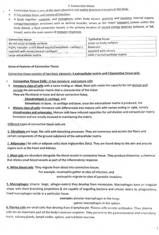
- 1. 7. Conn~ctive tissue • ~_q1_11wc.!_iy_e tissuP 15on_Q_ot the most abund_r111t.9!!!l_widely distribu_ted tissues in the body. • !l_has various forms .rnd variety of functions. • It binds together, ~!!P..P_.9rl~, and strengthen~ other body tissues; grotPcts and insulates internal organs; co111partmentr1lizes structures such as skeletal muscles; serves as the major transport system within the body (blood, a fluid connective tissue); Is the rrimary location of stored energy reserves (adipose, or fat, tissue); and is the main source of immune responses . c~~nc~tiv; tissue - --- ---- -- - - - ·- ___- J~Epit-h~llal ti~ ~e ___- --------- - - - - do not occur on body surfaces occur o~ojt ~:faces___ - - - - - - -- - -- ---< highly~rola~- a rich blood supply(Exceptions- cartllage,_L_ Avastulil~-- - - - - - - - ---------1 s~ppli~ with nerves(except cartilage) __. 7 ____ supplied with nerves . La rge extracellular matrix _ .,,. ___ UttlP. / _n_o_E_x_tr_a_c_e_llu_l_a_r_m_a_t_n_x________- General Features of Connective Tissue Connective tissue consists of two basic elements: II.extracellular matrix and I.Connective tissue cells. I. Connective Tissue Cells : It has immature and mature cells ► Immature class of cells with a name ending as -blast. Blast cells retain the capacity for cell division and secrete the extracellular matrix that is characteristic of the tissue They are fibroblasts in loose and dense connective tissue, Chondroblasts in cartilage, and Osteoblasts in bone.. In cartilage and bone, once the extracellular matrix is produced, the ).- Mature class of cells: immature cells differentiate into mature cells with names ending in -cyte, namely chondrocytes and osteocytes. Mature cells have reduced capacities for cell division and extracellular matrix formation and are mostly involved in maintaining the matrix. Different types of connective tissue cells are 1. Fibroblasts are large, flat cells with branching processes. They are numerous and secrets the fibers and certain components of the ground substance of the extracellular matrix. 2. Adipocytes ( fat cells or adipose cells) store triglycerides (fats). They are found deep to the skin and around organs such as the heart and kidneys. 3. Mast cells are abundant alongside the blood vessels in connective tissue. They produce histamine, a chemical that dilates small blood vessels as part of the inflammatory response. 4. White blood cells: They migrate from blood into connective tissues. For example, neutrophils gather at sites of infection, and eosinophils migrate to sites of parasitic invasions. 5. Macrophages: (macro- large; -phages-eaters) they develop from monocytes. Macrophages have an irregular shape with short branching projections & are capable of engulfing bacteria and cellular debris by phag-.xytosis. Fixed macrophages reside in a particular tissue; examples alveolar macrophages in the lungs splenic macrophages in the spleen. 6. Plasma cells are small cells that develop from B lymphocyte. PlasmJ cells srrrell' ,rntibodil's. Thus. olasma cells are an important part of the body's lmmu11<' responsf' . They prc~e11t in thC' r,a~trormestinJI ,rnd rr spir<'ltory tracts, salivary elands, lymph nodes, spleen, .:ind red bone marrow. 1 ...
- 2. 7. Connective tissue , II. .Connective tissue's extracelll,1lar matrix is the material located lJ0.tw.QQDit•,s:~•11·,. It i•, 1J',tJj1lly_•,<:< r,•t1 ·tl 11·1 the connective tissue cells and determines the tissue's qualities. For example in cartilage the extr11cellular matrix is firm but pliable. In bone the extra cellular matrix is hard and inflexible. The extracellular matrix consists of (1) ground substance and (2) fibers. {1) Ground substance: is the component of a connective tissue between the cells and fibers. ✓ It may be fluid, semifluid, gelatinous, or calcified. ✓ it supports cells, binds them together, stores water, and provides a medium through which substance•, are exchanged between the u:ood and cells. ✓ It plays an active role in how tissues develop, migrate, proliferate, and change shape, and in how th,!y carry out their metabolic functjons. ✓ It contains water and large organic molecules( hyaluronic acid, chondroitln sulphate) • GAGs is that they trap water, making the ground substance more jellylike. • Hyaluronic acid is a viscous, slippery substance that binds cells together, lubricates joints, and helps maintain the shape of the eyeballs • Chondroitin sulfate provides support and adhesiveness in cartilage, bone, skin, and blood vessels. • The skin, tendons, blood vessels, and heart valves contain dermatan sulfate; • bone, cartilage, and the cornea of the eye contain keratan sulfate. • proteoglycans( consists of protein and GAGs) is present in the ground substance are adhesion proteins • fibronectin is another adhesion protein of connective tissue which binds to both collagen fibers and ground substance and linkir.g them together. It also attaches cells to the ground substance. {2) fibers: Three types of fibers are embedded in the extracellular matrix between the cells: !.collagen fibers,2.elastic fibers, & 3.reticular fibers. They function to strengthen & support connective tissues. 1. Collagen fibers are very strong an~ resist pulling forces, but they are not stiff, which allows tissue flexibility. The properties of different types of collagen fibers vary from tissue to tissue. example, the collagen fibers fourid in cartilage attract more water molecules than those in bone, which gives cartilage a more cushioning effect Collagen fibers often occur in parallel bundles . The bundle arrangement adds great strength to the tissue. Collagen fibers are found in most types of connective tissues, especially bone, cartilage, tendons, and ligaments.-. ' . 2.Elastic fibers': which are smaller in diameter than collagen fibers, branch and join together to form a network within a tissue. An elastic fiber consists of molecules of the protein elastin surrounded by fibrillin, which adds strength and stability. Elastic fibers are strong but can be stretched up to 150% of their relaxed length without breaking. They have the ability to return to their original shape after being stretched, a property called elasticity. Elastic fibers are plentiful in skin, blood vessel walls, and lung tissue. 3.Reticular fibers; (reticul- net), consisting of collagen arranged in fine bundles with a coating of glycoprotein, provide support in the walls of blood vessels and form a network around the cells in some tissues, such as areolar connective tissue, adipose tissue, and smooth muscle tissue. Like collagen fibers, reticular fibers provide support 2
- 3. Cla,sification of Connective tissue I. Embryonic connective tissue A. Mesenchyme B. Mucous connective tissue II. Mature connective tissue A. Loose connective tissue 1. Areolar connective tissue 2. Adipose tissue 3. Reticular connective tissue B. Dense connective tissue 7. Connective tissue 1. Dense regular connective tissue 2. Dense irregular connective tissue 3. Elastic connective tissue C. Cartilage 1. Hyaline cartilage 2. Fibrocartilage 3. Elastic cartilage D. Bone tissue E. Liquid connective tissue 1. Blood tissue 2. Lymph Loose connective tissue : The fibers of loose connective tissue are loosely intertwined between cells. 1. Areolar connective tissue 2. Adipose tissue 3. Reticular connective tissue I. Embryonic connective Embryonic connective tissue is present primarily in the embryo,mesenchyme the tissue from which almost all other connective tissues eventually arise a) Mesenchyme is composed of irregularly shaped cells. a semi-fluid ground substance. and delicate reticular fibers. Location: Under skin and bones of embryo; b) Mucous connective tissue found mainly in the umbilical cord ofthe fetus. Mucous connective tissue is a form of mesenchyme that contains widely scattered fibroblasts, a more viscous jelly-like ground substance, and collagen fibers A. Loose connective tissue: The fibers of loose connective tissue are loosely intertwined between cells. The types of loose connective tissue are . "J. A-1.AREOIAR CONNECTIVE TISSUE D: tissues contains Several types of ce//s:fibroblasts. macrophages. plasma cells, mast cells, and adipocytes All three types offibers---collagen, elastic, and "'v,( reticularare arranged randomly throughout " ' 0 "'" ' the tissue. The ground substance contains / d µ0tf1o:1 hyaluronic acid, chondroitin sulfate, dermatan sulfate, and keratan sulfate. ~~ -- ..C _ c.,,_,.., ..... :: , - ~OC/IO ~ .... • [l;.Q&<: l'IY'='> ..' L:Combined with adipose tissue, areolar connective tissue forms the subcutaneous layer, the layer of tissue F:it attaches the skin to underlying tissues and organs. A-2.ADIPOSE TISSUE D: Adipose tissue the cells are called adipocytes (fat), are specialized for storage of triglycerides {fats) Adipocytes are derived from fibroblasts. Because the cell fills up with a single, large triglyceride droplet, the cytoplasm and nucleus are pushed to the periphery of the cell. L: it is found wherever areolar connective tissue is located and around organs d 'j(''' ' _..;.'--', ' ,, J
- 4. 7. Connective tissuC' F lt i, aCinsulator and can red_y~e he_c)__t l_Q;;2 through the skin. It is a m;:ijor ~11c'..[tY r~?I'!Y~: and !~•,n(•rd 1/ ,iJ1J1 ,,,, 1 :, aridprotects various organs. while adipose tissue present in in adults Br0 ~m adipose tissue(very rich blood supply+numerous pigmented mitochondria) present In fetus and infant, A-3.RETICUIAR CONNECTIVE TISSUE D: consists of fine Interlacing reticular fibers and reticular cells L: It forms the stroma (supporting framework) of the liver, spleen, and lymph nodes F:h~lps bind together smooth muscle cells. Additionally, reticular fibers in the spleen filter blood and remove worn-out blood cells, and reticular B. Dense Connective Tissue It contains more numerous, thicker and denser fibers (packed more closely) but considerably fewer cells than loose connective tissue. There are three types: dense regular connective tissue, dense Irregular connective tissue, and elastic Connective tissue. B 1.DENSE REGULAR CONNECTIVE TISSUE D:ln this bundles of collagen fibers are regularly arranged in parallel patterns that provide the tissue with great strength.Fibroblasts, appear in rows between the fibers. The tissue Is silvery white and tough, flexible F:The tissue withstands pulling along the axis of the fibers. L:. Tendons and ligaments. B.2 DENSE IRREGULAR CONNECTIVE TISSUE jl I ' ' ' 'l .,,.,.t, L,__ ,; ,•... D: It contains collagen fibers that are irregularly arranged F: Found in parts of the body where pulling forces are exerted in variou~ connective tissues directions L: it occurs In sheets, such as in the dermis of the skin, which is deep to the epidermis, or the fibrous pericardium around the . heart. Heart valves, the perichondrium (the tissue surrounding cartilage), and the periosteum (the tissue surrounding bone) are dense irregular B.3 ELASTIC CONNECTIVE TISSUE D:Yellow Branching ela·stic fibers predominate in elastic connective tissue Fibroblasts ,.,_,.,U#,, ~,A are present in the spaces between the fibers. F: strong and·can recoil to its original shape after being stretched. L: lung tissue-which recoils as you exhale, and elastic arteries- recoil between heartbeats to maintain blood flow. C. Cartilage It consists of a dense network of collagen fibers and elastic fibers with in chondroitin sulphate (a gel-like component of the ground substance). Cartilage can with stand stress than loose and dense connective tissues. It contains chondrocytes occur singly or in groups within spaces called lacunae (little lakes) in the extracellular matrix. cartilage has no blood vessels or nerves, except in the perichondrium. C1: HYAUNE CARTILAGE D:Hyaline cartilage contains a resilient gel as its ground substance and appears in the body as a bluish-white, shiny substance. The thin, fine collage.Q fibers are not visible andchondrocytes are found in lacunae. it has perichondrium. C 2: FIBROCARTILAGE L: the articular cartilage in joints and the cartilage of the epiphyseal F: provides flexibility and support at joints, reduces friction and absorbs shock. Hyaline cartilage is the weakest of the three types of cartilage. r ,tC; ~ ,. . r -"--:, , / ;.: ; di, .;. . /_I,.·'' "" ~ , ..- I. / ( ' ' 4
- 5. 7. Connective tissue I>. , 11.i1Hlit1cytc·, ,H P ~r,il!•·rnl ,irnonr, clc;irly visible thick bunQLP~.Qlsoll,1Gl'n fibers within the extracellular 111L111Ix of /1brotdr til.ir,L· f ,broc,irtd.:ir,c lacb a pPr1chondr1um. F: 11 P1ul.'(OD _~l..!_£n1~tl:i_,_1n-9.2_1R!.s:J.J.1Y, tlm tissue ,~ the ~trongest of th<' three types of cartilage. One l P1 c,c·11t in 111:c'rvt•rtl'bral discs, C 3 _llASTIC CARTILAGJ: n: l he.: chondrocrlL'S of el,1stic cartilage are located within a threadlike nl' r~q_rk of e l.1st~ bers within the> extracellular matrix A perichondrium is pr c·1c·nt F:_ Q_r_o!J..Q<'~('DJ.!Jh and elasticity and maintains the shape of certain trurturc>s~ ~ C:t tC'..[_11,II f'_il!, D. Bone tissue Bo1.1~ !i~l!.('_15-c_h}_~,.!f..'.£<.i_Js either comp~c_!_siJ:...i12.QDID', dt•pc•ndlng on its extracellular matrix and cells are •l , ollianlze::_d_, Description: DI.Compact bone tissue consists of osteons (haverslan systems) that contain IJmcllae, lacunae, osteocytes, canallculi, and central (haverslan) canals. 1.The lamellae (little plates) are concentric rings of extracellular matrix that consist of mln-eral salts (calcium and phosphates), which give bone its hardness, and collagen fibers, which give bone its strength. 2.Lacunae are small spaces between lamellae that contain mature bone cells called osteocytes. :~ "';:;:;: 3.Canalicull :Projecting from the lacunae are canaliculi (little canals), networks of ►•. •-vw-. ·- ·• mJnute canals containing the processes of osteocytes. Canaliculi provide routes for nutri<.'nts to reach osteocytes and for wastes to leave them. 4. Acentral (haverslan) canal contalm blood vessels and nerves. D 2. Spongy bone tissue consists of thin columns called trabeculae; spaces between trabeculae are filled with red bone marrow. Location: Both compact and spongy bone tissue make up the various parts of bone's of the body. Function: Support, protection, storage; houses blood-forming tissue; serves as ll'Vers that act with muscle tissue to enable movement. E. Liquid connective tissue lYMPH Lymph Is the extracellular fluid that flows In lymphatic vessels. It consists of several types of cells in a clear liquid extracellular matrix that Is similar to blood plasma but with much less protein. The composition of lymph varies from one part of the body to another. For example, lymph leaving lymph nodes includes many lymphocytes, in contrast to lymph from the small intestine, which has a high content of newly absorbed dietary lipids. 5