
Recommended
More Related Content
Similar to Presentation1.pptx
Similar to Presentation1.pptx (20)
More from MukhtarJamac3
More from MukhtarJamac3 (20)
Recently uploaded
Recently uploaded (20)
Presentation1.pptx
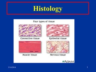
- 1. 1 3/14/2014
- 2. Connective tissue o Connective tissues function primarily to support the body and to bind or connect together all types of tissue. o This tissue also provide a mechanical framework (the skeleton) which plays an important role in locomotion. 3/14/2014 2
- 3. o This mechanical function is important in maintaining the form of the body, organs and tissues. o The tissue derives its name from its function in connecting or binding cells and tissues. Connective tissue is composed of: (a) cells (b) extracellular matrix. 3/14/2014 3
- 4. 3/14/2014 4 o Matrix consists of: 1.connective tissue fibers 2.Ground substance 3.Tissue Fluid
- 5. 3/14/2014 5 Cells of the connective tissue 1. Fibroblasts 2. Adipose cells 3. Macrophage or Histiocytes 4. Mast cells 5. Plasma cells 6. Leukocytes
- 6. Histiocyte: A tissue macrophage; the class includes hepatic Kupffer cells, alveolar macrophages, giant cells of granulomas, osteoclasts, and dermal Langerhans cells. 3/14/2014 6
- 7. oThe extracellular material of connective tissue, which plays a major role in the functioning of the tissue, is the dominant component of the tissue. o The dominance of the extracellular material is a special feature that distinguishes connective tissue from the other tissues of the body. 3/14/2014 7
- 8. o The extracellular matrix is composed of : 1. protein fibers (collagen fibers, reticular fibers, elastic fibers) 2. amorphous ground substance 3. tissue fluid (not preserved in histological preparations). The amount of tissue fluid is fairly constant and there is an equilibrium between the water entering and leaving the intercellular substance of the connective tissue. In pathological conditions (traumatic injury, inflammation) fluid may accumulate in the connective tissue, a condition known as edema. 3/14/2014 8
- 9. FUNCTIONS OF CONNECTIVE TISSUE 1. Structural support The connective tissues serve several functions, of which the most prominent function is structural support to enable maintenance of anatomical form of organs and organ systems. Examples include the connective tissue capsules surrounding organs (such as the kidney, lymph nodes). 3/14/2014 9
- 10. o The loose connective tissue acts to fill the spaces between organs. o The tendons (connecting muscles to bone) and the elastic ligaments (connecting bones to bones) are examples of specialized orderly forms of connective tissue. o The skeletal tissues (cartilage and bone) are special forms of connective tissue. 3/14/2014 10
- 11. 2. Metabolic functions o The connective tissues serve a nutritive role. All the metabolites from the blood pass from capillary beds and diffuse through the adjacent connective tissue to cells and tissues. o Similarly waste metabolites from the cells and tissues diffuse through the loose connective tissue before returning to the blood capillaries. 3/14/2014 11
- 12. o The adipose tissue (especially that of the hypodermis) serves as an energy store and also provides thermal insulation. o Surplus calories can be converted into lipid and stored in adipocytes. 3. Blood components and blood vessels o The hematopoietic tissues (blood-forming tissues) are a further specialized form of connective tissue. o These include the myeloid tissue (bone marrow) and the lymphoid (lymphatic) tissue. o The lining of the blood and lymphatic vessels (endothelial cells) as well as the peripheral blood, are also specialized forms of connective tissue. 3/14/2014 12
- 13. 4. Defensive functions o Various components of the connective tissue play roles in the defense or protection of the body including many of the components of the vascular and immune systems (plasma cells, lymphocytes, neutrophils, eosinophils, basophils, mast cells). 3/14/2014 13
- 14. o The various macrophages of the body are also categorized as connective tissue cells. o These all develop from monocytes and are grouped as part of the Mononuclear Phagocyte System of the body. o Macrophages are important in tissue repair as well as defense against bacterial invasion. 3/14/2014 14
- 15. Cell type Chief function Mesenchyme Embryonic source of all connective tissue cells Fibroblasts Chondroblasts Osteoblasts Structural support Plasma cells Lymphocytes Neutrophils Eosinophils Basophils Mast cells Macrophages Defense and immune Adipocytes Metabolic Energy storage Thermal insulation 3/14/2014 15
- 16. Between cells and fibers. o The intercellular ground substance is an amorphous, transparent material composed mainly of glycoproteins and proteoglycans, with a fairly high water content, that participate in binding cells to the fibers of connective tissue o Viscous clear substance that has a slippery feel o It acts as a lubricant and a barrier to the penetration to the tissues by foreign particles. 3/14/2014 16
- 17. o H+E staining: lost during preparation and appears empty. o Ground substance: Proteglycans and Hyaluronic acid o Proteoglycans: Proteins and Glucosamine glycans o The main proteoglycans consist of a core protein associated with sulfated glycosaminoglycan's (GAGs). The main GAGs include : chondroitin-4-sulfate, chondroitin-6-sulfate, keratin sulfate, heparan sulfate) and the non-sulfated hyaluronic acid. o All substances passing to and from cells must pass through the ground substance. 3/14/2014 17
- 18. CONNECTIVE TISSUE FIBERS Connective tissue fibers are composed of structural proteins. The three main types of fibers are: 1. collagen fibers 2. reticular fibers 3. elastic fibers. 3/14/2014 18
- 19. Collagen fibers o Collagen is the most abundant protein in the body (up to 30% dry weight). o There are more than 12 different types of collagen, though the most common types are Types I to V. o Collagen is synthesized by a wide number of cell types (including: fibroblasts, osteoblasts, Chondroblasts, odontoblasts, reticular cells, epithelial cells, endothelial cells, smooth muscle cells, Schwann cells). 3/14/2014 19
- 20. o The main amino acids of collagen are: 1. glycine (33.5%) 2. proline (12%) 3. hydroxyproline (10%) o The amino acids, hydroxyproline and hydroxylysine are characteristic of collagen. It is the only naturally occurring protein with both these amino-acids. 3/14/2014 20
- 21. Collagen type Main sites Special features Type I Bones, tendons, organ capsules, dentin Most abundant, Typical collagen fibers (64nm banding) Type II Hyaline cartilage Elastic cartilage Very thin fibrils Type III Reticular fibers ,smooth muscle,arteries,uterus ,liver,spleen,kidney and lungs Often associated with Type I Type IV Basal lamina associated with epithelial and endothelial cells Amorphous (non- fibrous) Type V Basal lamina associated with muscle Amorphous (non- fibrous) 3/14/2014 21
- 22. o Collagen fibers consist of closely packed orderly fibrils and when seen in bundles (as in tendons, aponeuroses) appear white. o In histological preparations after regular staining they are acidophilic (pink staining with eosin). o Collagen fibers are flexible, but very inelastic with extremely high tensile strength. 3/14/2014 22
- 23. Reticular fibers o Reticular fibers are very thin (diameters between 0.5 - 2m) and are not visible in normal histological preparations after regular staining (H & E), however they can be visualized and stained black after impregnation with silver salts. oThis affinity for silver is called argyrophilia. 3/14/2014 23
- 24. oReticular fibers are also stained with the PAS reaction due to the high content of glycoproteins associated with the fibers (6- 12% hexoses as opposed to 1% in collagen fibers). oIt is now recognized that reticular fibers are a special form of collagen (Type III). 3/14/2014 24
- 25. o Reticular fibers form fine-meshed networks around cells and cell groups. o in diverse organs. They are abundant in lymphatic organs (lymph nodes, spleen), smooth muscle (in the sheath surrounding each myocyte), in endoneurium (connective tissue surrounding peripheral nerve fibers), and supporting epithelial cells of several glands (liver, endocrine glands). 3/14/2014 25
- 27. 3/14/2014 27
- 28. Elastic fibers o Elastic fibers, as the name suggests, are highly elastic and stretch in response to tension. o In particular they are formed from the protein elastin. o The amino acid composition of elastin, similar to collagen, is rich in glycine and proline, but in addition has two unusual amino acids, desmosine and isodesmosine. o Elastic fibers also have a high content of valine. 3/14/2014 28
- 29. oElastic fibers are very prominent in elastic tissues such as the elastic ligaments. oWhen present in high concentration, the elastin imparts a yellow color to the tissue. The elastic laminae of arterial blood vessel walls are composed of a non- fibrillar form of elastin. oSites: Vertebral ligament, Larynx and Elastic arteries. 3/14/2014 29
- 30. 3/14/2014 30 Elastic Fibers Aorta is the example
- 31. CONNECTIVE TISSUE CELLS Fibroblasts o Fibroblasts are the most common cell type found in connective tissue. o The term "fibroblast" is commonly used to describe the active cell type, whereas the more mature form, which shows less active synthetic activity, is commonly described as the "fibrocyte". o Fibroblasts are elongated, spindle-shaped cells with many cell processes. 3/14/2014 31
- 32. o They have oval, pale-staining, regular nuclei with prominent nucleoli. o Abundant rough endoplasmic reticulum and active Golgi bodies are found in the cytoplasm. o Fibroblasts synthesize collagen, reticular and elastic fibers and the amorphous extracellular substance (including the glycosaminoglycans and glycoproteins). 3/14/2014 32
- 33. 3/14/2014 33 FIBROBLASTS o Fusiform shaped o Most common connective tissue cell o Produces Collagen i. Reticular fibers ii. Elastic fibers iii. Extra cellular matrix
- 35. 3/14/2014 35 Fibroblasts Fusiform cell with elliptical nuclei
- 36. Macrophages o Macrophages show pronounced phagocytotic activity. o This can be demonstrated following injection of vital dyes such as trypan blue or Indian ink and the uptake of the particulate matter. Macrophages originate from monocytes (from precursor cells in bone marrow), which migrate to connective tissue and differentiate into tissue macrophages. 3/14/2014 36
- 37. o the various macrophages of the body are grouped in a common system called the Mononuclear Phagocyte System (MPS). o A wide range of macrophages are included in the MPS and include : Kupffer cells of the liver, alveolar macrophages of the lung, osteoclasts, microglia etc. 3/14/2014 37
- 38. o The main functions of macrophages are ingestion by phagocytosis of microorganisms (bacteria, viruses, fungi), parasites, particulate matter such as dust, and they also participate in the breakdown of aged cells including erythrocytes. o The intracellular digestion occurs as a result of fusion of lysosomes with the phagosome (ingested body). o of the foreign body and sites of inflammation 3/14/2014 38
- 39. o Macrophages are normally long-lived and survive in the tissues for several months. o In some cases where a foreign body (such as a small splinter) has penetrated the inner tissues of the body, several macrophages may fuse together to form multinuclear foreign body giant cells. o These large cells accumulate at sites of invasion. 3/14/2014 39
- 40. 3/14/2014 40 Macrophages /Histiocytes o Resembles Fibroblasts o Phagocytic o Ingests Bacteria, cell debris and other foreign matter
- 43. Mast cells o Mast cells are oval or round cells (20-30m diameter) in connective tissue characterized by cytoplasm packed with large round basophilic granules (up to 2m diameter). o The granules are stained metachromatically (purple after toluidine blue staining). o Two of the main components of mast cell granules are histamine and heparin. 3/14/2014 43
- 44. o The granules of mast cells are released in inflammatory responses. o Mast cells are abundant in loose connective tissue (especially adjacent to blood vessels), in the dermis, and in the lamina propria of the respiratory and digestive tracts. 3/14/2014 44
- 45. 3/14/2014 45 MAST CELL o Spherical to round o Sites: CT of Skin, Digestive tract and respiratory tract o Functions: Secrets Heparin and Histamine Heparin is anticoagulant and Histamine is mediator of inflammation
- 47. Plasma cells o are responsible for antibody production. o These large cells have eccentric nuclei, basophilic cytoplasm (much rough endoplasmic reticulum associated with protein synthesis) and well- developed Golgi bodies. o Plasma cells are relatively short-lived (10-20 days) and are found in sites of chronic inflammation or sites of high risk of invasion by bacteria or foreign proteins (such as the lamina propria of the intestinal and respiratory tracts). 3/14/2014 47
- 48. 3/14/2014 48 o Sites :Respiratory and Digestive tract o Arises from Lymphocytes o Functions: Secretes antibodies into circulation Participate in the defense mechanism
- 49. 3/14/2014 49 Plasma cell Almost oval shape and large, offset nucleus with chromatin clumped in a "clockface" pattern, and an adjacent pale patch of clear cytoplasm.
- 50. Leukocytes o The white blood corpuscles are commonly found in connective tissue. o They migrate from the blood vessels to the connective tissue, especially to sites of injury or inflammation. 3/14/2014 50
- 51. 3/14/2014 51 o Neutrophils: Bacterial invasion o Eosnophils: Allergic reaction o Basophils:Heparin and histamines o Lymphocytes: Defense o Monocytes: Defense
- 52. 3/14/2014 52
- 53. 3/14/2014 53 Adipose cells o Single or in groups o Forms adipose tissue o Functions: 1. stores fat 2.Protective packing material
- 54. 3/14/2014 54