The document provides an overview of the contents of a biology textbook organized into 5 units covering various topics in biology. Unit 1 discusses diversity in the living world, including chapters on the living world, biological classification, plant kingdom, and animal kingdom. The summary provides the high-level structure and scope of the textbook without extensive detail on the document contents.


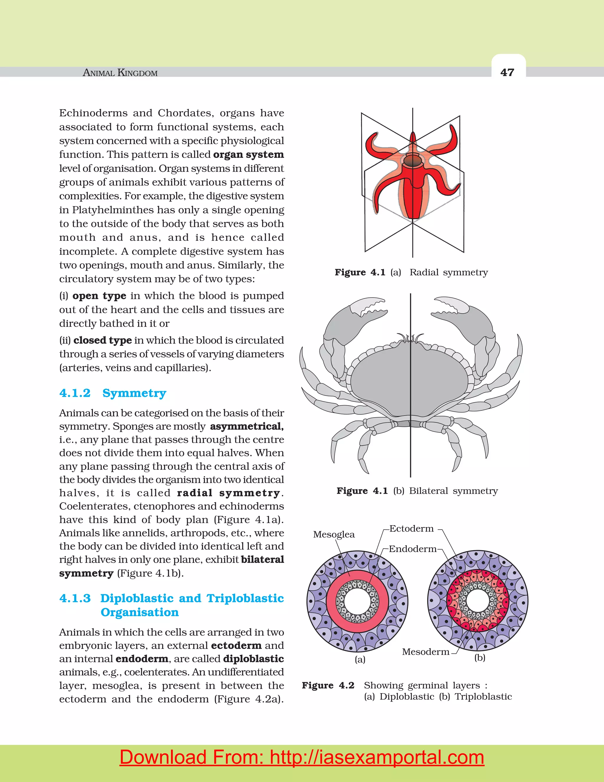


![STRUCTURAL ORGANISATION IN ANIMALS 115
glued to a suitable surface, usually in a crack or crevice of high relative
humidity near a food source. On an average, females produce 9-10
oothecae, each containing 14-16 eggs. The development of P. americana
is paurometabolous, meaning there is development through nymphal
stage. The nymphs look very much like adults. The nymph grows by
moulting about 13 times to reach the adult form. The next to last nymphal
stage has wing pads but only adult cockroaches have wings.
Many species of cockroaches are wild and are of no economic importance.
A few species thrive in and around human habitat. They are pests because
they destroy food and contaminate it with their smelly excreta. They can
transmit a variety of bacterial diseases by contaminating food material.
Testis
Phallic gland
Small tubules
Long tubules
Seminal vesicle
Vas deferens
Ejaculatory duct
Right phallomere
Ventral phallomere
Anal cercus
Caudal style
Pseudopenis
Titillator
Left phallomere
Ovary
Oviduct
Common oviduct
or vagina
Collaterial glands
Genital chamber
Vestibulum
Genital
pouch
Spermatheca
gonapophyses
]
Figure 7.18 Reproductive system of cockroach : (a) male (b) female
(a)
(b)
Download From: http://iasexamportal.com](https://image.slidesharecdn.com/biology-11th-ncert-151202101446-lva1-app6891/75/Biology-11th-ncert-117-2048.jpg)


![BIOMOLECULES 157
The catalytic cycle of an enzyme action can be described in the following
steps:
1. First, the substrate binds to the active site of the enzyme, fitting
into the active site.
2. The binding of the substrate induces the enzyme to alter its shape,
fitting more tightly around the substrate.
3. The active site of the enzyme, now in close proximity of the
substrate breaks the chemical bonds of the substrate and the
new enzyme- product complex is formed.
4. The enzyme releases the products of the reaction and the free
enzyme is ready to bind to another molecule of the substrate and
run through the catalytic cycle once again.
9.12.4 Factors Affecting Enzyme Activity
The activity of an enzyme can be affected by a change in the conditions
which can alter the tertiary structure of the protein. These include
temperature, pH, change in substrate concentration or binding of specific
chemicals that regulate its activity.
Temperature and pH
Enzymes generally function in a narrow range of temperature and pH
(Figure 9.7). Each enzyme shows its highest activity at a particular
temperature and pH called the optimum temperature and optimum pH.
Activity declines both below and above the optimum value. Low
temperature preserves the enzyme in a temporarily inactive state whereas
high temperature destroys enzymatic activity because proteins are
denatured by heat.
Figure 9.7 Effect of change in : (a) pH (b) Temperature and (c) Concentration of
substrate on enzyme activity
Vmax
Velocityofreaction(V)
[S]
V
2
max
Km
(a) (b) (c)
pH Temperature
Enzymeactivity
Download From: http://iasexamportal.com](https://image.slidesharecdn.com/biology-11th-ncert-151202101446-lva1-app6891/75/Biology-11th-ncert-159-2048.jpg)


![MINERAL NUTRITION 203
–
2 3 2 iN 8e 8H 16ATP 2NH H 16ADP 16P+
+ + + ⎯⎯→ + + +
Soil
particles
Root hair
Bacteria
Inner cortex and
pericycle cells
under division
Infection
thread
containing
bacteria
Mature nodule
Hook
Bacteria
Figure 12.4 Development of root nodules in soyabean : (a) Rhizobium bacteria contact
a susceptible root hair, divide near it, (b) Upon successful infection of
the root hair cause it to curl, (c) Infected thread carries the bacteria to
the inner cortex. The bacteria get modified into rod-shaped bacteroids
and cause inner cortical and pericycle cells to divide. Division and
growth of cortical and pericycle cells lead to nodule formation, (d) A
mature nodule is complete with vascular tissues continuous with those
of the root
(a)
N
+2 H
H
H
HH
H
H
H
H
HH
H
H
H
H
H
N
N N
N N
N
Enzyme
Substrate
[nitrogen gas (N )]2
Reduction Reduction
Reduction
Binding
of substrate(nitrogenase)
Product
[ammonia (NH )]3
Release
of products
Free nitrogenase
can bind another
molecule of N2
+2 H +2 H
N
N
H
HN
H
N
N
Figure 12.5 Steps of conversion of atmospheric nitrogen to ammonia by nitrogenase
enzyme complex found in nitrogen-fixing bacteria
Download From: http://iasexamportal.com](https://image.slidesharecdn.com/biology-11th-ncert-151202101446-lva1-app6891/75/Biology-11th-ncert-205-2048.jpg)


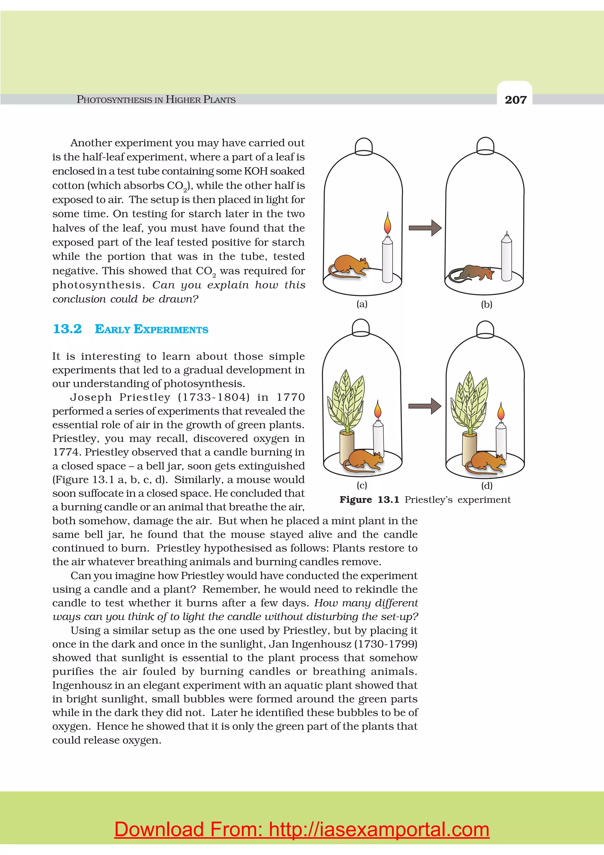
![208 BIOLOGY
It was not until about 1854 that Julius von Sachs provided evidence
for production of glucose when plants grow. Glucose is usually stored as
starch. His later studies showed that the green substance in plants
(chlorophyll as we know it now) is located in special bodies (later called
chloroplasts) within plant cells. He found that the green parts in plants is
where glucose is made, and that the glucose is usually stored as starch.
Now consider the interesting experiments done by T.W Engelmann
(1843 – 1909). Using a prism he split light into its spectral components
and then illuminated a green alga, Cladophora, placed in a suspension
of aerobic bacteria. The bacteria were used to detect the sites of O2
evolution. He observed that the bacteria accumulated mainly in the region
of blue and red light of the split spectrum. A first action spectrum of
photosynthesis was thus described. It resembles roughly the absorption
spectra of chlorophyll a and b (discussed in section 13.4).
By the middle of the nineteenth century the key features of plant
photosynthesis were known, namely, that plants could use light energy
to make carbohydrates from CO2
and water. The empirical equation
representing the total process of photosynthesis for oxygen evolving
organisms was then understood as:
CO H O CH O O
Light
2 2 2 2+ ⎯ →⎯⎯⎯⎯ +[ ]
where [CH2
O] represented a carbohydrate (e.g., glucose, a six-carbon
sugar).
A milestone contribution to the understanding of photosynthesis was
that made by a microbiologist, Cornelius van Niel (1897-1985), who,
based on his studies of purple and green bacteria, demonstrated that
photosynthesis is essentially a light-dependent reaction in which
hydrogen from a suitable oxidisable compound reduces carbon dioxide
to carbohydrates. This can be expressed by:
2 22 2 2 2H A CO A CH O H O
Light
+ ⎯ →⎯⎯⎯⎯ + +
In green plants H2
O is the hydrogen donor and is oxidised to O2
. Some
organisms do not release O2
during photosynthesis. When H2
S, instead
is the hydrogen donor for purple and green sulphur bacteria, the
‘oxidation’ product is sulphur or sulphate depending on the organism
and not O2
. Hence, he inferred that the O2
evolved by the green plant
comes from H2
O, not from carbon dioxide. This was later proved by using
radioisotopic techniques. The correct equation, that would represent the
overall process of photosynthesis is therefore:
6 12 6 62 2 6 12 6 2 2CO H O C H O H O O
Light
+ ⎯ →⎯⎯⎯⎯ + +
where C6
H12
O6
represents glucose. The O2
released is from water; this
was proved using radio isotope techniques. Note that this is not a single
Download From: http://iasexamportal.com](https://image.slidesharecdn.com/biology-11th-ncert-151202101446-lva1-app6891/75/Biology-11th-ncert-210-2048.jpg)


![212 BIOLOGY
system consisting of cytochromes (Figure
13.5). This movement of electrons is downhill,
in terms of an oxidation-reduction or redox
potential scale. The electrons are not used up
as they pass through the electron transport
chain, but are passed on to the pigments of
photosystem PS I. Simultaneously, electrons
in the reaction centre of PS I are also excited
when they receive red light of wavelength 700
nm and are transferred to another accepter
molecule that has a greater redox potential.
These electrons then are moved downhill
again, this time to a molecule of energy-rich
NADP+
. The addition of these electrons reduces
NADP+
to NADPH + H+
. This whole scheme of
transfer of electrons, starting from the PS II,
uphill to the accepter, down the electron
transport chain to PS I, excitation of electrons,
transfer to another accepter, and finally down hill to NADP+
causing it to
be reduced to NADPH + H+
is called the Z scheme, due to its characterstic
shape (Figure 13.5). This shape is formed when all the carriers are placed
in a sequence on a redox potential scale.
13.6.1 Splitting of Water
You would then ask, How does PS II supply electrons continuously? The
electrons that were moved from photosystem II must be replaced. This is
achieved by electrons available due to splitting of water. The splitting of
water is associated with the PS II; water is split into H+
, [O] and electrons.
This creates oxygen, one of the net products of photosynthesis. The
electrons needed to replace those removed from photosystem I are provided
by photosystem II.
2 4 42 2H O H O e⎯ →⎯ + ++ −
We need to emphasise here that the water splitting complex is associated
with the PS II, which itself is physically located on the inner side of the
membrane of the thylakoid. Then, where are the protons and O2
formed
likely to be released – in the lumen? or on the outer side of the membrane?
13.6.2 Cyclic and Non-cyclic Photo-phosphorylation
Living organisms have the capability of extracting energy from oxidisable
substances and store this in the form of bond energy. Special substances
like ATP, carry this energy in their chemical bonds. The process of which
Electron
transport
system
-
-
e acceptor
e acceptor
Light
Photosystem II Photosystem I
NADPH
NADP+
LHC
LHC
H O 2e + 2H + [O]2 - +
ADP+iP ATP
Figure 13.5 Z scheme of light reaction
Download From: http://iasexamportal.com](https://image.slidesharecdn.com/biology-11th-ncert-151202101446-lva1-app6891/75/Biology-11th-ncert-214-2048.jpg)


![222 BIOLOGY
13.10 FACTORS AFFECTING PHOTOSYNTHESIS
An understanding of the factors that affect photosynthesis is necessary.
The rate of photosynthesis is very important in determining the yield of
plants including crop plants. Photosynthesis is under the influence of
several factors, both internal (plant) and external. The plant factors include
the number, size, age and orientation of leaves, mesophyll cells and
chloroplasts, internal CO2
concentration and the amount of chlorophyll.
The plant or internal factors are dependent on the genetic predisposition
and the growth of the plant.
The external factors would include the availability of sunlight,
temperature, CO2
concentration and water. As a plant photosynthesises,
all these factors will simultaneously affect its rate. Hence, though several
factors interact and simultaneously affect photosynthesis or CO2
fixation,
usually one factor is the major cause or is the one that limits the rate.
Hence, at any point the rate will be determined by the factor available at
sub-optimal levels.
When several factors affect any [bio] chemical process, Blackman’s
(1905) Law of Limiting Factors comes into effect. This states the following:
If a chemical process is affected by more than one factor, then its
rate will be determined by the factor which is nearest to its minimal
value: it is the factor which directly affects the process if its quantity is
changed.
For example, despite the presence of a green
leaf and optimal light and CO2
conditions, the
plant may not photosynthesise if the temperature
is very low. This leaf, if given the optimal
temperature, will start photosynthesising.
13.10.1 Light
We need to distinguish between light quality, light
intensity and the duration of exposure to light,
while discussing light as a factor that affects
photosynthesis. There is a linear relationship
between incident light and CO2
fixation rates at
low light intensities. At higher light intensities,
gradually the rate does not show further increase
as other factors become limiting (Figure 13.10).
What is interesting to note is that light saturation
occurs at 10 per cent of the full sunlight. Hence,
except for plants in shade or in dense forests, light
is rarely a limiting factor in nature. Increase in
Figure 13.10 Graph of light intensity on the
rate of photosynthesis
Rateofphotosynthesis
Light intensity
A
B C
D
E
Download From: http://iasexamportal.com](https://image.slidesharecdn.com/biology-11th-ncert-151202101446-lva1-app6891/75/Biology-11th-ncert-224-2048.jpg)


