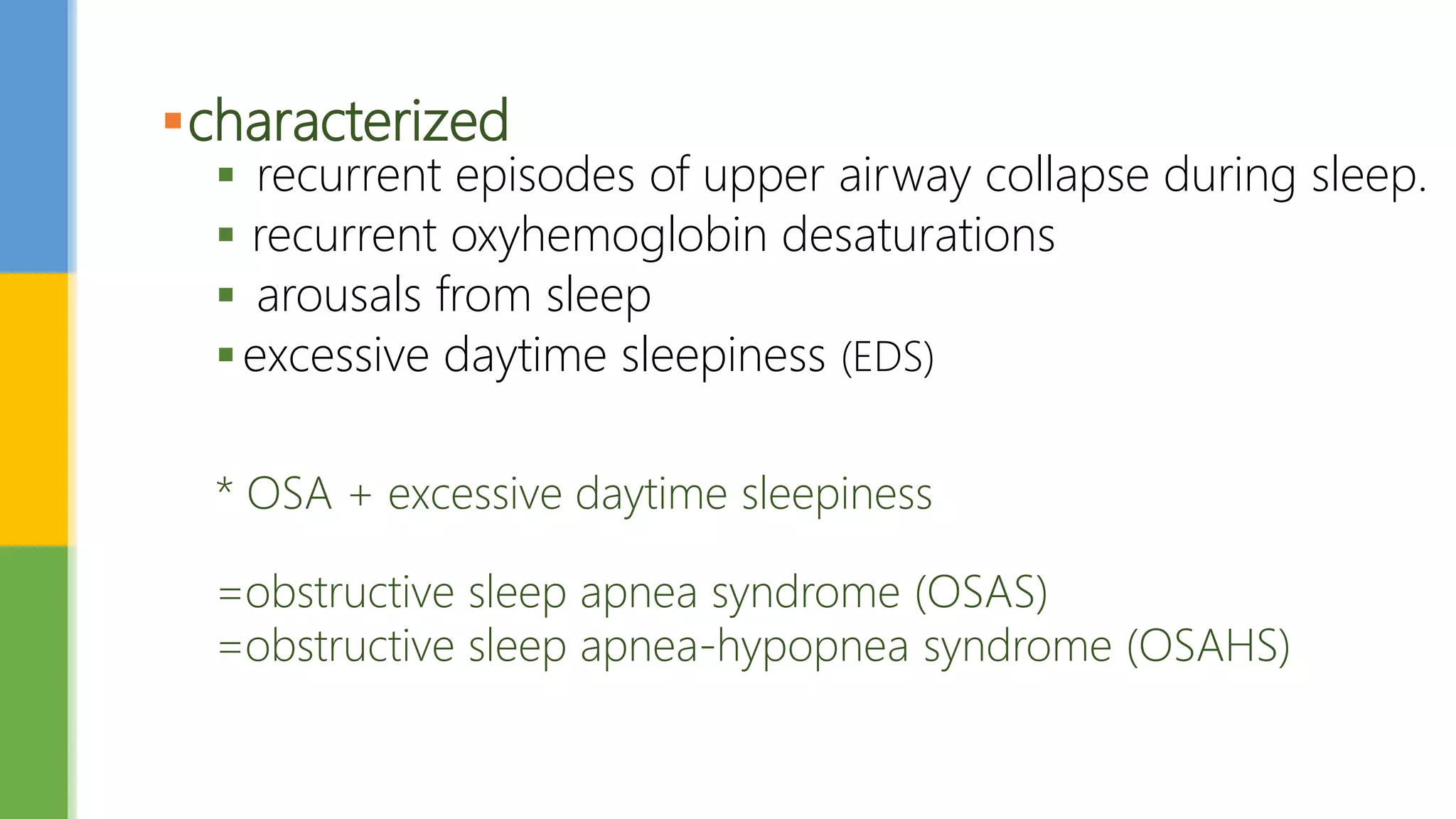
พญ.สุภลักษณ์ ตันติทวีโชค
แพทย์ประจำบ้ำนเวชศำสตร์ครอบครัว ชั้นปีที่3
ENT Topic presentation:
Obstructive Sleep Apnea
 Obstructive sleep apnea (OSA)
Obstructive sleep apnea-hypopnea (OSAH)
 a sleep disorder that involves cessation or significant decrease in
airflow in the presence of breathing effort.
 most common type of sleep-disordered breathing(SDB)
Obstructive sleep apnea
No disease Disease
characterized
 recurrent episodes of upper airway collapse during sleep.
 recurrent oxyhemoglobin desaturations
 arousals from sleep
excessive daytime sleepiness (EDS)
* OSA + excessive daytime sleepiness
=obstructive sleep apnea syndrome (OSAS)
=obstructive sleep apnea-hypopnea syndrome (OSAHS)
Signs and symptoms
Nocturnal symptoms
Snoring
Witnessed apneas
Gasping and choking
Apneas
Nocturia
Insomnia
Daytime symptoms
Morning headache
Dry / sore throat
Excessive daytime
sleepiness
Daytime fatigue/tiredness
Cognitive deficits
Personality/mood changes
Sexual dysfunction
The cardinal symptoms
3 S :
 S noring
 S leepiness
 S ignificant-other report of sleep apnea episodes
The clinical presentation of a child with obstructive sleep apnea is
nonspecific and requires increased awareness by the primary care
physician.
OSA symptoms in children can include the following:
Obstructive sleep apnea in children
the typical pediatricOSA patient was one with adenotonsillar hypertrophy
Abnormal breathing during sleep
Frequent awakenings or restlessness
Frequent nightmares
Enuresis
Difficulty awakening
Excessive daytime sleepiness
Hyperactivity/behavior problems
Daytime mouth breathing
 Naresh M. Punjabi,et al.The Epidemiology of Adult Obstructive Sleep Apnea. Proc Am Thorac Soc. 2008
Feb 15; 5(2): 136–143. pubmed
3.2%
The prevalence of SDB (AHI > or = 5) was 27% and 16% in men and women
The prevalence of OSAS (AHI > or = 5 + excessive daytime sleepiness) was 4.5% in men and 3.2% in
women
 healthy children younger than 8 years, the prevalence of obstructive sleep
apnea is estimated to be 1-3%.
 Habitual snoring is common during childhood and affects approximately
10% of children aged 2-8 years
 the frequency of habitual snoring decreases after age 9-10 years.
 Obesity confers 4-fold to 5-fold added risk for sleep-disordered breathing.
Epidemiology of OSA in children
 Most children with obstructive sleep apnea are aged 2-10 years
(coinciding with adenotonsillar lymphatic tissue growth).
 Children with severe obstructive apnea are likely to present
when aged 3-5 years.
The etiology of OSA involves
Structural factors
nonstructural factors
Etiology
Structural factors related to craniofacial bony anatomy that
predispose to pharyngeal collapse during sleep
 Retrognathia and micrognathia
 Inferior displacement of the hyoid
 Adenotonsillar hypertrophy
 High, arched palate
 Pierre Robin syndrome
 Down syndrome
 Prader-Willi syndrome
Structural factors
Retrognathia micrognathia
• Micrognathia
• glossoptosis
• Horsehoe-shaped
cleft palate
• Autosomal
dominant
• new (de novo)
genetic changes
 Obesity
 Age
 Supine sleep position
 Hypothyroidism
 Stroke
Nonstructural factors
 Apnea
 Hypopnea
 Respiratory effort–related arousal (RERA)
Definitions of respiratory events
Apnea
defined by the American Academy of Sleep Medicine (AASM) as
 the cessation of airflow for 10 seconds
Hypopnea
 a recognizable transient reduction (but not complete
of breathing 10 seconds
 a decrease of greater than 30% in the amplitude of a
measure of breathing,
 or a reduction in amplitude of less than 30%
+with oxygen desaturation of 4% or more
Respiratory effort–related arousal (RERA)
 an event characterized by increasing respiratory effort for 10
seconds or longer
 an arousal from sleep but one that does not fulfill the criteria
for a hypopnea or apnea.
 The criterion standard to measure RERAs is esophageal
manometry, as the AASM recommends. but impractical
 The overnight polysomnography(PSG) is the standard diagnosis
 It involves simultaneous recordings of multiple physiologic signals
during sleep, including
 Electroencephalogram (EEG)
 Electrooculogram (EOG)
 Electromyogram (EMG)
 Electrocardiogram (ECG)
 oronasal airflow
 oxyhemoglobin saturation.
 chest wall effort
 body position
 snore microphone
Diagnosis
Polysomnogram = gold-standard diagnosis
 Apneic episodes occur in the presence of respiratory muscle effort.
 Apneic episodes lasting 10 seconds or longer
 Apneic episodes are most prevalent during REM sleep.
 Patients may have a combination of apneas and hypopneas
 Mixed apneas may occur.
 Sleep disruption due to arousals is usually seen at the termination of an
episode of apnea.
PSG findings characteristic of OSA
Polysomnographic Criteria for OSA
The AHI is the number apnea and hypopnea of recorded during the
study per hour of sleep.
AHI = ( Apneas + Hypopneas)
the severity of OSA is classified as follows:
 None/Minimal: AHI < 5 per hour
 Mild: AHI ≥ 5, but < 15 per hour
 Moderate: AHI ≥ 15, but < 30 per hour
 Severe: AHI ≥ 30 per hour
Apnea Hypopnea Index (AHI)
Children
Normal AHI< 1 per hour
Very mild AHI 1,< 5 per hour
Mild AHI  5,<10 per hour
Moderate AHI  10,< 20 per hour
Severely AHI  20 per hour
the average number of episodes of apnea, hypopnea, and
respiratory event-related arousal per hour of sleep
 RDI = (RERAs + Hypopneas + apneas)
The respiratory disturbance index (RDI)
Epworth Sleepiness Scale(ESS)
> 10 = ESD
Score >10 is reported to have
93.5% sensitivity
100% specificity
The multiple sleep latency test (MSLT)
• Is tests for excessive daytime sleepiness by measuring how quickly
you fall asleep in a quiet environment during the day.
• Also known as a daytime nap study.
Cardiovascular comorbidities
 The most severe cardiovascular consequence of OSA is pulmonary
hypertension, and the resultant cor pulmonale
OSA and inflammation
 There is emerging evidence that OSA is a disease with chronic
grade systemic inflammation and increased oxidative stress
likely lead to the end organ morbidities
 Systemically, the pro-inflammatory cytokines, TNF-α, IL-6, IL-8
and C-reactive protein have been found to be elevated in the
serum of OSA patients.
 Levels of regulatory cytokines such as IL-10 are decreased
Consequences of OSA
Metabolic comorbidities
 Elevations in low density lipoprotein (LDL) cholesterol
 reduced levels in high density lipoprotein (HDL) cholesterol
 Mild apnea have a wider variety of options,
 Moderate-to-severe apnea should be treated with nasal
continuous positive airway pressure (CPAP).
Treatment
Behavior modification
 Weight loss and exercise
 Sleep position :Sleeping in a non-supine position (eg, lateral
recumbent) may correct or improve OSA
 Alcohol avoidance
 Medications avoidance
 Sedative effect medication: benzodiazepine receptor agonists, barbiturates,
other antiepileptic drugs, sedating antidepressants, antihistamines, and opiates
 Weight gain side effect :Antidepressants that cause weight gain
(eg, mirtazapine )
conservative nonsurgical treatment
 The mechanism of continuous positive airway pressure (CPAP) involves
maintenance of a positive pharyngeal transmural pressure
 In a meta-analysis of 35 randomized trials, CPAP compared with sham resulted in
a significant reduction in the apnea-hypopnea index
-AHI; mean difference -33.8 events/hour
-improved daytime sleepiness as assessed by the Epworth Sleepiness
Scale (mean difference -2 points)
-reduce systolic and diastolic blood pressure
-imorove sleep-related quality of life
POSITIVE AIRWAY PRESSURE THERAPY
Indications for treatment of CPAP
 The American Academy of Sleep Medicine (AASM) recommends offering positive
airway pressure therapy to all patients who have been diagnosed OSA by
 respiratory disturbance index (RDI) ≥15 events per hour with or without symptoms
 RDI between 5 and 14 events per hour that is accompanied by any of the following:
- sleepiness, nonrestorative sleep, fatigue
- insomnia symptoms; waking up with breath holding, gasping, or choking
- habitual snoring and/or breathing interruptions
- hypertension
- mood disorder
- cognitive dysfunction
- coronary artery disease
- stroke
- congestive heart failure
- atrial fibrillation
- type 2 diabetes
Oral appliances
 mandibular advancement devices
 tongue retaining devices)
ALTERNATIVE THERAPIES
Oropharyngeal :Structure tonsillar hypertrophy, adenoid hypertrophy
craniofacial abnormalities
 Tonsillectomy ± adenoidectomy
 Uvulopalatopharyngoplasty (UPPP)
 maxillomandibular advancement surgery
 Hyoid Suspension
Upper airway surgery
maxillomandibular advancement
 http://www.atsjournals.org/doi/full/10.1513/pats.200708-118MG
 http://emedicine.medscape.com/article/295807-overview
 http://rcot.org/datafile/_file/_doctor/3b03465492591d3f97333cec47ce490a.pdf
 http://www.rcot.org/pdf/MR!RC9M9!C9%20(Snoring)%20%20aEP@RGPKBX4KRBc(_3P9M9
KEQ_%20(Obstructive%20Sleep%20Apnea).pdf
 http://rcot.org/datafile/_file/_doctor/67e4c100dd2a9cc4051a5c9112eea741.pdf
 http://slideplayer.com/slide/6109008/
 http://advan.physiology.org/content/32/3/196
 http://reference.medscape.com/medline/abstract/8464434
Reference
Osa topic presentation

Osa topic presentation

  • 1.
  • 2.
     Obstructive sleepapnea (OSA) Obstructive sleep apnea-hypopnea (OSAH)  a sleep disorder that involves cessation or significant decrease in airflow in the presence of breathing effort.  most common type of sleep-disordered breathing(SDB) Obstructive sleep apnea
  • 3.
  • 5.
    characterized  recurrent episodesof upper airway collapse during sleep.  recurrent oxyhemoglobin desaturations  arousals from sleep excessive daytime sleepiness (EDS) * OSA + excessive daytime sleepiness =obstructive sleep apnea syndrome (OSAS) =obstructive sleep apnea-hypopnea syndrome (OSAHS)
  • 6.
    Signs and symptoms Nocturnalsymptoms Snoring Witnessed apneas Gasping and choking Apneas Nocturia Insomnia Daytime symptoms Morning headache Dry / sore throat Excessive daytime sleepiness Daytime fatigue/tiredness Cognitive deficits Personality/mood changes Sexual dysfunction
  • 7.
    The cardinal symptoms 3S :  S noring  S leepiness  S ignificant-other report of sleep apnea episodes
  • 8.
    The clinical presentationof a child with obstructive sleep apnea is nonspecific and requires increased awareness by the primary care physician. OSA symptoms in children can include the following: Obstructive sleep apnea in children the typical pediatricOSA patient was one with adenotonsillar hypertrophy Abnormal breathing during sleep Frequent awakenings or restlessness Frequent nightmares Enuresis Difficulty awakening Excessive daytime sleepiness Hyperactivity/behavior problems Daytime mouth breathing
  • 9.
     Naresh M.Punjabi,et al.The Epidemiology of Adult Obstructive Sleep Apnea. Proc Am Thorac Soc. 2008 Feb 15; 5(2): 136–143. pubmed 3.2% The prevalence of SDB (AHI > or = 5) was 27% and 16% in men and women The prevalence of OSAS (AHI > or = 5 + excessive daytime sleepiness) was 4.5% in men and 3.2% in women
  • 10.
     healthy childrenyounger than 8 years, the prevalence of obstructive sleep apnea is estimated to be 1-3%.  Habitual snoring is common during childhood and affects approximately 10% of children aged 2-8 years  the frequency of habitual snoring decreases after age 9-10 years.  Obesity confers 4-fold to 5-fold added risk for sleep-disordered breathing. Epidemiology of OSA in children
  • 11.
     Most childrenwith obstructive sleep apnea are aged 2-10 years (coinciding with adenotonsillar lymphatic tissue growth).  Children with severe obstructive apnea are likely to present when aged 3-5 years.
  • 12.
    The etiology ofOSA involves Structural factors nonstructural factors Etiology
  • 13.
    Structural factors relatedto craniofacial bony anatomy that predispose to pharyngeal collapse during sleep  Retrognathia and micrognathia  Inferior displacement of the hyoid  Adenotonsillar hypertrophy  High, arched palate  Pierre Robin syndrome  Down syndrome  Prader-Willi syndrome Structural factors
  • 14.
  • 15.
    • Micrognathia • glossoptosis •Horsehoe-shaped cleft palate • Autosomal dominant • new (de novo) genetic changes
  • 18.
     Obesity  Age Supine sleep position  Hypothyroidism  Stroke Nonstructural factors
  • 19.
     Apnea  Hypopnea Respiratory effort–related arousal (RERA) Definitions of respiratory events
  • 20.
    Apnea defined by theAmerican Academy of Sleep Medicine (AASM) as  the cessation of airflow for 10 seconds
  • 21.
    Hypopnea  a recognizabletransient reduction (but not complete of breathing 10 seconds  a decrease of greater than 30% in the amplitude of a measure of breathing,  or a reduction in amplitude of less than 30% +with oxygen desaturation of 4% or more
  • 22.
    Respiratory effort–related arousal(RERA)  an event characterized by increasing respiratory effort for 10 seconds or longer  an arousal from sleep but one that does not fulfill the criteria for a hypopnea or apnea.  The criterion standard to measure RERAs is esophageal manometry, as the AASM recommends. but impractical
  • 23.
     The overnightpolysomnography(PSG) is the standard diagnosis  It involves simultaneous recordings of multiple physiologic signals during sleep, including  Electroencephalogram (EEG)  Electrooculogram (EOG)  Electromyogram (EMG)  Electrocardiogram (ECG)  oronasal airflow  oxyhemoglobin saturation.  chest wall effort  body position  snore microphone Diagnosis Polysomnogram = gold-standard diagnosis
  • 27.
     Apneic episodesoccur in the presence of respiratory muscle effort.  Apneic episodes lasting 10 seconds or longer  Apneic episodes are most prevalent during REM sleep.  Patients may have a combination of apneas and hypopneas  Mixed apneas may occur.  Sleep disruption due to arousals is usually seen at the termination of an episode of apnea. PSG findings characteristic of OSA
  • 28.
  • 29.
    The AHI isthe number apnea and hypopnea of recorded during the study per hour of sleep. AHI = ( Apneas + Hypopneas) the severity of OSA is classified as follows:  None/Minimal: AHI < 5 per hour  Mild: AHI ≥ 5, but < 15 per hour  Moderate: AHI ≥ 15, but < 30 per hour  Severe: AHI ≥ 30 per hour Apnea Hypopnea Index (AHI) Children Normal AHI< 1 per hour Very mild AHI 1,< 5 per hour Mild AHI  5,<10 per hour Moderate AHI  10,< 20 per hour Severely AHI  20 per hour
  • 30.
    the average numberof episodes of apnea, hypopnea, and respiratory event-related arousal per hour of sleep  RDI = (RERAs + Hypopneas + apneas) The respiratory disturbance index (RDI)
  • 31.
    Epworth Sleepiness Scale(ESS) >10 = ESD Score >10 is reported to have 93.5% sensitivity 100% specificity
  • 32.
    The multiple sleeplatency test (MSLT) • Is tests for excessive daytime sleepiness by measuring how quickly you fall asleep in a quiet environment during the day. • Also known as a daytime nap study.
  • 33.
    Cardiovascular comorbidities  Themost severe cardiovascular consequence of OSA is pulmonary hypertension, and the resultant cor pulmonale
  • 34.
    OSA and inflammation There is emerging evidence that OSA is a disease with chronic grade systemic inflammation and increased oxidative stress likely lead to the end organ morbidities  Systemically, the pro-inflammatory cytokines, TNF-α, IL-6, IL-8 and C-reactive protein have been found to be elevated in the serum of OSA patients.  Levels of regulatory cytokines such as IL-10 are decreased Consequences of OSA
  • 35.
    Metabolic comorbidities  Elevationsin low density lipoprotein (LDL) cholesterol  reduced levels in high density lipoprotein (HDL) cholesterol
  • 36.
     Mild apneahave a wider variety of options,  Moderate-to-severe apnea should be treated with nasal continuous positive airway pressure (CPAP). Treatment
  • 37.
    Behavior modification  Weightloss and exercise  Sleep position :Sleeping in a non-supine position (eg, lateral recumbent) may correct or improve OSA  Alcohol avoidance  Medications avoidance  Sedative effect medication: benzodiazepine receptor agonists, barbiturates, other antiepileptic drugs, sedating antidepressants, antihistamines, and opiates  Weight gain side effect :Antidepressants that cause weight gain (eg, mirtazapine ) conservative nonsurgical treatment
  • 38.
     The mechanismof continuous positive airway pressure (CPAP) involves maintenance of a positive pharyngeal transmural pressure  In a meta-analysis of 35 randomized trials, CPAP compared with sham resulted in a significant reduction in the apnea-hypopnea index -AHI; mean difference -33.8 events/hour -improved daytime sleepiness as assessed by the Epworth Sleepiness Scale (mean difference -2 points) -reduce systolic and diastolic blood pressure -imorove sleep-related quality of life POSITIVE AIRWAY PRESSURE THERAPY
  • 40.
    Indications for treatmentof CPAP  The American Academy of Sleep Medicine (AASM) recommends offering positive airway pressure therapy to all patients who have been diagnosed OSA by  respiratory disturbance index (RDI) ≥15 events per hour with or without symptoms  RDI between 5 and 14 events per hour that is accompanied by any of the following: - sleepiness, nonrestorative sleep, fatigue - insomnia symptoms; waking up with breath holding, gasping, or choking - habitual snoring and/or breathing interruptions - hypertension - mood disorder - cognitive dysfunction - coronary artery disease - stroke - congestive heart failure - atrial fibrillation - type 2 diabetes
  • 41.
    Oral appliances  mandibularadvancement devices  tongue retaining devices) ALTERNATIVE THERAPIES
  • 42.
    Oropharyngeal :Structure tonsillarhypertrophy, adenoid hypertrophy craniofacial abnormalities  Tonsillectomy ± adenoidectomy  Uvulopalatopharyngoplasty (UPPP)  maxillomandibular advancement surgery  Hyoid Suspension Upper airway surgery
  • 46.
  • 48.
     http://www.atsjournals.org/doi/full/10.1513/pats.200708-118MG  http://emedicine.medscape.com/article/295807-overview http://rcot.org/datafile/_file/_doctor/3b03465492591d3f97333cec47ce490a.pdf  http://www.rcot.org/pdf/MR!RC9M9!C9%20(Snoring)%20%20aEP@RGPKBX4KRBc(_3P9M9 KEQ_%20(Obstructive%20Sleep%20Apnea).pdf  http://rcot.org/datafile/_file/_doctor/67e4c100dd2a9cc4051a5c9112eea741.pdf  http://slideplayer.com/slide/6109008/  http://advan.physiology.org/content/32/3/196  http://reference.medscape.com/medline/abstract/8464434 Reference

Editor's Notes

  • #3 Sleep-related disordered breathing continuum ranging from simple snoring to-- obstructive sleep apnea (OSA)-- Upper airway resistance syndrome (UARS) 
  • #14 https://ghr.nlm.nih.gov/condition/prader-willi-syndrome
  • #15 https://www.infantlaserdentistry.com/infant-birth-and-medical-complications.html https://www.slideshare.net/marwanmouakeh/obstructive-sleep-apnea-52853234
  • #22 using nasal pressure (diagnostic study), positive airway pressure device flow (titration study), or an alternative hypopnea sensor (diagnostic study);
  • #23 With either method, the respiratory disturbance index (RDI) is greater than 5 and the normal RDI cutoff is greater than 15.
  • #24 https://www.ncbi.nlm.nih.gov/pmc/articles/PMC2645248/#bib12