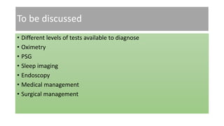
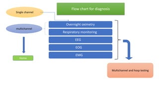
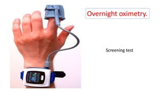
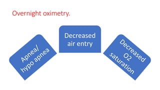
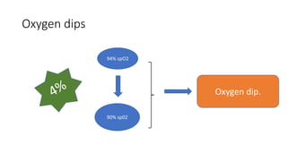
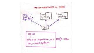
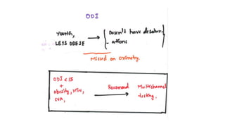
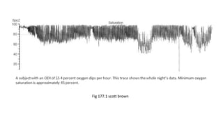
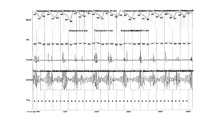
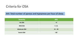
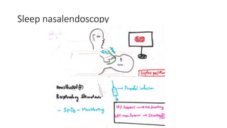
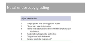
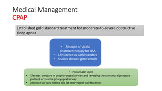
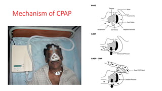
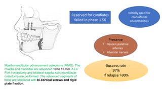
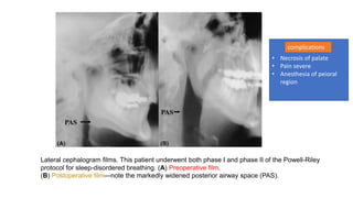
Diagnosis and management of OSA
Dr. Sharath Chandra.C
JR ENT,
Dept of ENT,
AIIMS Rishikesh.
To be discussed
• Different levels of tests available to diagnose
• Oximetry
• PSG
• Sleep imaging
• Endoscopy
• Medical management
• Surgical management
Overnight oximetry
Respiratory monitoring
EEG
EOG
EMG
Single channel
multichannel
Multichannel and hosp testing
Home
Flow chart for diagnosis
Screening test
Overnight oximetry.
Overnight oximetry.
Decreased
air entry
Oxygen dips
94% spO2
90% sp02
Oxygen dip.
Fig 177.1 scott brown
Home multichannel testing.
Advantages
• Patient comfort
• Cost savings
• Prevent hospital stay
Disadvantages
• Sensor failure at home
• Fewer signals channels used
leads to less info..
Oronasal cannula
Thorasic band
Abdominal band
Finger oximetry probe
Home multichannel testing
Central apnoea
Not a/c raised effort chest /
abdominal movements
OSAH
Raised efforts of abd/ thoracic
muscles
Multi channel hosp testing(PSG)
• Used in complex cases
• More respiratory monitoring required
• If assisted ventilation required
• If other sleep disorders suspected
narcolepsy ; parasomnias
Polysomnography
• AAS( American academy of sleep medicine) indicated PSG as
investigation of choice for sleep disordered breathing.
Respiratory channels Nasal, oral airflow& thorasic and
abdomen
Sleep staging EEG
Eye movement EOG
Arousal Electromyogram(chin)
Cardiac monitoring ECG
Leg movements B/L tibialis anterior electrodes
Accompanied by sleep technician for over night monitoring
Criteria for OSA
Severity AHI
No OSA <5
Mild OSA 5- 15
Moderate OSA 15 – 30
Severe OSA >30
AHI: Total number of apneas and hypopneas per hour of sleep.
Sleep nasalendoscopy
Nasal endoscopy grading
Imaging in OSA
• Cephalometric radiography
• Conventional CT
• 3D CT
• MRI/volumetric MRI
• Optical coherence tomography
Optical coherence tomography
OCT
Qualitative real time
images
3mm optical probe
360 rotation
Longitudinal movement
Advantages Disadvantages
Sleep state Limited view at
places
Min discomfort No extraluminal
factors examined
No radiation
Accurate determination of
shape and size
Hypopharynx (a), oropharynx (b) and (c), and nasopharynx (d). 3-D rendering
profile view (e) and front-on view (f) from in vivo data. Epiglottis (E), base of
tongue (BT), soft palate (SP), adenoidal tissue (AT), right nasal cavity (NC) are
labeled.
MRI/VOLUMETRIC MRI
Best current mode of imaging for assessment of the upper airway and
surrounding soft tissue and craniofacial structures
MRI studies helped us in under standing
• Pathophysiology of sleep(OSA)
• Mechanisms undelynig treatments like
• Weight loss
• CPAP
• Oral appliances
• Upper airway surgery
Mid-sagittal magnetic resonance imaging of a normal subject (left) and a patient
with sleep apnea (right). The upper airway is smaller in both the retropalatal and
retroglossal region in the apneic patient. The soft palate is longer in the apneic
patient. The amount of subcutaneous fat (white area at the back of the neck) is
greater in the apneic.
Volumetric MRI
Volumetric state-dependent airway imaging in a normal subject using magnetic
resonance imaging. Airway volume during sleep is smaller in the retropalatal (RP)
region but not the retroglossal (RG) region. Such images suggest that the upper
airway during sleep does not narrow as a homogenous tube.
Cephalometry
Cephalometry
Simple and standardized
techRadiographs of head and
neck
Study measurements
of setpoints, planes
and distances
• 2D images
• Static images
• Cannot cooment on transverse ,
volumetric dimensions
• Sleep state assessment not possible
1) The genioglossus (GG) part that advances the tongue; 2) the GG part that depresses
and draws down the tongue; 3) oropharynx; 4) velopharynx; 5) soft palate; 6) distance
between the hyoid bone and the mandible; and 7) angle of the chin-nose base-maxilla.
The heavy black quadrangle delineates the mouth-box.
Medical Management
CPAP
Established gold standard treatment for moderate-to-severe obstructive
sleep apnea
• Absence of viable
pharmacotherapy for OSA
• Considered as Gold standard
• Studies showed good results
• Pneumatic splint
• Elevates pressure in oropharyngeal airway and reversing the transmural pressure
gradient across the pharyngeal airway
• Decrease air way edema and lat pharyngeal wall thickness
Mechanism of CPAP
Side effects
Common
• Nasal congestion
• Facemask Interface problems
• Leak related
• claustrophobic feeling
• Uncommon
• pulmonary barotrauma,
• pneumocephalus,
• increased intraocular pressure,
• tympanic membrane rupture,
cerebrospinal fluid (CSF) leak and
meningitis
post-neuro, -airway, or -facial surgery
COMPARISON WITH OTHER TREATMENTS
• Great advantages of nasal CPAP is that it
is immediately and demonstrably
efficacious in relieving OSA
• Can be used as trail
Volumetric magnetic resonance imaging reconstruction of the
upper airway in a normal subject with progressively greater continuous positive airway pressure
(CPAP) (0 to 15 cm H2O) settings. Upper airway volume increases significantly in both the retropalatal
(RP) and retro glossal (RG) regions with higher levels of CPAP.
Surgery indications
• Apnea-hypopnea index ≥ 20 events per hour of sleep
• Oxygen desaturation nadir < 90%
• Esophageal pressure (Pes) more negative than −10 cm H2O
• Cardiovascular derangements (arrhythmia, hypertension)
• Neurobehavioral symptoms (excessive daytime sleepiness)
• Failure of medical management
• Anatomical sites of obstruction (nose, palate, tongue base)
• Surgery may be indicated with an AHI < 20 if accompanied by excessive daytime
fatigue
Poor candidates for surgery
• Severe pulmonary disease
• Unstable cardiovascular disease
• Morbid obesity
• Alcohol or drug abuse
• Psychiatric instability
• Unrealistic expectations
Preoperative evaluation
• General examinations( vitals, weight , neck, nose etc)
• Polysomnography
• Fiberoptic nasopharyngolaryngoscopy
• Cephalometric analysis
Powell-Riley Definition of Surgical Responders
• Apnea-hypopnea index < 20 events per hour of sleep
• Oxygen desaturation nadir ≥ 90%
• Excessive daytime sleepiness alleviated
• Response equivalent to CPAP on full-night titration
• Reduction of the apnea-hypopnea index by 50% or more is
considered a cure if the preoperative apnea-hypopnea index is less
than 20
Powell-Riley Protocol Surgical
Procedures
• Phase I
• Nasal surgery (septoplasty, turbinate reduction, nasal valve grafting)
• Tonsillectomy
• Uvulopalatopharyngoplasty or uvulopalatal flap
• Mandibular osteotomy with genioglossus advancement
• Hyoid myotomy and suspension
• Temperature-controlled radiofrequency—turbinate's, palate, tongue base
• Phase II
• Maxillomandibular advancement osteotomy
• Temperature-controlled radiofrequency—tongue base
Uvulopalatopharyngoplasty
Ikematsu
1950
Fugita
1985
Later
developed
• Excellent technique to alleviate isolated retropalatal
obstruction
• Conservative surgery
• Performed under GA
Uvulopalatopharyngoplasty
This patient demonstrates tonsillar hypertrophy, an elongated uvula and redundant tissue of the lateral pharyngeal
wall resulting in a narrowed airway space.
(B) Removal of the tonsils, lateral pharyngeal wall mucosa, and soft palate
mucosa has enlarged the airway.
(C) Excised surgical specimen.
9%
15%
15%
11%14%
11%
10%
10%
5%
1st Qtr
post op pain
hemorrage
laryngospasm
nasal regurgiatation
dry throat
swallowing prob
voice cahnges
taste distub
Post op complications
Laser-Assisted Uvulopalatoplasty
• Office procedure
• Bloodless field
• No GA needed
• Less post op regurgitation
• CO2 laser used
• shorten and stiffen the soft palate via a series of
• carbon dioxide laser incisions
• Globus symptoms are common
Mandibular Osteotomy with Genioglossus
Advancement
• Indication: type II and III fujita grade(hypopharyngealobstruction)
• Enlarge post airway by preventing prolapse of tongue posteriorly
• Genial tubercle & attached genioglossus muscle are advanced
anteriorly
• Depends on anterior mand. thickness and compliance of genioglossus
muscle
10 x14mm
Secure margins
Superiorly: 5mm below roots
Inferiorly: 10mm inf border
Lateral: medial to canines
Success rate
Over all 61%
Mild dis: 77%
Severe dis: 42%
Riley et al 1986
Complications
• Post op hematoma and edema
• Mandible fracture
• Root injury
• Wound infection
Hyoid Myotomy and Suspension(Kaya 1984)
Alleviate hypopharyngeal obstruction by advancing the hyoid complex in
anterior direction.
Hyoid myotomy and suspension. The hyoid bone and thyroid cartilage
are exposed via a small neck incision. The hyoid bone is advanced
anteriorly and secured to the thyroid cartilage with three or four
permanent sutures.
75 success rate with
combination of
UPPP and GGAO
Mostly used as
adjuvant surgery
rather than primary
SX
Transient aspiration
and dysphagia
Maxillomandibular Advancement
Osteotomy(Kuo et al).
• phase II of the Powell-Riley two-phase surgical protocol
• Ind: Refractory hypopharyngeal obstruction
• Expands hypopharyngeal and pharyngeal airway
• Results equal to medical therapy (CPAP)
Maxillomandibular advancement osteotomy (MMO). The
maxilla and mandible are advanced 10 to 15 mm. A Le
Fort I osteotomy and bilateral sagittal split mandibular
osteotomy are performed. The advanced segments of
bone are stabilized with bi-cortical screws and rigid
plate fixation.
Reserved for candiates
failed in phase 1 SX
Initially used for
craniofacial
abnormalities
Preserve
• Descen palatine
arteries
• Alveolar nerves
Success rate
97%
If relapse >90%
Lateral cephalogram films. This patient underwent both phase I and phase II of the Powell-Riley
protocol for sleep-disordered breathing. (A) Preoperative film.
(B) Postoperative film—note the markedly widened posterior airway space (PAS).
• Necrosis of palate
• Pain severe
• Anesthesia of peioral
region
complications
Radiofrequency tissue volume reduction
• Comparable to diathermy with low power and
low temperatures
• Thermal injury to specific submucosal sites
• Results fibrosis and volume reduction
• Day care procedure
• Less post op pain
Tongue based procedures
• Tracheostomy required
• Midline Laser glossectomy - laser is used to extirpate a rectangular
strip (2.5 into 5 cms) of the posterior portion of tongue, useful in
Down’s syndrome, Mucopolysaccharidosis
• Lingualplasty - modification of laser glossectomy, involves additional
excision of lateral tongue tissue
• Radiofrequency tissue ablation of tongue base - RF probe with 465
KHZ
• Injection snoreplasty
• Palatal implant
• Mandibular positioning device – in non obese patients with
micrognathia / retrognathia, advances the mandible and increases
posterior airway space, has success rate of 50 % & compliance rate of
25%
• Tongue retaining device
• Positional devices
• Nasal splints
Thank you

Diagnois obstructive sleep apnoea

  • 1.
    Diagnosis and managementof OSA Dr. Sharath Chandra.C JR ENT, Dept of ENT, AIIMS Rishikesh.
  • 2.
    To be discussed •Different levels of tests available to diagnose • Oximetry • PSG • Sleep imaging • Endoscopy • Medical management • Surgical management
  • 3.
    Overnight oximetry Respiratory monitoring EEG EOG EMG Singlechannel multichannel Multichannel and hosp testing Home Flow chart for diagnosis
  • 4.
  • 5.
  • 6.
    Oxygen dips 94% spO2 90%sp02 Oxygen dip.
  • 9.
  • 10.
    Home multichannel testing. Advantages •Patient comfort • Cost savings • Prevent hospital stay Disadvantages • Sensor failure at home • Fewer signals channels used leads to less info..
  • 11.
    Oronasal cannula Thorasic band Abdominalband Finger oximetry probe Home multichannel testing
  • 12.
    Central apnoea Not a/craised effort chest / abdominal movements OSAH Raised efforts of abd/ thoracic muscles
  • 13.
    Multi channel hosptesting(PSG) • Used in complex cases • More respiratory monitoring required • If assisted ventilation required • If other sleep disorders suspected narcolepsy ; parasomnias
  • 15.
    Polysomnography • AAS( Americanacademy of sleep medicine) indicated PSG as investigation of choice for sleep disordered breathing. Respiratory channels Nasal, oral airflow& thorasic and abdomen Sleep staging EEG Eye movement EOG Arousal Electromyogram(chin) Cardiac monitoring ECG Leg movements B/L tibialis anterior electrodes Accompanied by sleep technician for over night monitoring
  • 18.
    Criteria for OSA SeverityAHI No OSA <5 Mild OSA 5- 15 Moderate OSA 15 – 30 Severe OSA >30 AHI: Total number of apneas and hypopneas per hour of sleep.
  • 19.
  • 20.
  • 21.
    Imaging in OSA •Cephalometric radiography • Conventional CT • 3D CT • MRI/volumetric MRI • Optical coherence tomography
  • 22.
    Optical coherence tomography OCT Qualitativereal time images 3mm optical probe 360 rotation Longitudinal movement Advantages Disadvantages Sleep state Limited view at places Min discomfort No extraluminal factors examined No radiation Accurate determination of shape and size
  • 23.
    Hypopharynx (a), oropharynx(b) and (c), and nasopharynx (d). 3-D rendering profile view (e) and front-on view (f) from in vivo data. Epiglottis (E), base of tongue (BT), soft palate (SP), adenoidal tissue (AT), right nasal cavity (NC) are labeled.
  • 24.
    MRI/VOLUMETRIC MRI Best currentmode of imaging for assessment of the upper airway and surrounding soft tissue and craniofacial structures MRI studies helped us in under standing • Pathophysiology of sleep(OSA) • Mechanisms undelynig treatments like • Weight loss • CPAP • Oral appliances • Upper airway surgery
  • 26.
    Mid-sagittal magnetic resonanceimaging of a normal subject (left) and a patient with sleep apnea (right). The upper airway is smaller in both the retropalatal and retroglossal region in the apneic patient. The soft palate is longer in the apneic patient. The amount of subcutaneous fat (white area at the back of the neck) is greater in the apneic.
  • 27.
  • 28.
    Volumetric state-dependent airwayimaging in a normal subject using magnetic resonance imaging. Airway volume during sleep is smaller in the retropalatal (RP) region but not the retroglossal (RG) region. Such images suggest that the upper airway during sleep does not narrow as a homogenous tube.
  • 29.
    Cephalometry Cephalometry Simple and standardized techRadiographsof head and neck Study measurements of setpoints, planes and distances • 2D images • Static images • Cannot cooment on transverse , volumetric dimensions • Sleep state assessment not possible
  • 30.
    1) The genioglossus(GG) part that advances the tongue; 2) the GG part that depresses and draws down the tongue; 3) oropharynx; 4) velopharynx; 5) soft palate; 6) distance between the hyoid bone and the mandible; and 7) angle of the chin-nose base-maxilla. The heavy black quadrangle delineates the mouth-box.
  • 32.
    Medical Management CPAP Established goldstandard treatment for moderate-to-severe obstructive sleep apnea • Absence of viable pharmacotherapy for OSA • Considered as Gold standard • Studies showed good results • Pneumatic splint • Elevates pressure in oropharyngeal airway and reversing the transmural pressure gradient across the pharyngeal airway • Decrease air way edema and lat pharyngeal wall thickness
  • 33.
  • 34.
    Side effects Common • Nasalcongestion • Facemask Interface problems • Leak related • claustrophobic feeling • Uncommon • pulmonary barotrauma, • pneumocephalus, • increased intraocular pressure, • tympanic membrane rupture, cerebrospinal fluid (CSF) leak and meningitis post-neuro, -airway, or -facial surgery
  • 35.
    COMPARISON WITH OTHERTREATMENTS • Great advantages of nasal CPAP is that it is immediately and demonstrably efficacious in relieving OSA • Can be used as trail
  • 36.
    Volumetric magnetic resonanceimaging reconstruction of the upper airway in a normal subject with progressively greater continuous positive airway pressure (CPAP) (0 to 15 cm H2O) settings. Upper airway volume increases significantly in both the retropalatal (RP) and retro glossal (RG) regions with higher levels of CPAP.
  • 38.
    Surgery indications • Apnea-hypopneaindex ≥ 20 events per hour of sleep • Oxygen desaturation nadir < 90% • Esophageal pressure (Pes) more negative than −10 cm H2O • Cardiovascular derangements (arrhythmia, hypertension) • Neurobehavioral symptoms (excessive daytime sleepiness) • Failure of medical management • Anatomical sites of obstruction (nose, palate, tongue base) • Surgery may be indicated with an AHI < 20 if accompanied by excessive daytime fatigue
  • 39.
    Poor candidates forsurgery • Severe pulmonary disease • Unstable cardiovascular disease • Morbid obesity • Alcohol or drug abuse • Psychiatric instability • Unrealistic expectations
  • 40.
    Preoperative evaluation • Generalexaminations( vitals, weight , neck, nose etc) • Polysomnography • Fiberoptic nasopharyngolaryngoscopy • Cephalometric analysis
  • 41.
    Powell-Riley Definition ofSurgical Responders • Apnea-hypopnea index < 20 events per hour of sleep • Oxygen desaturation nadir ≥ 90% • Excessive daytime sleepiness alleviated • Response equivalent to CPAP on full-night titration • Reduction of the apnea-hypopnea index by 50% or more is considered a cure if the preoperative apnea-hypopnea index is less than 20
  • 42.
    Powell-Riley Protocol Surgical Procedures •Phase I • Nasal surgery (septoplasty, turbinate reduction, nasal valve grafting) • Tonsillectomy • Uvulopalatopharyngoplasty or uvulopalatal flap • Mandibular osteotomy with genioglossus advancement • Hyoid myotomy and suspension • Temperature-controlled radiofrequency—turbinate's, palate, tongue base • Phase II • Maxillomandibular advancement osteotomy • Temperature-controlled radiofrequency—tongue base
  • 43.
    Uvulopalatopharyngoplasty Ikematsu 1950 Fugita 1985 Later developed • Excellent techniqueto alleviate isolated retropalatal obstruction • Conservative surgery • Performed under GA
  • 44.
  • 45.
    This patient demonstratestonsillar hypertrophy, an elongated uvula and redundant tissue of the lateral pharyngeal wall resulting in a narrowed airway space. (B) Removal of the tonsils, lateral pharyngeal wall mucosa, and soft palate mucosa has enlarged the airway. (C) Excised surgical specimen.
  • 46.
    9% 15% 15% 11%14% 11% 10% 10% 5% 1st Qtr post oppain hemorrage laryngospasm nasal regurgiatation dry throat swallowing prob voice cahnges taste distub Post op complications
  • 47.
    Laser-Assisted Uvulopalatoplasty • Officeprocedure • Bloodless field • No GA needed • Less post op regurgitation • CO2 laser used • shorten and stiffen the soft palate via a series of • carbon dioxide laser incisions • Globus symptoms are common
  • 48.
    Mandibular Osteotomy withGenioglossus Advancement • Indication: type II and III fujita grade(hypopharyngealobstruction) • Enlarge post airway by preventing prolapse of tongue posteriorly • Genial tubercle & attached genioglossus muscle are advanced anteriorly • Depends on anterior mand. thickness and compliance of genioglossus muscle
  • 49.
    10 x14mm Secure margins Superiorly:5mm below roots Inferiorly: 10mm inf border Lateral: medial to canines Success rate Over all 61% Mild dis: 77% Severe dis: 42% Riley et al 1986 Complications • Post op hematoma and edema • Mandible fracture • Root injury • Wound infection
  • 50.
    Hyoid Myotomy andSuspension(Kaya 1984) Alleviate hypopharyngeal obstruction by advancing the hyoid complex in anterior direction.
  • 51.
    Hyoid myotomy andsuspension. The hyoid bone and thyroid cartilage are exposed via a small neck incision. The hyoid bone is advanced anteriorly and secured to the thyroid cartilage with three or four permanent sutures. 75 success rate with combination of UPPP and GGAO Mostly used as adjuvant surgery rather than primary SX Transient aspiration and dysphagia
  • 52.
    Maxillomandibular Advancement Osteotomy(Kuo etal). • phase II of the Powell-Riley two-phase surgical protocol • Ind: Refractory hypopharyngeal obstruction • Expands hypopharyngeal and pharyngeal airway • Results equal to medical therapy (CPAP)
  • 53.
    Maxillomandibular advancement osteotomy(MMO). The maxilla and mandible are advanced 10 to 15 mm. A Le Fort I osteotomy and bilateral sagittal split mandibular osteotomy are performed. The advanced segments of bone are stabilized with bi-cortical screws and rigid plate fixation. Reserved for candiates failed in phase 1 SX Initially used for craniofacial abnormalities Preserve • Descen palatine arteries • Alveolar nerves Success rate 97% If relapse >90%
  • 54.
    Lateral cephalogram films.This patient underwent both phase I and phase II of the Powell-Riley protocol for sleep-disordered breathing. (A) Preoperative film. (B) Postoperative film—note the markedly widened posterior airway space (PAS). • Necrosis of palate • Pain severe • Anesthesia of peioral region complications
  • 55.
    Radiofrequency tissue volumereduction • Comparable to diathermy with low power and low temperatures • Thermal injury to specific submucosal sites • Results fibrosis and volume reduction • Day care procedure • Less post op pain
  • 56.
    Tongue based procedures •Tracheostomy required • Midline Laser glossectomy - laser is used to extirpate a rectangular strip (2.5 into 5 cms) of the posterior portion of tongue, useful in Down’s syndrome, Mucopolysaccharidosis • Lingualplasty - modification of laser glossectomy, involves additional excision of lateral tongue tissue • Radiofrequency tissue ablation of tongue base - RF probe with 465 KHZ
  • 57.
  • 58.
    • Mandibular positioningdevice – in non obese patients with micrognathia / retrognathia, advances the mandible and increases posterior airway space, has success rate of 50 % & compliance rate of 25% • Tongue retaining device • Positional devices • Nasal splints
  • 60.