
Recommended
More Related Content
What's hot
What's hot (20)
Similar to Reza Khorramirouz
Similar to Reza Khorramirouz (20)
More from Reza Khorramirouz
More from Reza Khorramirouz (6)
Recently uploaded
Recently uploaded (20)
Reza Khorramirouz
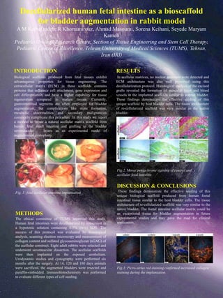
- 1. Decellularized human fetal intestine as a bioscaffold for bladder augmentation in rabbit model A M Kajbafzadeh, R Khorramirouz, Ahmad Masoumi, Sorena Keihani, Seyede Maryam Kameli Pediatric Urology Research Centre, Section of Tissue Engineering and Stem Cell Therapy, Pediatric Center of Excellence, Tehran University of Medical Sciences (TUMS), Tehran, Iran (IRI) INTRODUCTION Biological scaffolds produced from fetal tissues exhibit advantageous properties for tissue engineering. The extracellular matrix (ECM) in these scaffolds contains proteins that influence cell attachment, gene expression and cell differentiation and have a higher capability for tissue regeneration compared to mature tissues. Currently, gastrointestinal segments are often employed for bladder augmentation, but complications like stone formation, metabolic abnormalities and secondary malignancies commonly complicate this procedure .In this study we report a method to create a natural acellular matrix scaffold from human fetal small intestine and grafting to the bladder seromuscular layers as an experimental model of augmentation cystoplasty. RESULTS In acellular matrices, no nuclear remnants were detected and ECM architecture was also well preserved using this decellularization protocol. Histological analysis of the excised grafts revealed the formation of muscular layer and blood vessels in the implanted scaffolds similar to normal bladder. These findings demonstrate the effective seeding of this unique scaffold by host bladder cells. The tissue architecture of re-cellularized scaffold was very similar to the native bladder. METHODS The ethical committee of TUMS approved this study. Human fetal intestines were decellularized by immersion in a hypotonic solution containing 0.5% (w/v) SDS. The success of this protocol was evaluated by histological analysis, scanning electron microscopy and measurement of collagen content and sulfated glycosaminoglycan (sGAG) of the acellular construct. Eight adult rabbits were selected and underwent seromuscular dissection. The acellular scaffolds were then implanted on the exposed urothelium. Urodynamic studies and cystography were performed six months after the surgery. At 14, 120 and 180 days animals were sacrificed, the augmented bladders were resected and paraffin-embedded. Immunohistochemistry was performed to evaluate different types of cell seeding. DISCUSSION & CONCLUSIONS These findings demonstrate the effective seeding of this unique biological scaffold produced from human foetal intestinal tissue similar to the host bladder cells. The tissue architecture of re-cellularized scaffold was very similar to the native bladder. The foetal intestine acellular matrix could be an exceptional tissue for bladder augmentation in future experimental studies and may pave the road for clinical application. Fig. 1: fetal acellular intestine implantation Fig.3: Picro-sirius red staining confirmed increased collagen staining during the implantation. Fig.2. Movat pentachrome staining of control and acellular fetal intestine