Recommended

Recommended
More Related Content
Similar to Reza Khorramirouz
Similar to Reza Khorramirouz (20)
More from Reza Khorramirouz
Recently uploaded
Recently uploaded (20)
Reza Khorramirouz
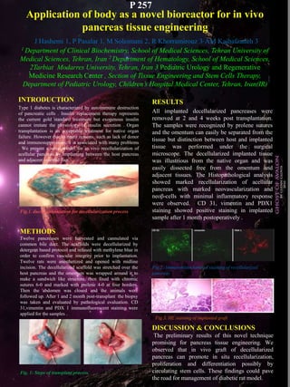
- 1. Application of body as a novel bioreactor for in vivo pancreas tissue engineering J Hashemi 1, P Pasalar 1, M Soleimani 2, R Khorramirouz 3, AM Kajbafzadeh 3 1 Department of Clinical Biochemistry, School of Medical Sciences, Tehran University of Medical Sciences, Tehran, Iran 2 Department of Hematology, School of Medical Sciences, 2Tarbiat Modarres University, Tehran, Iran 3 Pediatric Urology and Regenerative Medicine Research Center , Section of Tissue Engineering and Stem Cells Therapy, Department of Pediatric Urology, Children’s Hospital Medical Center, Tehran, Iran(IR) INTRODUCTION Type 1 diabetes is characterized by autoimmune destruction of pancreatic cells . Insulin replacement therapy represents the current gold standard treatment but exogenous insulin cannot imitate the physiology of insulin secretion . Organ transplantation is an acceptable treatment for native organ failure. However due to many reasons, such as lack of donor and immunosuppression, it is associated with many problems . We present a novel model for in vivo recellularization of acellular pancreas by implanting between the host pancreas and adjacent omental flap. RESULTS All implanted decellularized pancreases were removed at 2 and 4 weeks post transplantation. The samples were recognized by prolene sutures and the omentum can easily be separated from the tissue but distinction between host and implanted tissue was performed under the surgical microscope. The decellularized implanted tissue was illustrious from the native organ and was easily dissected free from the omentum and adjacent tissues. The Histopathological analysis showed marked recellularization of acellular pancreas with marked neovascularization and neoβ-cells with minimal inflammatory response were observed. CD 31, vimentin and PDX1 staining showed positive staining in implanted sample after 1 month postoperatively . METHODS Twelve pancreases were harvested and cannulated via common bile duct. The scaffolds were decellularized by detergent based protocol and infused with methylene blue in order to confirm vascular integrity prior to implantation. Twelve rats were anesthetized and opened with midline incision. The decellularized scaffold was stretched over the host pancreas and the omentum was wrapped around it to make a sandwich like structure, then fixed with chromic sutures 6-0 and marked with prolene 4-0 at four borders. Then the abdomen was closed and the animals were followed up. After 1 and 2 month post-transplant the biopsy was taken and evaluated by pathological evaluation. CD 31,vimentin and PDX 1 immunofluorescent staining were applied for the samples . Fig.2: Immunohistochemical staining of recellularized pancreas DISCUSSION & CONCLUSIONS The preliminary results of this novel technique promising for pancreas tissue engineering. We observed that in vivo graft of decellularized pancreas can promote in situ recellularization, proliferation and differentiation possibly by circulating stem cells. These findings could pave the road for management of diabetic rat model. P 257 Fig.3. HE staining of implanted graft Fig. 1: Steps of transplant process. Fig.1. ductal cannulation for decellularization process