Recommended

Recommended
More Related Content
Similar to Reza Khorramirouz
Similar to Reza Khorramirouz (20)
More from Reza Khorramirouz
More from Reza Khorramirouz (7)
Recently uploaded
Recently uploaded (20)
Reza Khorramirouz
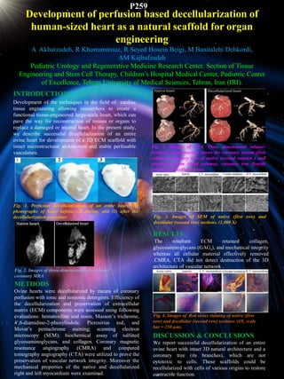
- 1. Development of perfusion based decellularization of human-sized heart as a natural scaffold for organ engineering A Akbarzadeh, R Khorramirouz, R Seyed Hosein Beigi, M Banitalebi Dehkordi, AM Kajbafzadeh Pediatric Urology and Regenerative Medicine Research Center, Section of Tissue Engineering and Stem Cell Therapy, Children’s Hospital Medical Center, Pediatric Center of Excellence, Tehran University of Medical Sciences, Tehran, Iran (IRI). INTRODUCTION Development of the techniques in the field of cardiac tissue engineering allowing researchers to create a functional tissue-engineered large-scale heart, which can pave the way for reconstruction of tissues or organs to replace a damaged or injured heart. In the present study, we describe successful decellularization of an entire ovine heart for development of a 3D ECM scaffold with intact microstructural architecture and stable perfusable vasculature. Fig. 1. Perfusion decellularization of an ovine heart; (1) photographs of heart before, (2) during, and (3) after the decellularization procedure Fig. 3. Images of CTA. Three dimensional volume- rendered reconstruction shows the coronary system (first column), coronary tree of native (second column ) and coronary system (third column), coronary tree (fourth column)of decellular heart. METHODS Ovine hearts were decellularized by means of coronary perfusion with ionic and nonionic detergents. Efficiency of the decellularization and preservation of extracellular matrix (ECM) components were assessed using following evaluations: hematoxiline and eosin, Masson’s trichrome, 4′,6-diamidino-2-phenylindole, Picrosirius red, and Movat’s pentachrome staining; scanning electron microscopy (SEM); biochemical assay of sulfated glycosaminoglycans, and collagen. Coronary magnetic resonance angiography (CMRA) and computed tomography angiography (CTA) were utilized to prove the preservation of vascular network integrity. Moreover the mechanical properties of the native and decellularized right and left myocardium were examined. Fig. 3. Images of SEM of native (first row) and decellular (second row) sections. (1,000 X) RESULTS The resultant ECM retained collagen, glycosaminoglycans (GAGs), and mechanical integrity whereas all cellular material effectively removed .CMRA, CTA did not detect destruction of the 3D architecture of vascular network . DISCUSSION & CONCLUSIONS We report successful decellularization of an entire ovine heart with intact 3D natural architecture and a coronary tree (its branches), which are not cytotoxic to cells. These scaffolds could be recellularized with cells of various origins to restore contractile function. Fig. 4. Images of Red sirius staining of native (first row) and decellular (second row) sections. (4X, scale bar = 250 μm). Fig. 2. Images of three-dimensional whole-heart coronary MRA P259