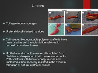
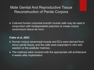
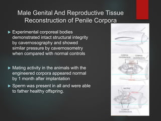
Tissue engineering applications in urology include organ transplantation, reconstructive procedures, and novel therapies for chronic illness. Studies have reconstructed tissues of the urethra, bladder, and male genitalia using cell-seeded matrices. For the urethra, tubular matrices seeded with autologous cells generated neourethral segments of 5-15cm. For the bladder, acellular matrices and cell-seeded matrices showed regeneration of transitional layers. Reconstructing penile corpora used smooth muscle cells on biodegradable scaffolds, generating intact structures. Tissue engineering offers alternatives to gastrointestinal tissues currently used for reconstruction and potential treatments for conditions like erectile dysfunction and infertility.
Introduction
Definition
History
Principle
Cell sources
What cells can be used?
Scaffolds
Biomaterials
Bioreactor
How tissue engineering is done?
How does tissue engineering differ from cloning?
Tissue engineering of specific structures
Application of tissue engineering
Limitations
Conclusion
References
The document summarizes the past, present, and future of regenerative tissue engineering. It discusses how the field began in the 1950s-60s by combining cell biology with new materials to generate living tissue components. Major advances included the use of stem cells and development of biocompatible scaffolds. The future of the field involves improved biomaterials that mimic natural extracellular matrix, bioprinting of complex tissues, and using various stem cell sources for cell therapy and organ regeneration to treat aging populations. The market for tissue engineering is estimated to grow substantially in coming years.
This document provides an overview of tissue engineering. It discusses the process of tissue engineering which involves using a scaffold material, seeding it with living cells, using growth factors, and implanting the new tissue. It also describes different types of stem cells, materials used for scaffolds, and methods to synthesize tissue engineered scaffolds. Applications of tissue engineering include bioartificial organs and tissues like skin, bone, and blood vessels. Both advantages and disadvantages of the field are mentioned.
Tissue engineering is the use of a combination of cells, engineering and materials methods, and suitable biochemical and physicochemical factors to improve or replace biological functions.
The term has also been applied to efforts to perform specific biochemical functions using cells within an artificially-created support system (e.g. an artificial pancreas, or a bio artificial liver).
A commonly applied definition of tissue engineering, as stated by Langer and Vacanti is “An interdisciplinary field that applies the principles of engineering and life sciences toward the development of biological substitutes that restore, maintain, or improve [Biological tissue] function or a whole organ”
This document discusses tissue engineering principles and their application to periodontal regeneration. It outlines that tissue engineering involves enhancing biologic processes or developing implantable products to modify deficient tissues. For periodontal regeneration specifically, the goal is to restore the original architecture and function of periodontal tissues affected by disease. Various techniques for periodontal regeneration are discussed, including guided tissue regeneration using membranes, root surface conditioning, and use of regenerative materials like ceramics, growth factors, and stem cells. Successful regeneration requires balancing cells, signaling molecules, and scaffolds in both in vitro and in vivo contexts.
This editorial discusses urine-derived stem cells (USCs) as a novel cell source for urethral tissue regeneration. USCs can be obtained non-invasively from urine and have characteristics similar to mesenchymal stem cells, including the ability to differentiate into multiple cell types. USCs provide advantages over other cell sources as they can be easily obtained in large quantities without morbidity. Studies show USCs seeded onto biomaterial scaffolds can form tissue with urothelial and smooth muscle layers resembling urethra when implanted. USCs therefore show promise as a cell source for engineering urethral tissues to treat urethral strictures.
A feature run by the monthly magazine for the polo community highlighting the latest in cutting edge regenerative therapy and how it has been translated for equine veterinary use from the human medical world.
Human fetal intestine was decellularized to create a natural scaffold for bladder augmentation. The decellularization protocol successfully removed cellular material while preserving the extracellular matrix. The scaffolds were implanted in rabbit bladders, where host bladder cells effectively repopulated the scaffolds over time. Six months later, the tissue architecture of the repopulated scaffolds resembled the native bladder, demonstrating the potential of this approach for bladder tissue engineering applications.
Tissue engineering involves combining living cells with biomaterials to generate new living tissue. It aims to regenerate damaged or diseased tissues and organs. The process involves taking cells from a patient and growing them on a biodegradable scaffold. Once the new tissue forms, it is implanted to replace the damaged tissue. This allows tissue to be grown with the patient's own cells, avoiding rejection. Successful applications include growing skin to treat burns and cartilage to repair joints. Tissue engineering could solve the shortage of donor organs and offer permanent solutions for many medical conditions.
Tissue engineering involves using cells, biomaterials, and growth factors to regenerate damaged tissues and organs. There are several strategies for tissue engineering, including injecting stem cells, using scaffolds to guide cell growth, and inducing cell differentiation. Ideal scaffolds are biocompatible, porous, and gradually degrade as new tissue forms. Common scaffold materials include natural polymers, ceramics, and synthetic polymers. Tissue-engineered dental tissues are being developed by harvesting patient cells and growing them on scaffolds or as cell sheets to regenerate the periodontal ligament.
This document provides an overview of principles of tissue engineering. It discusses why tissue engineering is needed due to limited organ transplantation availability. Tissue engineering uses regenerative medicine approaches including cell therapies, biomaterials, and tissue engineering to repair or replace damaged tissues. Various cell sources for therapy are described, including stem cells (embryonic, adult, perinatal), somatic cell nuclear transfer, and induced pluripotent stem cells. Biomaterials are discussed that can be used as scaffolds to support cell growth. The importance of vascularization for tissue volumes over 3mm is also highlighted.
The robotic implant is designed to induce lengthening of tubular organs like the esophagus and intestines through computer-controlled application of traction forces. Testing in swine demonstrated the applied forces can induce cell proliferation and lengthening of the esophagus without reducing diameter, allowing normal eating. The implant establishes that precise, controlled lengthening can be achieved while maintaining organ geometry, exploiting mechanostimulation to regenerate tissue without traditional engineering challenges.
The cell and its evolution. Camila DuncanCamila Duncan
The document discusses two studies related to cell regeneration and evolution. The first study successfully grew new cartilage tissue in the lab using cartilage cells from cow knee joints, which could help treatments for osteoarthritis. The second study found that macrophages, important immune cells, have the ability to self-renew through turning off two genes, showing potential for tissue regeneration. Both studies indicate advances in regenerative medicine techniques that may help patients with tissue degeneration diseases in the future.
Cultured skin substitutes prepared from cultured skin cells and biopolymers can reduce the need for donor skin grafts and have been shown to effectively treat excised burns, burn scars, and congenital skin lesions. Cultured skin substitutes generate skin phenotypes in the lab and restore tissue function and systemic homeostasis when implanted. Healed skin from cultured skin substitutes is smooth, soft and strong, though pigmentation may be irregular. Cultured skin substitutes close 67 times the area of donor skin compared to less than 4 times for split-thickness skin grafts, and result in similar qualitative outcomes.
Tissue engineering and regenerative medicine aim to regenerate human tissues and organs. Tissue engineering involves seeding cells onto scaffolds to create tissues, while regenerative medicine focuses on cell therapies. The field is multidisciplinary and requires collaboration across various areas. Applications have included skin, blood vessels, heart valves, cartilage, bones and whole organs. Challenges remain around ethics, quality control, understanding tissue differentiation, and meeting clinical demand. While still early, the field is making progress in translating technologies to treat conditions like burns, heart disease, arthritis and diabetes.
Tissue engineering and stem cell by regenerative medicine.pptx badal 2014Pradeep Kumar
The document discusses the history and applications of tissue engineering using stem cells for regenerative medicine. It provides background on the field of tissue engineering and milestones from the 1960s to present. It describes different types of stem cells like hematopoietic, mesenchymal, embryonic and their uses. Applications discussed include using stem cells to treat diseases like cardiovascular disease, diabetes, and neurological disorders. Recent advances mentioned are growing tissues like ears, noses, kidneys and pancreatic islets using 3D printing and scaffolds. The document concludes by noting both the promise and challenges of tissue engineering for regenerative medicine.
The Indian Dental Academy is the Leader in continuing dental education , training dentists in all aspects of dentistry and
offering a wide range of dental certified courses in different formats.
stemcells treatment on Neurogenic bladder repair using ms csDr Pradeep Mahajan
This case report describes the successful treatment of a 65-year-old man's neurogenic bladder using autologous mesenchymal stem cells. The man developed a neurogenic bladder following a laminectomy procedure for lumbar spinal stenosis and from long-standing diabetes mellitus with neuropathy. Tests showed his bladder had reduced compliance and an inability to voluntarily empty. He was treated with mesenchymal stem cells derived from his own bone marrow and fat tissue, which were injected into his bladder. Within 10 days he was able to voluntarily urinate, and further improvements continued over the following month. The report discusses how mesenchymal stem cells may help repair bladder function through differentiation, reducing cell death, and stimulating regeneration.
Tissue engineering and regenerative medicine Suman Nandy
Tissue engineering is the use of a combination of cells, engineering and materials methods, and suitable biochemical and physicochemical factors to improve or replace biological tissues. Tissue engineering involves the use of a scaffold for the formation of new viable tissue for a medical purpose.
Tissue engineering is an interdisciplinary field that applies engineering and life science principles to develop biological substitutes that restore and maintain normal function. The goal is to repair, replace, and regenerate diseased tissue. Stem cells are undifferentiated cells that can differentiate into specialized cell types and divide to produce more stem cells. They have potential uses in tissue engineering but also risks like tumor formation that require further research.
This document discusses tissue engineering techniques in endodontics, specifically root canal revascularization. It begins by defining regenerative endodontics and its goal of regenerating damaged dental tissues. The key elements of tissue engineering are then explained: stem cells, growth factors, and scaffolds. Various techniques are described, including revascularization via blood clotting, postnatal stem cell therapy, scaffold and pulp implantation. The document outlines the objectives and components of regenerative endodontic treatment, including a case selection and disinfection protocol for revascularization. In under 3 sentences, this document discusses regenerative endodontic techniques for tissue engineering and regenerating damaged dental tissues, outlining the key elements and various approaches, including a protocol for revascularization
This study assessed the feasibility of generating colon tissue from decellularized colon scaffolds using tissue engineering techniques. Rat colon was successfully decellularized and the scaffolds were implanted either in the mesentery or in situ in the colon. Histological analysis after 9 months found that scaffolds implanted in the mesentery developed features of small intestine, while those implanted in situ in the colon developed features of colon tissue. The study highlights how the microenvironment influences tissue development from decellularized scaffolds and suggests this technique may be useful for generating intestinal tissues, though further studies are needed to evaluate its potential for producing longer tissue segments required for clinical applications.
This document provides an overview of the principles of tissue engineering. It defines tissue engineering and regenerative medicine, and traces the history from early experiments in the 1970s to the development of organizational structures like TERMIS in the 2000s. The key components of tissue engineering are described as cells, scaffolds, and the cellular environment. The document discusses sources of cells including adult stem cells and challenges in obtaining and expanding cells for tissue engineering applications.
Similar to tissueengineeringapplicationsinurology-200716071252.pdf (20)
This document summarizes a seminar on rabies. It discusses the history, epidemiology, pathogenesis, clinical features, diagnosis and prevention of rabies. Rabies is a fatal viral disease transmitted through animal bites, primarily from dogs. It has affected humans for thousands of years. Current prevention strategies focus on mass dog vaccination programs, post-exposure prophylaxis for bite victims, and surveillance to identify outbreaks. Early diagnosis is difficult but important for effective treatment.
This document discusses stone disease in pregnancy. It notes that while the incidence of urolithiasis is similar in pregnant and non-pregnant women, stones can cause complications like premature birth in up to 40% of cases. Imaging choices are limited by concerns over radiation exposure to the fetus, but ultrasound is usually first-line. While most stones will pass spontaneously with conservative management, uncontrolled pain, infection, or obstruction of the solitary kidney may require interventions like ureteroscopy, with precautions taken to minimize radiation exposure to the fetus.
This document discusses the use of marginal or expanded criteria donors for kidney transplantation. It notes that there is a large gap between the number of patients needing kidney transplants and the availability of organs. Using marginal donors, such as those older than 60 years of age, deceased donors with hypertension, or living donors with medical risks can help increase the donor pool by 20-25%. Outcomes are inferior to normal criteria donors but provide recipients with improved survival over remaining on dialysis. Careful screening and optimization of allocation can help maximize outcomes when using marginal kidney donors.
A 68-year-old male farmer presented with complaints of dysuria and poor urine stream for 6 months. He had a suprapubic catheter inserted 2 months ago. On examination, his prostate was grade 1 and firm. Urinalysis showed plenty of white blood cells and 6-8 red blood cells. Urine culture showed no growth. Imaging showed a thickened bladder wall and post-void residual of 234cc. The patient was diagnosed with benign prostatic hyperplasia.
Surgical management of primary penile carcinoma includes biopsy for histologic confirmation, followed by organ-conserving or amputation procedures depending on the stage and grade. For low-stage Tis, Ta, T1 grade 1-2 tumors, organ-conserving options like circumcision, laser therapy, Mohs microsurgery, local excision or partial glansectomy are preferred to preserve sexual and urinary function. For more advanced or high-grade tumors, partial or total penectomy may be required. Radiation therapy is an alternative for small, early-stage lesions but is associated with higher risks of complications like necrosis, stenosis and the potential need for salvage surgery.
This document discusses urine cytology specimen collection and preparation techniques. There are three types of specimens that can be collected - voided urine, catheter specimens, and bladder washings. Voided urine is the simplest but early morning samples should be avoided due to poor cell morphology. Catheter specimens avoid contamination but can mimic tumors. Bladder washings use saline to irrigate the bladder and have good cellularity. Preparation methods include centrifugation with Esposito's fixative or cytocentrifugation, direct smears, membrane filters, and monolayer techniques. Normal urinary cytology shows a range of superficial and deep urothelial cell types with characteristic features.
This document presents a case report of a 70-year-old diabetic male patient who presented with right leg pain for 3 months and a raw area on his right foot for 1 month. On examination, he was found to have signs of peripheral vascular disease in his right leg including skin changes, delayed pulses, and inability to move toes. He was diagnosed with peripheral arterial occlusive disease in his right leg and a grade 3 diabetic foot ulcer on his right foot, likely due to atherosclerosis.
The CT scan showed a 40-year-old male patient complaining of abdominal pain for 3 days. The liver, gallbladder, pancreas, spleen, kidneys, bladder, prostate, bones, and bowel loops were normal. A 6.4mm renal calculus was seen in the lower pole calyx of the right kidney. A 1cm cortical cyst was seen in the midpole of the right kidney. Some fibroatelectatic strands and ground glass opacities were seen in the posterior basal segments of the lower lobes of both lungs. The impression was of a right renal calculus and right simple renal cortical cyst.
Wound is defined as a break in continuity of tissue that can be caused by transfer of energy either externally or internally. Wounds are classified as mechanical, chemical, thermal, or radiation-induced. Special wounds are classified by origin or bacterial contamination. Rabies is a fatal viral disease spread through infected saliva that causes encephalitis. It is endemic in parts of Asia and Africa where dog bites are a major transmission route, especially in children. Symptoms include bizarre behavior, hydrophobia, paralysis and death. Diagnosis involves antigen detection in tissues or PCR. Prevention focuses on wound cleansing, vaccination, and immunoglobulin administration based on exposure category. Post-exposure prophylaxis includes vaccination and immunoglobulin over 28-
This patient is a 32-year-old male with a history of hypertension and chronic kidney disease who has been on maintenance hemodialysis for 1 year. He has a neurogenic bladder with a history of urinary tract abnormalities and is being evaluated for a renal transplant. His wife is willing to be a living donor. On evaluation, he was found to have grade 2-3 bladder trabeculations, dilated ureters and renal pelvis, and a bladder capacity of 250ml. Cystoscopy and other tests confirmed his neurogenic bladder and suitability for a renal transplant.
Surgical management of primary penile carcinoma includes biopsy for histologic confirmation, followed by organ-conserving or amputational surgery depending on the stage and grade. Organ-conserving options for early stage low-grade tumors include circumcision, laser therapy, Mohs microsurgery, local excision and partial glansectomy. More advanced tumors may require partial or total penectomy to achieve adequate surgical margins while preserving sexual and urinary function when possible. Radiation therapy is an alternative for small early tumors but is associated with higher risks of complications like necrosis, stenosis and the potential need for salvage surgery.
Peritoneal dialysis solutions have evolved over time. Originally, scientists experimented with various solutions instilled into the peritoneal cavity to treat uremia. Commercially available solutions in the 1950s contained lactate. Newer solutions aim to be pH neutral with low glucose degradation products and better preserve residual renal function compared to high glucose and lactate solutions. Clinical trials show neutral pH solutions result in slower decline in renal function and longer time to anuria with similar ultrafiltration and fluid status but lower peritonitis rates.
This document discusses emphysematous pyelonephritis (EPN), a severe necrotizing infection of the renal parenchyma that causes gas accumulation. EPN most often occurs in diabetics, especially women, and presents similarly to acute pyelonephritis but has a more fulminating course. Diagnosis is made through CT scan and treatment involves aggressive antibiotics, fluid resuscitation, and either percutaneous drainage or emergency nephrectomy depending on severity and patient stability. Prognosis has improved with current treatments but mortality remains around 20-25% if not promptly recognized and treated.
Urinary biomarkers are being developed and used as noninvasive alternatives to cystoscopy for bladder cancer diagnosis and monitoring. While no single biomarker has shown better specificity than urine cytology, many have higher sensitivity. Commonly used biomarkers include NMP22, BTA stat, ImmunoCyt/uCyt+, and UroVysion, with UroVysion and telomerase showing promise in sensitivity and specificity. An ideal biomarker would be noninvasive, highly sensitive and specific, easy to perform, rapid, reproducible, cost-effective, and able to detect cancer before it becomes visible on cystoscopy. Currently, biomarkers are used alongside cystoscopy for surveillance of non-muscle invasive bladder cancer, and may anticipate
This document summarizes the etiopathogenesis of renal tumors. It discusses benign renal tumors such as renal cysts, oncocytoma, angiomyolipoma and others. It describes genetic factors associated with various tumors. Risk factors for renal cell carcinoma include tobacco use, obesity, hypertension and familial syndromes like Von Hippel Lindau disease, hereditary papillary renal cell carcinoma, Birt-Hogg-Dubé syndrome and more. Various familial syndromes are outlined along with their associated genes and tumor types.
This document discusses urine cytology and urinary markers for detecting bladder cancer. It notes that urine cytology has high specificity but low sensitivity for both high and low grade tumors. Several urinary markers are discussed, including BTA, ImmunoCyt, NMP-22, UroVysion, microsatellite analysis, Lewis antigen X, CK20, CYFRA 21.1, survivin, hyaluronic acid and TRAP, along with their reported sensitivities and specificities. However, none meet the 90% sensitivity threshold needed to replace cystoscopy, so the conclusion is that a combination of cystoscopy and urine markers is currently the best approach for bladder cancer surveillance.
Local Advanced Lung Cancer: Artificial Intelligence, Synergetics, Complex Sys...Oleg Kshivets
Overall life span (LS) was 1671.7±1721.6 days and cumulative 5YS reached 62.4%, 10 years – 50.4%, 20 years – 44.6%. 94 LCP lived more than 5 years without cancer (LS=2958.6±1723.6 days), 22 – more than 10 years (LS=5571±1841.8 days). 67 LCP died because of LC (LS=471.9±344 days). AT significantly improved 5YS (68% vs. 53.7%) (P=0.028 by log-rank test). Cox modeling displayed that 5YS of LCP significantly depended on: N0-N12, T3-4, blood cell circuit, cell ratio factors (ratio between cancer cells-CC and blood cells subpopulations), LC cell dynamics, recalcification time, heparin tolerance, prothrombin index, protein, AT, procedure type (P=0.000-0.031). Neural networks, genetic algorithm selection and bootstrap simulation revealed relationships between 5YS and N0-12 (rank=1), thrombocytes/CC (rank=2), segmented neutrophils/CC (3), eosinophils/CC (4), erythrocytes/CC (5), healthy cells/CC (6), lymphocytes/CC (7), stick neutrophils/CC (8), leucocytes/CC (9), monocytes/CC (10). Correct prediction of 5YS was 100% by neural networks computing (error=0.000; area under ROC curve=1.0).
These lecture slides, by Dr Sidra Arshad, offer a quick overview of the physiological basis of a normal electrocardiogram.
Learning objectives:
1. Define an electrocardiogram (ECG) and electrocardiography
2. Describe how dipoles generated by the heart produce the waveforms of the ECG
3. Describe the components of a normal electrocardiogram of a typical bipolar lead (limb II)
4. Differentiate between intervals and segments
5. Enlist some common indications for obtaining an ECG
6. Describe the flow of current around the heart during the cardiac cycle
7. Discuss the placement and polarity of the leads of electrocardiograph
8. Describe the normal electrocardiograms recorded from the limb leads and explain the physiological basis of the different records that are obtained
9. Define mean electrical vector (axis) of the heart and give the normal range
10. Define the mean QRS vector
11. Describe the axes of leads (hexagonal reference system)
12. Comprehend the vectorial analysis of the normal ECG
13. Determine the mean electrical axis of the ventricular QRS and appreciate the mean axis deviation
14. Explain the concepts of current of injury, J point, and their significance
Study Resources:
1. Chapter 11, Guyton and Hall Textbook of Medical Physiology, 14th edition
2. Chapter 9, Human Physiology - From Cells to Systems, Lauralee Sherwood, 9th edition
3. Chapter 29, Ganong’s Review of Medical Physiology, 26th edition
4. Electrocardiogram, StatPearls - https://www.ncbi.nlm.nih.gov/books/NBK549803/
5. ECG in Medical Practice by ABM Abdullah, 4th edition
6. Chapter 3, Cardiology Explained, https://www.ncbi.nlm.nih.gov/books/NBK2214/
7. ECG Basics, http://www.nataliescasebook.com/tag/e-c-g-basics
Integrating Ayurveda into Parkinson’s Management: A Holistic ApproachAyurveda ForAll
Explore the benefits of combining Ayurveda with conventional Parkinson's treatments. Learn how a holistic approach can manage symptoms, enhance well-being, and balance body energies. Discover the steps to safely integrate Ayurvedic practices into your Parkinson’s care plan, including expert guidance on diet, herbal remedies, and lifestyle modifications.
Here is the updated list of Top Best Ayurvedic medicine for Gas and Indigestion and those are Gas-O-Go Syp for Dyspepsia | Lavizyme Syrup for Acidity | Yumzyme Hepatoprotective Capsules etc
ABDOMINAL TRAUMA in pediatrics part one.drhasanrajab
Abdominal trauma in pediatrics refers to injuries or damage to the abdominal organs in children. It can occur due to various causes such as falls, motor vehicle accidents, sports-related injuries, and physical abuse. Children are more vulnerable to abdominal trauma due to their unique anatomical and physiological characteristics. Signs and symptoms include abdominal pain, tenderness, distension, vomiting, and signs of shock. Diagnosis involves physical examination, imaging studies, and laboratory tests. Management depends on the severity and may involve conservative treatment or surgical intervention. Prevention is crucial in reducing the incidence of abdominal trauma in children.
Recomendações da OMS sobre cuidados maternos e neonatais para uma experiência pós-natal positiva.
Em consonância com os ODS – Objetivos do Desenvolvimento Sustentável e a Estratégia Global para a Saúde das Mulheres, Crianças e Adolescentes, e aplicando uma abordagem baseada nos direitos humanos, os esforços de cuidados pós-natais devem expandir-se para além da cobertura e da simples sobrevivência, de modo a incluir cuidados de qualidade.
Estas diretrizes visam melhorar a qualidade dos cuidados pós-natais essenciais e de rotina prestados às mulheres e aos recém-nascidos, com o objetivo final de melhorar a saúde e o bem-estar materno e neonatal.
Uma “experiência pós-natal positiva” é um resultado importante para todas as mulheres que dão à luz e para os seus recém-nascidos, estabelecendo as bases para a melhoria da saúde e do bem-estar a curto e longo prazo. Uma experiência pós-natal positiva é definida como aquela em que as mulheres, pessoas que gestam, os recém-nascidos, os casais, os pais, os cuidadores e as famílias recebem informação consistente, garantia e apoio de profissionais de saúde motivados; e onde um sistema de saúde flexível e com recursos reconheça as necessidades das mulheres e dos bebês e respeite o seu contexto cultural.
Estas diretrizes consolidadas apresentam algumas recomendações novas e já bem fundamentadas sobre cuidados pós-natais de rotina para mulheres e neonatos que recebem cuidados no pós-parto em unidades de saúde ou na comunidade, independentemente dos recursos disponíveis.
É fornecido um conjunto abrangente de recomendações para cuidados durante o período puerperal, com ênfase nos cuidados essenciais que todas as mulheres e recém-nascidos devem receber, e com a devida atenção à qualidade dos cuidados; isto é, a entrega e a experiência do cuidado recebido. Estas diretrizes atualizam e ampliam as recomendações da OMS de 2014 sobre cuidados pós-natais da mãe e do recém-nascido e complementam as atuais diretrizes da OMS sobre a gestão de complicações pós-natais.
O estabelecimento da amamentação e o manejo das principais intercorrências é contemplada.
Recomendamos muito.
Vamos discutir essas recomendações no nosso curso de pós-graduação em Aleitamento no Instituto Ciclos.
Esta publicação só está disponível em inglês até o momento.
Prof. Marcus Renato de Carvalho
www.agostodourado.com
TEST BANK For Community Health Nursing A Canadian Perspective, 5th Edition by...Donc Test
TEST BANK For Community Health Nursing A Canadian Perspective, 5th Edition by Stamler, Verified Chapters 1 - 33, Complete Newest Version Community Health Nursing A Canadian Perspective, 5th Edition by Stamler, Verified Chapters 1 - 33, Complete Newest Version Community Health Nursing A Canadian Perspective, 5th Edition by Stamler Community Health Nursing A Canadian Perspective, 5th Edition TEST BANK by Stamler Test Bank For Community Health Nursing A Canadian Perspective, 5th Edition Pdf Chapters Download Test Bank For Community Health Nursing A Canadian Perspective, 5th Edition Pdf Download Stuvia Test Bank For Community Health Nursing A Canadian Perspective, 5th Edition Study Guide Test Bank For Community Health Nursing A Canadian Perspective, 5th Edition Ebook Download Stuvia Test Bank For Community Health Nursing A Canadian Perspective, 5th Edition Questions and Answers Quizlet Test Bank For Community Health Nursing A Canadian Perspective, 5th Edition Studocu Test Bank For Community Health Nursing A Canadian Perspective, 5th Edition Quizlet Test Bank For Community Health Nursing A Canadian Perspective, 5th Edition Stuvia Community Health Nursing A Canadian Perspective, 5th Edition Pdf Chapters Download Community Health Nursing A Canadian Perspective, 5th Edition Pdf Download Course Hero Community Health Nursing A Canadian Perspective, 5th Edition Answers Quizlet Community Health Nursing A Canadian Perspective, 5th Edition Ebook Download Course hero Community Health Nursing A Canadian Perspective, 5th Edition Questions and Answers Community Health Nursing A Canadian Perspective, 5th Edition Studocu Community Health Nursing A Canadian Perspective, 5th Edition Quizlet Community Health Nursing A Canadian Perspective, 5th Edition Stuvia Community Health Nursing A Canadian Perspective, 5th Edition Test Bank Pdf Chapters Download Community Health Nursing A Canadian Perspective, 5th Edition Test Bank Pdf Download Stuvia Community Health Nursing A Canadian Perspective, 5th Edition Test Bank Study Guide Questions and Answers Community Health Nursing A Canadian Perspective, 5th Edition Test Bank Ebook Download Stuvia Community Health Nursing A Canadian Perspective, 5th Edition Test Bank Questions Quizlet Community Health Nursing A Canadian Perspective, 5th Edition Test Bank Studocu Community Health Nursing A Canadian Perspective, 5th Edition Test Bank Quizlet Community Health Nursing A Canadian Perspective, 5th Edition Test Bank Stuvia
- Video recording of this lecture in English language: https://youtu.be/kqbnxVAZs-0
- Video recording of this lecture in Arabic language: https://youtu.be/SINlygW1Mpc
- Link to download the book free: https://nephrotube.blogspot.com/p/nephrotube-nephrology-books.html
- Link to NephroTube website: www.NephroTube.com
- Link to NephroTube social media accounts: https://nephrotube.blogspot.com/p/join-nephrotube-on-social-media.html
NVBDCP.pptx Nation vector borne disease control programSapna Thakur
NVBDCP was launched in 2003-2004 . Vector-Borne Disease: Disease that results from an infection transmitted to humans and other animals by blood-feeding arthropods, such as mosquitoes, ticks, and fleas. Examples of vector-borne diseases include Dengue fever, West Nile Virus, Lyme disease, and malaria.
3. Normal wound healing has been studied extensively and is
helpful in maximizing success for the engineering of tissues
At the time of tissue injury, cell ingrowth is initiated from the
wound edges to cover the tissue defect
Cells are able to traverse short distances without any
detrimental effects
wound is large, more than a few millimeters in distance or
depth, increased collagen deposition, fibrosis, and scar
formation ensue
4. Matrices implanted in wound beds are able to lengthen the
distances that cells can traverse
Tissue defects greater than 1 cm that are treated with a matrix
alone, without cells, usually have increased collagen deposition
Cell-seeded matrices implanted in wound beds are able to
further lengthen the distance for normal tissue formation without
initiating an adverse fibrotic response
5. Urethra
Naturally derived collagen-based materials such as
Woven meshes of PGA without cells and with cells
bladder-derived acellular submucosa
acellular urethral submucosa
collagen gels
bladder submucosa matrix proved to be a suitable graft for
repair of urethral defects
Nonseeded acellular matrices, were applied in a successful
manner for onlay urethral repairs
6. Urethra
Atala et al, 1999
Neourethras were created by anastomosing the matrix in an on-
lay fashion to the urethral plate
size of the created neourethra ranged from 5 to 15 cm
After a 3-year follow-up,
3 or 4 patients had a successful outcome with regard to
cosmetic appearance and function
One patient who had a 15-cm neourethra created developed a
sub-glanular fistula
7. Urethra
Acellular collagen-based matrix
Eliminated the necessity of additional surgical procedures for
graft harvesting,
Potential morbidity from the harvest procedure were
decreased
Reduced operative time
But when tubularized urethral repairs were attempted
experimentally, adequate urethral tissue regeneration was
not achieved and complications ensued(contracture &
Stricture).
8. Urethra
To overcome this bladder epithelial and smooth muscle cells
were grown and seeded onto preconfigured tubular matrices
Entire urethra segments were resected and
urethroplasties were performed with tubularized collagen
matrices seeded with cells
9. Urethra
Raya-Rivera et al, 2011
5 patients with urethral injuries
had a small tissue biopsy specimen retrieved
cells were expanded in vitro and seeded in two layers on
tubularized scaffolds that were implanted surgically
engineered urethras were able to show adequate anatomy,
both by urethroscopy and with urethrography and function
long term
10.
11. Bladder
Currently, gastrointestinal segments are commonly used as
tissues for bladder replacement or repair
GI tissues are designed to absorb specific solutes
whereas bladder tissue is designed for the excretion of solutes
Thus, multiple complications may ensue, such as infection,
metabolic disturbances, urolithiasis, perforation, increased
mucus production, and malignancy
12. Bladder
Matrices
Synthetic materials that have been tried include polyvinyl
sponge, Teflon, collagen matrices, Vicryl (PGA) matrices, and
silicone
Permanent synthetic materials used for bladder reconstruction
succumb to mechanical failure and urinary stone formation
use of degradable materials leads to fibroblast deposition,
scarring, graft contracture, and a reduced reservoir volume over
time
13. Bladder
Non-seeded allogeneic acellular bladder matrices have
served as scaffolds for the ingrowth of host bladder wall
components serve as vehicles for partial bladder regeneration
Acellular collagen matrices can be enhanced with growth
factors to improve bladder regeneration
Cell-seeded allogeneic acellular bladder matrices showed
better tissue regeneration
14. Bladder
SIS, a biodegradable, acellular, xenogeneic collagen-based
tissue matrix graft, was first described in early 1960s
derived from pig small intestine in which mucosa is
mechanically removed from inner surface and serosa and
muscular layer are removed from outer surface
Non seeded SIS matrix used for bladder augmentation is able
to regenerate in vivo
transitional layer was the same as that of the native bladder
tissue but muscle layer was not fully developed
15. Bladder
Regenerative medicine with selective cell transplantation in SIS
may provide a means to create functional new bladder
segments.
Native cells are currently preferable because they can be used
without rejection
Amniotic fluid– and bone marrow–derived stem cells have the
potential to differentiate into bladder tissue and urothelium.
Embryonic stem cells also have the potential to differentiate into
bladder tissue.
16. Bladder
A study using engineered bladder tissue for cystoplasty
reconstruction was conducted starting in 1998
pilot study of seven patients was reported , Atala et al, 2006
Patients underwent reconstruction with the engineered bladder
tissue created with the PGA-collagen cell-seeded scaffolds
with omental coverage
showed increased compliance, decreased end-filling pressures,
increased capacities, and longer dry periods over time
18. Bladder cell therapies
Injectable therapy within the bladder may be useful for SUI and
VUR
Injection of chondrocytes for the correction of VUR in children
At 1-year follow-up, reflux correction was maintained in 70%
SUI in adults have been attempted
After 1 year, 1/8 women achieved total continence and 5 reported
improvement
19. Bladder cell therapies
autologous smooth muscle cells was explored for urinary
incontinence, SUI and VUR applications
myoblasts isolated from the abdominal wall vasculature were
injected in a series of bladder exstrophy patients with urinary
incontinence.
88% of patients were socially dry
The patients were also on a pelvic floor electrical stimulation and
pelvic floor exercise program
20. Ureters
Collagen tubular sponges
Ureteral decellularized matrices
Cell-seeded biodegradable polymer scaffolds have
been used as cell transplantation vehicles to
reconstruct ureteral tissues
Urothelial and smooth muscle cells isolated from
bladders and expanded in vitro were seeded onto
PGA scaffolds with tubular configurations and
implanted subcutaneously resulted in the eventual
formation of natural urothelial tissues
21. Male Genital And Reproductive Tissue
One of the major limitations of genital reconstructive surgery is
the availability of sufficient autologous tissue
Phallic reconstruction was initially attempted in the late 1930s,
with rib cartilage but discouraged because of the unsatisfactory
functional and cosmetic results
Silicone rigid prostheses were popularized in the 1970s and
have been used widely but biocompatibility issues have been a
problem
22. Male Genital And Reproductive Tissue
Reconstruction of Penile Corpora
Cultured human corporeal smooth muscle cells may be used in
conjunction with biodegradable polymers to create corpus
cavernosum tissue de novo
Falke et al, 2003
Human corpus cavernosal muscle and ECs were derived from
donor penile tissue, and the cells were expanded in vitro and
seeded on the acellular matrices.
The matrices were covered with the appropriate cell architecture
4 weeks after implantation
23. Male Genital And Reproductive Tissue
Reconstruction of Penile Corpora
Experimental corporeal bodies
demonstrated intact structural integrity
by cavernosography and showed
similar pressure by cavernosometry
when compared with normal controls
Mating activity in the animals with the
engineered corpora appeared normal
by 1 month after implantation
Sperm was present in all and were able
to father healthy offspring.
24. Male Genital And Reproductive Tissue
Penile Cell Therapy
Various cell lines have been used in animal models in an
attempt to reverse erectile dysfunction in animal models
Endothelial cells
Mesenchymal stem cells either alone or with matrices
human bone marrow–derived stem cells
Muscle-derived stem cells
Long-term studies are needed to gauge the full impact of
these therapies
25. Penile transplant
Such a scaffold could represent a new solution in cases of total penile loss after
cancer or trauma or in transgender surgeries, cases where the incidence is
increasing rapidly.
26. Male Genital And Reproductive Tissue
Testis – Leydig cells
Patients with testicular dysfunction require androgen
replacement for somatic development in form of
periodic intramuscular injections
Skin patch applications
Long-term non-pulsatile testosterone therapy is not
optimal and can cause multiple problems
27. Male Genital And Reproductive Tissue
Testis – Leydig cells
Leydig cells were microencapsulated in an alginate-poly-L-
lysine solution for controlled testosterone replacement
Provides a barrier between the transplanted cells and
the host’s immune system, as well as allowing for the
long-term physiologic release of testosterone
On similar principles, testicular prostheses have been
created with chondrocytes and loaded with testosterone
28. Male Genital And Reproductive Tissue
Testis – Spermatogenesis
Spermatogenesis for infertility purposes has been a major area of
interest
First successful isolation of human spermatogonial stem cells in
2002 showed that the cells were able to colonize and survive for 6
months in mice recipient testes
Successful autologous and allogeneic spermatogonial stem cell
transplantation has been demonstrated
In vitro propagation of human spermatogonial stem cells from both
adult and pubertal testes has been established
29. Female Genital And Reproductive Tissue
Uterus
Congenital malformations of the uterus may have profound
implications clinically
possibility of engineering functional uterine tissue using
autologous cells was investigated
Autologous uterine smooth muscle and epithelial cells were
harvested, grown, and expanded in culture.
These cells were seeded onto preconfigured uterine-shaped
biodegradable polymer scaffolds, which were then used for
subtotal uterine tissue replacement
30. Female Genital And Reproductive Tissue
Vagina
Many techniques and materials can be used successfully for
vaginal reconstruction
most common, creating a canal by dissecting the potential
neovaginal space and subsequently lining the pelvic canal with a
graft
Vaginal epithelial and smooth muscle cells were harvested,
expanded, and seeded on biodegradable polymer scaffolds
31.
32. Female Genital And Reproductive Tissue
Ovary
Ovarian tissue is essential for fertility.
Recent studies have shown that ovarian cells can be derived
from stem cell populations. The cells can lead to the
production of oocytes and embryos
Cell therapies have also been used to enhance the
functionality of the ovary
Adipose-, amniotic fluid–, umbilical cord–, and bone
marrow–derived stem cells have all resulted in a return of
experimentally damaged ovarian function in animal models
33. Renal Structures
Isolation of particular cell types that produce specific factors
may be a good approach for selective cell therapies
Cells that produce erythropoietin have been isolated in culture,
and these cells could eventually be used to treat anemia that
results from ESRD
More ambitious approaches involve working toward the goal of
total renal function replacement
34. Renal Structures
Nuclear material from bovine dermal fibroblasts was transferred into
unfertilized enucleated eggs.
Renal cells from the cloned embryos were harvested, expanded in
vitro, and seeded onto three-dimensional renal devices.
The devices were implanted into the back into the steer and were
retrieved 12 weeks later.
This process produced functioning renal units
Cells derived from nuclear transfer can be successfully harvested,
expanded in culture, and transplanted in vivo with the use of
biodegradable scaffolds on which the single suspended cells can
organize into tissue
35.
36. Renal Structures
These studies were the first demonstration of the use of
therapeutic cloning for regeneration of tissues in vivo
Renal cells seeded on the matrix adhered to the inner surface
and proliferated to confluency by 7 days after seeding. Renal
tubular and glomerulus-like structures were observed 8 weeks
after implantation
More recent data has confirmed that the creation of larger
kidney structures using decellularized kidney matrices and
repopulated with cells is possible