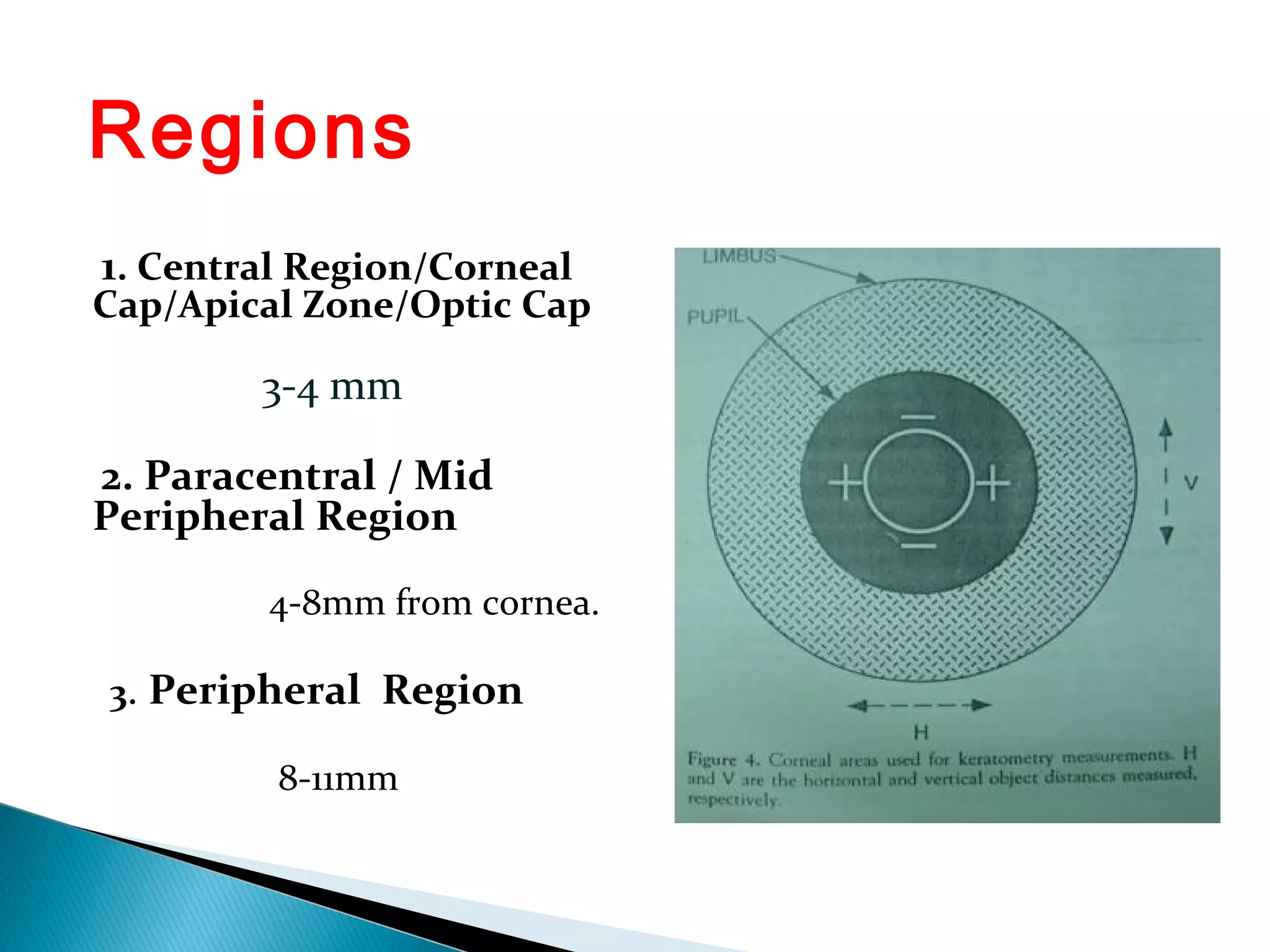
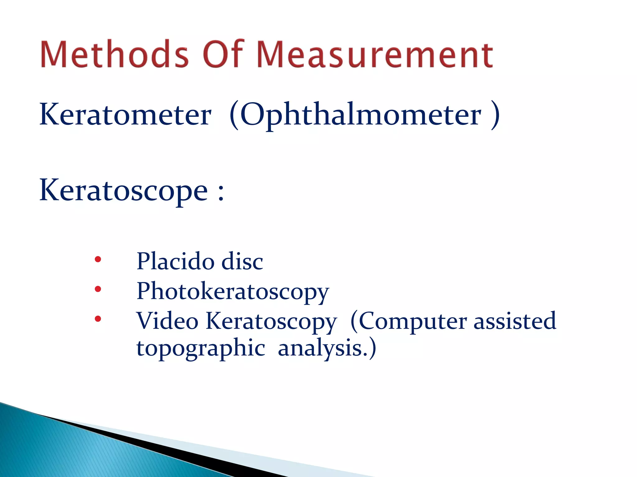
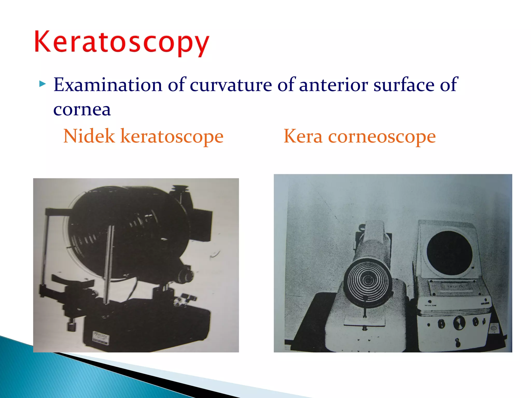
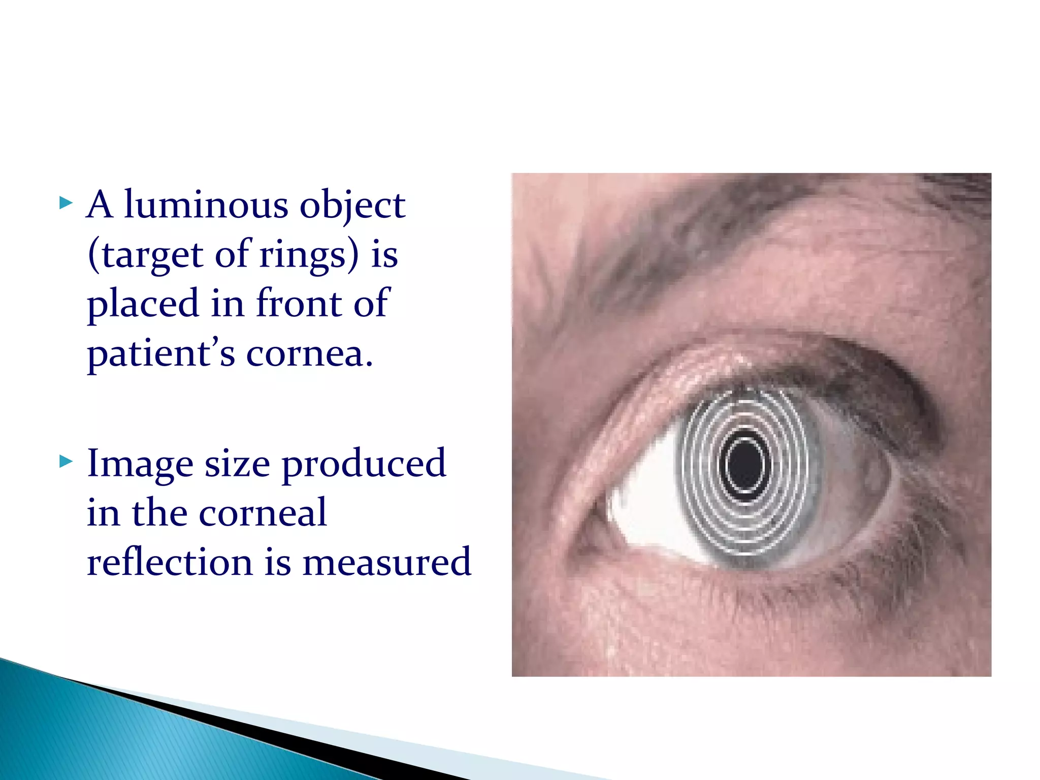
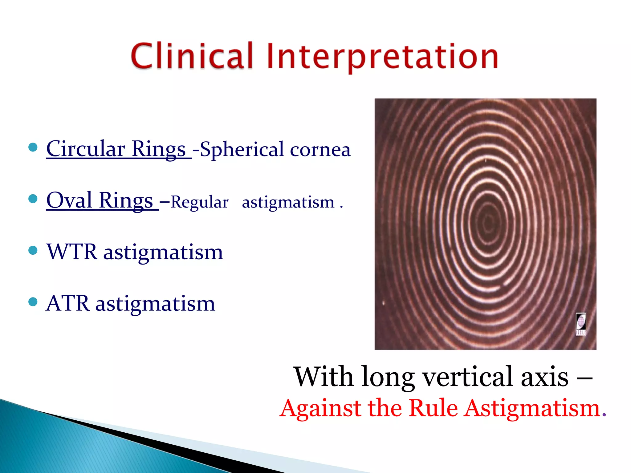
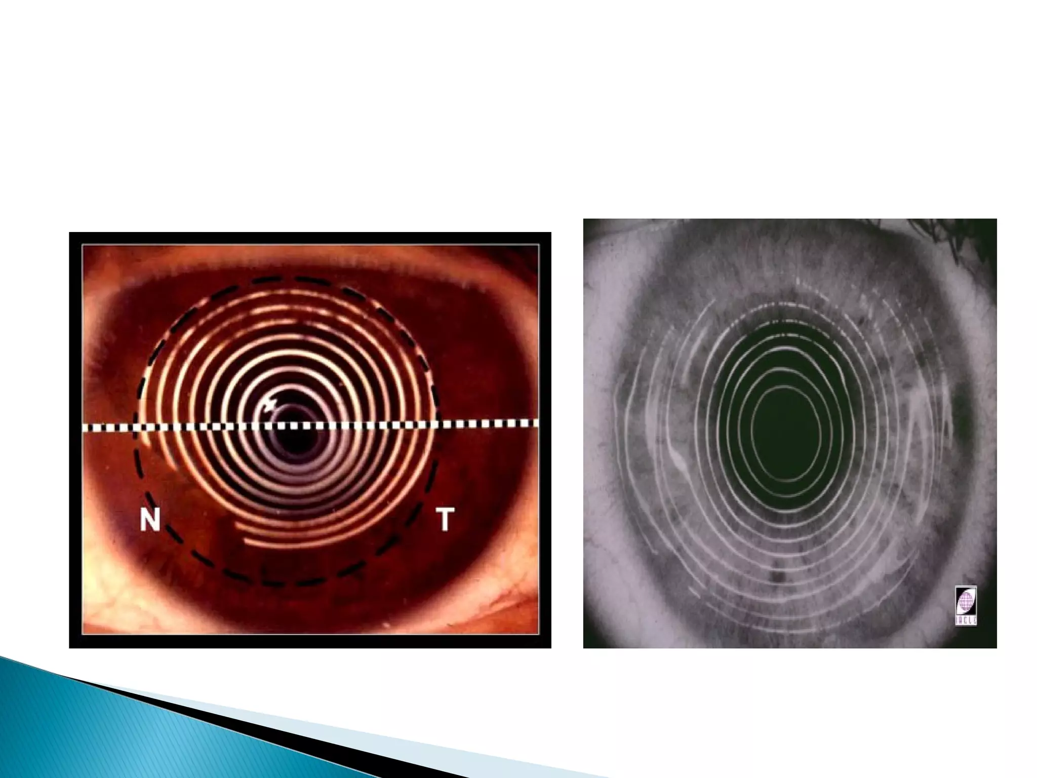
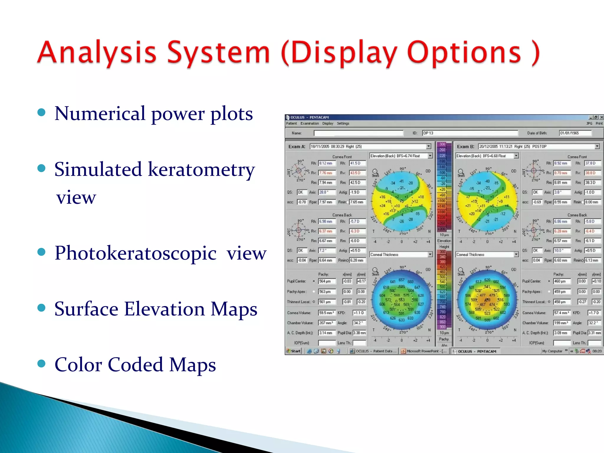
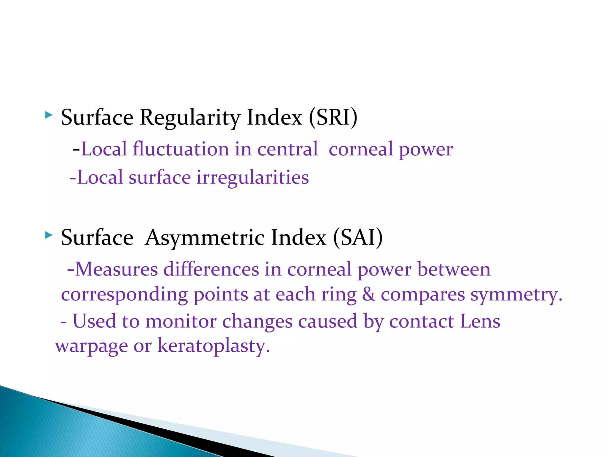
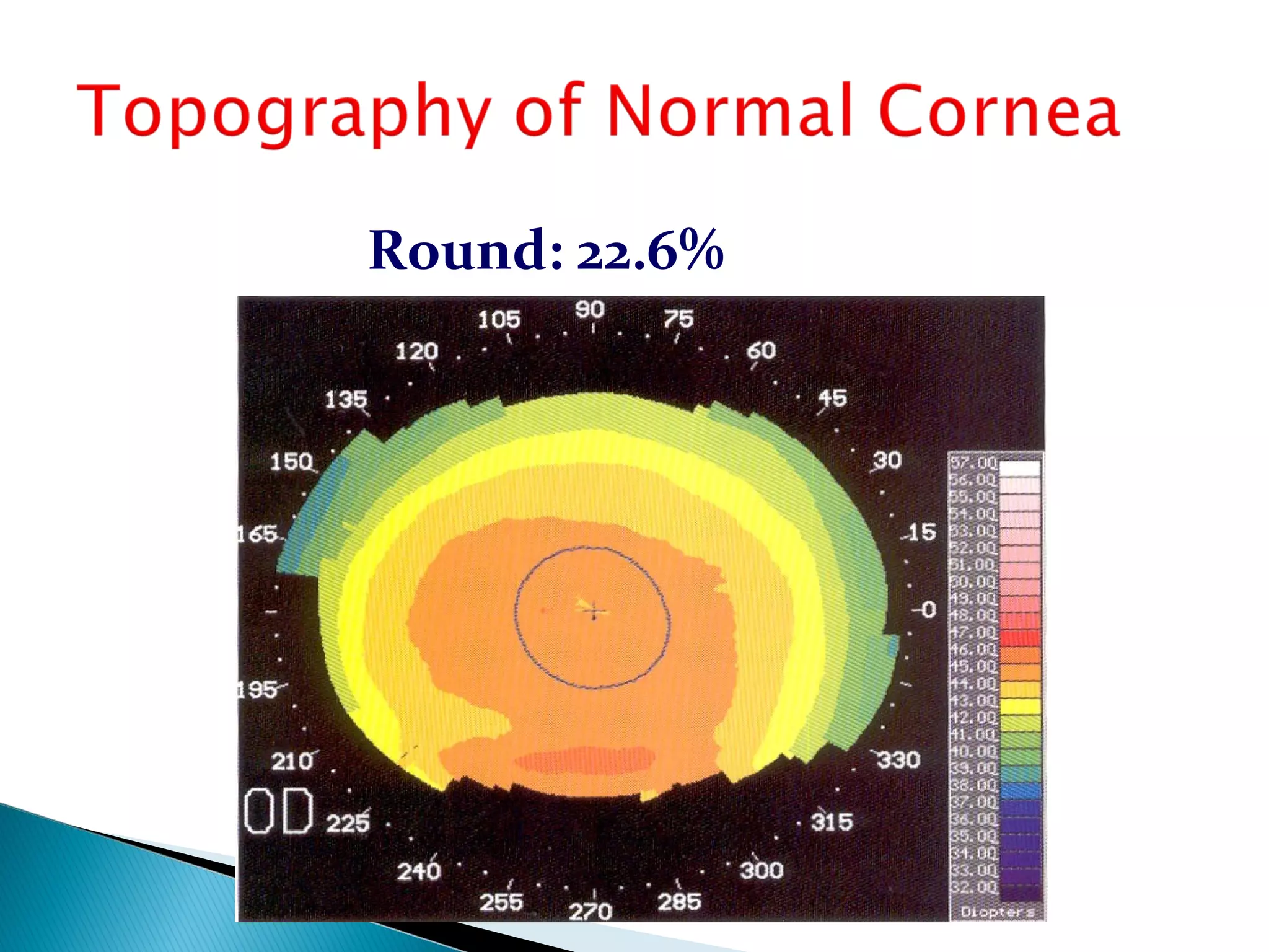
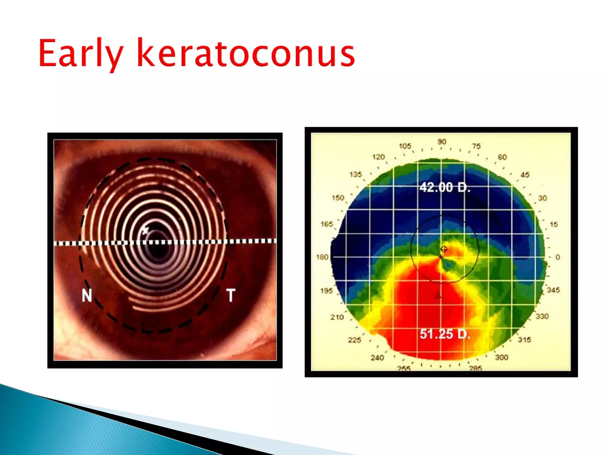
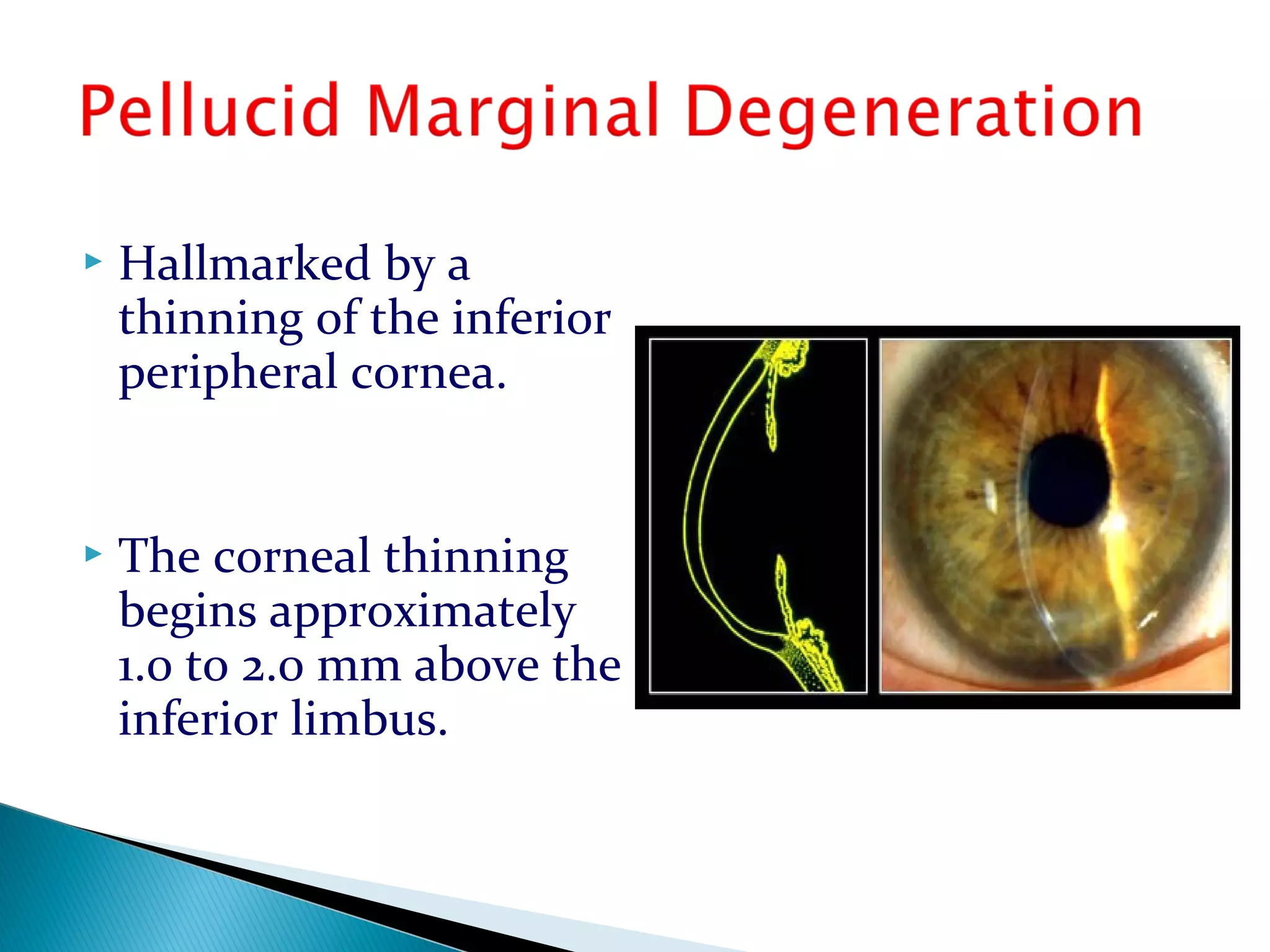
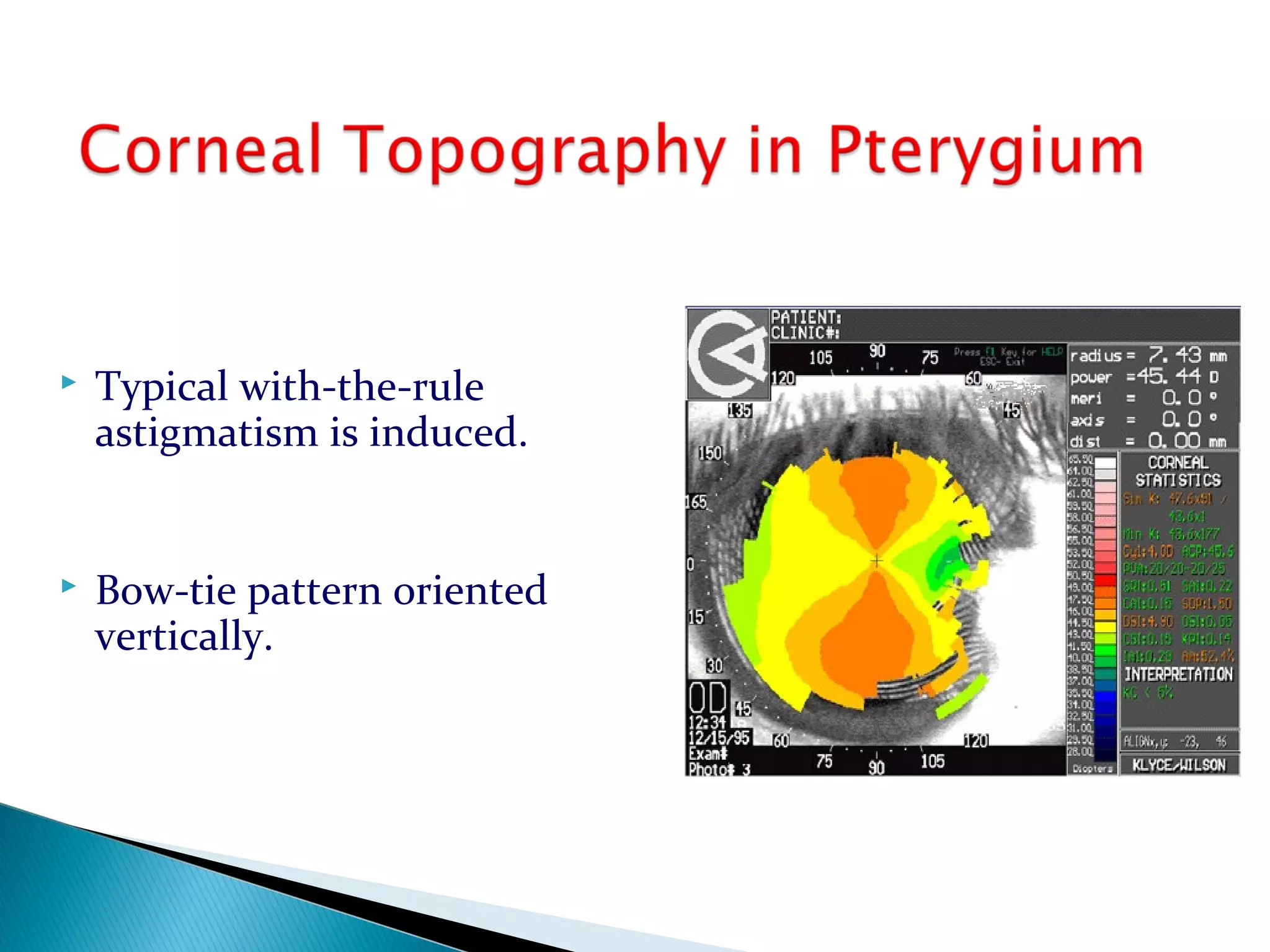
Raman Prasad Sah
Optometrist
B.P. Koirala Lions Centre for Ophthalmic
Studies
Institute of Medicine, Kathmandu, Nepal
Study of shape 0f corneal surface.
Configuration or description of surface.
Aspheric – elliptical shape.
Eccentricity: 0.41 – o.58 ( Aver:0.47 )
1. Central Region/Corneal
Cap/Apical Zone/Optic Cap
3-4 mm
2. Paracentral / Mid
Peripheral Region
4-8mm from cornea.
3. Peripheral Region
8-11mm
Regions
Keratometer (Ophthalmometer )
Keratoscope :
• Placido disc
• Photokeratoscopy
• Video Keratoscopy (Computer assisted
topographic analysis.)
 Examination of curvature of anterior surface of
cornea
Nidek keratoscope Kera corneoscope
Principle
Use of the first Purkinje
image.
 Consists of equally
spaced alternating black
& white lines.
 A luminous object
(target of rings) is
placed in front of
patient’s cornea.
 Image size produced
in the corneal
reflection is measured
 Circular Rings -Spherical cornea
 Oval Rings –Regular astigmatism .
 WTR astigmatism
 ATR astigmatism
With long vertical axis –
Against the Rule Astigmatism.
 Irregular astigmatism
 Corneal anomaly–
irregular cornea
distorted cornea
scar
 Keratoconus
 Small degrees of abnormalities of corneal shape are
not easily identifiable.
 Not used in corneas with epithelial defects and
stromal ulcers .
 Clinically significant amounts of corneal
cylinder (up to 3.00 D) may not be diagnosed.
 Subtle irregularities cannot be detected.
 Measurement is restricted to a small central
corneal area (3-3.5mm) in keratometry.
 Videokeratoscopes evaluate several thousands of
points from nearly the entire corneal surface.
They measure the entire corneal contour.
 LSU Corneal Topographic System
(LSUCTS)
 PAR CTS
 Topographic Modelling
System (TMS)
 Corneal Lens Analysis
System (CLAS)II Unit
 Computerized Corneal
Topographic EH-270
 The EyeSys 2000 Corneal
Analysis System
 ORBSCAN
 PENTACAM
 Process of building a topographic map of cornea
from keratoscopic data .
Capture video images of the keratoscope rings.
Measure angular size of points on the rings.
Reconstruct the corneal surface point by point.
Assign dioptric or other descriptors for each surface.
Present surface descriptors in a color topographic map.
 Numerical power plots
 Simulated keratometry
view
 Photokeratoscopic view
 Surface Elevation Maps
 Color Coded Maps
 1 .Color coding
 2. Scale
 3. Quantitative indices
Cool colors (black, blue, azure)
Flatter surfaces
Warm colors (orange, red, white)
Steeper surfaces
Normal (green, yellow)
Normal surfaces
Range: 28.o0 D to 65.00 D
Interval : 1.5 D
Simulated keratometry (Sim K )
Surface Regularity Index (SRI)
Surface Asymmetric Index (SAI)
Irregular astigmatic index (IAI )
Differential sector index(DSI)
Opposite sector index(OSI)
Centre/surround index (CSI )
Analyzed area (AA)
- Measure differences in corneal power & compares
asymmetry.
-Provides the power and location of the steepest and
flattest meridians
- Equivalent to Conventional Keratometry Reading.

 Surface Regularity Index (SRI)
-Local fluctuation in central corneal power
-Local surface irregularities
 Surface Asymmetric Index (SAI)
-Measures differences in corneal power between
corresponding points at each ring & compares symmetry.
- Used to monitor changes caused by contact Lens
warpage or keratoplasty.
Round: 22.6%
Bow- tie patterns indicate astigmatism
Small, near central ectasia,
less than 5.0 mm in cord
diameter
May manifest as moderate to
high with-the-rule corneal
astigmatism
In advanced keratoconus.
Corneal apex is displaced
well below the midline
resulting in varying
degrees of inferior mid-
peripheral steepening.
Kissing pigeon pattern
 Hallmarked by a
thinning of the inferior
peripheral cornea.
 The corneal thinning
begins approximately
1.0 to 2.0 mm above the
inferior limbus.
 High against the rule
astigmatism.
 Inferior mid-peripheral
steepening at 4 & 8
o’clock position.
 Kissing pigeon pattern
(diagnostic of PMD)
 Typical with-the-rule
astigmatism is induced.
 Bow-tie pattern oriented
vertically.
 Depends upon
◦ Location
◦ Severity(extent & depth)
◦ Type of trauma
 Flattening along the meridian
of laceration & steepening
along 90* away.
Photorefractive Surgery
Preoperative screening
Surgical planning
Assessment of surgical outcomes
Detection & management of complications
Refinement & development of surgical outcome
Expected shape of cornea after refractive surgery
Evaluation Of intraocular Surgery
 Cataract
 Penetrating keratoplasty
 Radial Keratotomy
 Epikeratophakia
Diagnosis of Corneal degenerations &
dystrophies.
Keratoconus
Keratoglobus
Pellucid Marginal Degeneration
Terrien’s Marginal degenerations
 Determination Of
◦ Refraction esp. for poor quality retinoscopic reflex.
◦ Curvature of cornea
◦ Amount and direction of corneal astigmatism
◦ Quality of corneal refracting surface
◦ Stability of corneal refracting surface.
 In cases of trauma
 Progressive Myopia
 Aphakia or high ametropia
 To calculate the power of Intraocular Lens
Contact lens fitting
Choice of trial lens.
Verification of contact lens parameters
Monitoring corneal shape
Orthokeratology
Borish’s Clinical Refraction
Benjamin Franklin
Principle and practice of refractive surgery
Elander Robin
Corneal surgery, 3rd
edition
Fredrich Brightbill
Refractive Eye Surgery ,2nd
Edition
Leo D. Bores
Optics - Refraction & Contact Lens
AAO-3
IACLE Contact lens module 1 & 9
Primary care optometry
Theodore & Grosvenor
Corneal Topography

Corneal Topography

  • 1.
    Raman Prasad Sah Optometrist B.P.Koirala Lions Centre for Ophthalmic Studies Institute of Medicine, Kathmandu, Nepal
  • 2.
    Study of shape0f corneal surface. Configuration or description of surface. Aspheric – elliptical shape. Eccentricity: 0.41 – o.58 ( Aver:0.47 )
  • 3.
    1. Central Region/Corneal Cap/ApicalZone/Optic Cap 3-4 mm 2. Paracentral / Mid Peripheral Region 4-8mm from cornea. 3. Peripheral Region 8-11mm Regions
  • 4.
    Keratometer (Ophthalmometer ) Keratoscope: • Placido disc • Photokeratoscopy • Video Keratoscopy (Computer assisted topographic analysis.)
  • 7.
     Examination ofcurvature of anterior surface of cornea Nidek keratoscope Kera corneoscope
  • 9.
    Principle Use of thefirst Purkinje image.  Consists of equally spaced alternating black & white lines.
  • 10.
     A luminousobject (target of rings) is placed in front of patient’s cornea.  Image size produced in the corneal reflection is measured
  • 11.
     Circular Rings-Spherical cornea  Oval Rings –Regular astigmatism .  WTR astigmatism  ATR astigmatism With long vertical axis – Against the Rule Astigmatism.
  • 14.
     Irregular astigmatism Corneal anomaly– irregular cornea distorted cornea scar  Keratoconus
  • 16.
     Small degreesof abnormalities of corneal shape are not easily identifiable.  Not used in corneas with epithelial defects and stromal ulcers .  Clinically significant amounts of corneal cylinder (up to 3.00 D) may not be diagnosed.  Subtle irregularities cannot be detected.
  • 17.
     Measurement isrestricted to a small central corneal area (3-3.5mm) in keratometry.  Videokeratoscopes evaluate several thousands of points from nearly the entire corneal surface. They measure the entire corneal contour.
  • 18.
     LSU CornealTopographic System (LSUCTS)  PAR CTS  Topographic Modelling System (TMS)
  • 19.
     Corneal LensAnalysis System (CLAS)II Unit  Computerized Corneal Topographic EH-270  The EyeSys 2000 Corneal Analysis System  ORBSCAN  PENTACAM
  • 22.
     Process ofbuilding a topographic map of cornea from keratoscopic data . Capture video images of the keratoscope rings. Measure angular size of points on the rings. Reconstruct the corneal surface point by point. Assign dioptric or other descriptors for each surface. Present surface descriptors in a color topographic map.
  • 23.
     Numerical powerplots  Simulated keratometry view  Photokeratoscopic view  Surface Elevation Maps  Color Coded Maps
  • 24.
     1 .Colorcoding  2. Scale  3. Quantitative indices
  • 25.
    Cool colors (black,blue, azure) Flatter surfaces Warm colors (orange, red, white) Steeper surfaces Normal (green, yellow) Normal surfaces
  • 26.
    Range: 28.o0 Dto 65.00 D Interval : 1.5 D
  • 27.
    Simulated keratometry (SimK ) Surface Regularity Index (SRI) Surface Asymmetric Index (SAI) Irregular astigmatic index (IAI ) Differential sector index(DSI) Opposite sector index(OSI) Centre/surround index (CSI ) Analyzed area (AA)
  • 28.
    - Measure differencesin corneal power & compares asymmetry. -Provides the power and location of the steepest and flattest meridians - Equivalent to Conventional Keratometry Reading. 
  • 29.
     Surface RegularityIndex (SRI) -Local fluctuation in central corneal power -Local surface irregularities  Surface Asymmetric Index (SAI) -Measures differences in corneal power between corresponding points at each ring & compares symmetry. - Used to monitor changes caused by contact Lens warpage or keratoplasty.
  • 31.
  • 36.
    Bow- tie patternsindicate astigmatism
  • 38.
    Small, near centralectasia, less than 5.0 mm in cord diameter May manifest as moderate to high with-the-rule corneal astigmatism
  • 40.
    In advanced keratoconus. Cornealapex is displaced well below the midline resulting in varying degrees of inferior mid- peripheral steepening. Kissing pigeon pattern
  • 42.
     Hallmarked bya thinning of the inferior peripheral cornea.  The corneal thinning begins approximately 1.0 to 2.0 mm above the inferior limbus.
  • 43.
     High againstthe rule astigmatism.  Inferior mid-peripheral steepening at 4 & 8 o’clock position.  Kissing pigeon pattern (diagnostic of PMD)
  • 44.
     Typical with-the-rule astigmatismis induced.  Bow-tie pattern oriented vertically.
  • 45.
     Depends upon ◦Location ◦ Severity(extent & depth) ◦ Type of trauma  Flattening along the meridian of laceration & steepening along 90* away.
  • 46.
    Photorefractive Surgery Preoperative screening Surgicalplanning Assessment of surgical outcomes Detection & management of complications Refinement & development of surgical outcome Expected shape of cornea after refractive surgery
  • 47.
    Evaluation Of intraocularSurgery  Cataract  Penetrating keratoplasty  Radial Keratotomy  Epikeratophakia
  • 48.
    Diagnosis of Cornealdegenerations & dystrophies. Keratoconus Keratoglobus Pellucid Marginal Degeneration Terrien’s Marginal degenerations
  • 49.
     Determination Of ◦Refraction esp. for poor quality retinoscopic reflex. ◦ Curvature of cornea ◦ Amount and direction of corneal astigmatism ◦ Quality of corneal refracting surface ◦ Stability of corneal refracting surface.
  • 50.
     In casesof trauma  Progressive Myopia  Aphakia or high ametropia  To calculate the power of Intraocular Lens
  • 51.
    Contact lens fitting Choiceof trial lens. Verification of contact lens parameters Monitoring corneal shape Orthokeratology
  • 52.
    Borish’s Clinical Refraction BenjaminFranklin Principle and practice of refractive surgery Elander Robin Corneal surgery, 3rd edition Fredrich Brightbill Refractive Eye Surgery ,2nd Edition Leo D. Bores Optics - Refraction & Contact Lens AAO-3 IACLE Contact lens module 1 & 9 Primary care optometry Theodore & Grosvenor