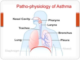
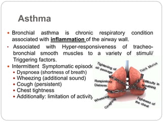
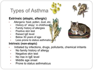
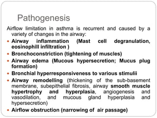
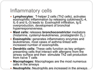
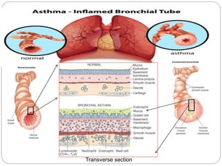
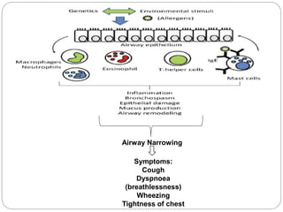
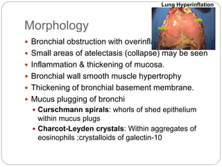
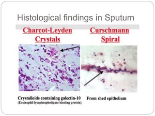
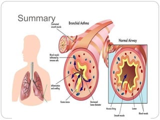
2. Asthma
Bronchial asthma is chronic respiratory condition
associated with inflammation of the airway wall.
• Associated with Hyper-responsiveness of tracheo-
bronchial smooth muscles to a variety of stimuli/
Triggering factors.
Intermittent Symptomatic episodes of
Dyspnoea (shortness of breath)
Wheezing (additional sound)
Cough (persistent)
Chest tightness
Additionally: limitation of activity
3. Etiology/ Triggering factors
Tobacco smoke
Infections such as colds, flu, or pneumonia
Allergens such as food, pollen, mold, dust mites, and
pet dander
Exercise
Air pollution and toxins
Weather, especially extreme changes in temperature
Drugs (such as aspirin, NSAID, and beta-blockers)
Food additives
Emotional stress and anxiety
Singing, laughing, or crying
Smoking, perfumes, or sprays
Acid reflux
4. Types of Asthma
Extrinsic (atopic, allergic)
• Allergens: food, pollen, dust, etc.
• History of `atopy` in childhood
• Family history of allergies
• Positive skin test
• Raised IgE level
• Below 30 years of age
• Less prone to status asthmaticus
Intrinsic (non-atopic)
• Initiated by infections, drugs, pollutants, chemical irritants
• No family history of allergy
• Negative skin test
• No rise in IgE level
• Middle age onset
• Prone to status asthmaticus
5. Pathogenesis
Airflow limitation in asthma is recurrent and caused by a
variety of changes in the airway:
Airway inflammation (Mast cell degranulation,
eosinophill infiltration )
Bronchoconstriction (tightening of muscles)
Airway edema (Mucous hypersecretion; Mucus plug
formation)
Bronchial hyperresponsiveness to various stimulii
Airway remodelling (thickening of the sub-basement
membrane, subepithelial fibrosis, airway smooth muscle
hypertrophy and hyperplasia, angiogenesis and
vasodilation, and mucous gland hyperplasia and
hypersecretion)
Airflow obstruction (narrowing of air passage)
6. Inflammatory cells
Lymphocytes: T-helper 2 cells (Th2 cells), activates
eosinophillic inflammation by releasing cytokines(IL-4,
IL-5 and IL-3) leads to: Eosinophill infiltration, Ig E
overproduction, development of bronchial
hyperresponsiveness.
Mast cells: releases bronchoconstrictor mediators
(histamine, cysteinyl-leukotrienes, prostaglandin D2 )
Eosinophils: generates inflammatory enzymes and
leukotrienes, most cases of asthma linked with
increased number of eosinophills.
Dendritic cells: These cells function as key antigen-
presenting cells that interact with allergens from the
airway surface and then stimulate Th2 cell production
from naïve T cells
Macrophages: Macrophages are the most numerous
cells in the airways
Neutrophils: Neutrophils are increased in the airways
9. Morphology
Bronchial obstruction with overinflation
Small areas of atelectasis (collapse) may be seen
Inflammation & thickening of mucosa.
Bronchial wall smooth muscle hypertrophy
Thickening of bronchial basement membrane.
Mucus plugging of bronchi
Curschmann spirals: whorls of shed epithelium
within mucus plugs
Charcot-Leyden crystals: Within aggregates of
eosinophils ;crystalloids of galectin-10
Lung Hyperinflation