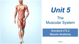
2. Muscular System Tissue Types
•Muscles are responsible for all types of body movement
•There are three basic muscle tissue types found in the
body
•Cardiac Muscle
•Smooth Muscle
•Skeletal (or Striated) Muscle
3. Cardiac Muscle Tissue
The characteristics of cardiac muscle
include:
• Makes up myocardium of heart
• Tissue is unconsciously (involuntarily)
controlled
• Microscopically appears striated
• Cells are short, branching and have a single
nucleus
• Cells connect to each other at intercalated
discs
4. Smooth Muscle Tissue
The characteristics of smooth muscle include:
• Found in the lining of internal organs (GI tract,
uterus, blood vessels, eyes, etc.)
• It controls the involuntary constriction of these
regions (e.g., peristalsis, vasoconstriction, pupil
dilation)
• Cells (fibers) are non-striated, have a short
spindle-shape and each fiber contains a single
nucleus
• Tissue is extremely extensible, while still
retaining ability to contract
5. Skeletal Muscle Tissue
The characteristics of skeletal muscle
include:
• Associated with and attached to the skeleton
• Responsible for moving parts of the body,
such as the limbs, trunk, and face
• Under our conscious (voluntary) control
• Cells (fibers) are long and cylindrical
• Cells (fibers) run in parallel tracts and are
multinucleated and heavily striated
6. Comparison of Muscle Tissue Types
Characteristic Skeletal Cardiac Smooth
Body Location
attached to bone (by
tendons) or skin (for
some facial muscles)
walls of the heart
mostly in walls of
visceral organs (other
than the heart)
Cell Shape and
Appearance
single, very long,
cylindrical,
multinucleate cells with
very obvious striations
branching chains of
cells, uninucleate,
striations, intercalated
discs
single, fusiform,
uninucleate, no
striations
Connective
Tissue
Components
endomysium,
perimysium, and
epimysium
endomysium endomysium
8. Comparison of Muscle Tissue Types
Characteristic Skeletal Cardiac Smooth
Regulation of
Contraction
voluntary involuntary involuntary
Speed of
Contraction
slow to fast slow very slow
Rhythmic
Contractions
no yes yes, in some
9. Skeletal Muscle Structure
Although muscle fiber makes up most of the muscle tissue,
a large amount of connective tissue, blood vessels, and
nerves are also present.
• Connective tissue covers and supports each muscle fiber and
reinforces the muscle as a whole.
• The health of muscle depends on a sufficient nerve and blood supply.
Each skeletal muscle has a nerve ending that controls its activity.
• Active muscles use a lot of energy and require a continuous supply of
oxygen and nutrients, which are supplied by arteries. Muscles produce
large amounts of metabolic waste that must be removed by veins.
10. Gross Anatomy of Skeletal Muscles
Cells are surrounded and bundled by connective tissue!
Connective Tissue Organization:
• Endomysium – encloses a single muscle fiber
• Perimysium – wraps around a fascicle (bundle) of
muscle fibers
• Epimysium – covers the entire skeletal muscle
• Fascia – outer layer of the epimysium
12. Arrangement of Fascicles
The fascicles of muscles can be arranged in a variety of
ways – the most common are:
• Parallel – fascicles are parallel to the long axis of the muscle. Some
of these are also classified as fusiform muscles.
• Pennate – the fascicles are short and attach to a central tendon.
Can be unipennate, bipennate or multipennate.
• Convergent – the muscle has a broad origin but the fascicles
converge to one tendon.
• Circular – muscle has the fascicles arranged in concentric circles.
13.
14. Skeletal Muscle Attachments
Skeletal muscles attach to bones, cartilage, and
connective tissue coverings.
•Epimysium blends into a connective tissue attachment
•Tendons – cord-like structures
•Mostly collagen fibers
•Often cross a joint due to toughness and small size
•Aponeuroses – sheet-like structures
•Attach muscles indirectly to bones, cartilages, or
connective tissue coverings
15. Microanatomy of Skeletal Muscles
• Sarcolemma – specialized cell membrane
• Sarcoplasm – muscle cell cytoplasm
• Sarcoplasmic Reticulum – specialized smooth
endoplasmic reticulum
• used to store and maintain calcium ion levels
• surrounds the myofibril
16. Microanatomy of Skeletal Muscles
• T Tubules – tubules running through the muscle fibers
through which calcium ions flow
• Sarcomeres – repeating contractile unit of the myofibril
• Myofibril – long contraction organelle aligned to
give distinct bands
• I band = light band that contains
only thin filaments called actin.
• A band = dark band that contains
thick filaments called myosin.
• Z-lines at each end provide anchors for thin
filaments
Editor's Notes
The skeleton and its joints support, protect and provide flexibility for the body, but the skeleton CANNOT move itself. That job is performed by the muscle tissue that makes up the muscular system.
The only place in the body where cardiac muscle is found is in the heart.
Cardiac cells are striated but they are not under voluntary control. Cardiac muscles contract without direct stimulation by the nervous system. A bundle of specialized muscle cells in the upper part of the heart sends electrical signals through cardiac muscle tissue, causing the heart to rhythmically contract and pump blood through the body.
The cardiac muscle cell contains one nucleus located near the center, adjacent cells form branching fibers that allow nerve impulses to pass from cell to cell.
Smooth muscles are usually not under voluntary control.
Smooth muscle cells are spindle-shaped and have a single nucleus. They are not striated and interlace to form sheets of smooth muscle tissue.
Smooth muscles are found in many internal organs, such as the stomach, intestines, and in the walls of blood vessels.
Most smooth muscle cells can contract without nervous stimulation. Because most of its movements cannot be consciously controlled, smooth muscle is referred to as involuntary muscle.
The contractions in smooth muscles move food through our digestive tract, control the way blood flows through the cardiovascular system, and increases the size of the pupils of our eyes in bright light.
The skeletal muscles are responsible for moving parts of the body, such as the limbs, trunk, and face.
Skeletal muscles are generally attached to bones via tendons, and are at work every time we make a move.
Skeletal muscles are responsible for voluntary (conscious) movement, and are consciously controlled by the central nervous system.
A skeletal muscle is made of elongated cells called muscle fibers. Varying movements require contraction of variable numbers of muscle fibers in a muscle. Skeletal muscle fibers are grouped into dense bundles called fascicles. A group of fascicles are bound together by connective tissue to form a muscle.
When viewed under a microscope, skeletal muscles appear to have striations (bands or stripes). This gives skeletal muscle the name of striated muscle. Skeletal muscle cells are large and have more than one nucleus. They vary in length from 1 mm to 60 cm (just under 24 inches)
We will discuss these later, just wanted you to see what the chart was referring to!
Endo MY sium. Peri MY sium. Epi MY sium
Fascia is the wrapping around the entire muscle. Fascia is to muscle like the clear casing is on a sausage; It is a connective tissue that is keeping it all wrapped together as one unit.
Every muscle has the same basic setup…attaches in 2, 3, or 4 places!
Every muscle is attached to bones in AT LEAST two locations (via tendons)
Where does the bicep attach? Humerus and scapula
Think about a bungee cord as the muscle – attaches in 2 places (bridge, person)
Muscle fibers consist of bundles of threadlike structures called myofibrils.
Each myofibril is made up of two types of protein filaments – thick ones and thin ones.
The thick filaments are made up of a protein called myosin.
The thin filaments are made up of a protein called actin.
Myosin and actin filaments are arranged to form overlapping patterns, which are responsible for the light and dark bands that can be seen in skeletal muscles (the striated appearance of the muscle).
The thin actin filaments are anchored at their midpoints to a structure called the Z line. The region from one Z-line to the next is called a sarcomere, which is the functional unit of muscle contractions.