SlideShare a Scribd company logo
1 of 49
Study Question 
You are given a shoe box full of an assortment of small objects 
including: 
Ping Pong balls 
Sugar cubes 
Paper clips 
1/2” brass screws 
Iron filings 
1. List the properties of each of these components that might 
help you fractionate them. 
2. Devise the most efficient method you can for getting pure 
paper clips.
Techniques of Protein 
Purification
Protein isolation 
Selection of protein source 
1. Tissues from animals 
2. Microorganisms (E. coli or yeast) 
3. Molecular cloning techniques 
Methods of solubilization 
1. Osmosis lysis (with hypotonic solution) 
2. Use of lysozyme (enzyme that degrades cell wall) 
3. French press or sonication
Stabilization of proteins 
1. pH (think buffers!) 
2. Temperature (close to 0oC) 
Thermal stability could be used for purification 
3. Addition of protease inhibitors 
4. Gentle handling (no frothing) 
Assay of proteins 
1. If purifying an enzyme, use the 
reaction it catalyzes as an assay 
2. If metalloprotein use the metals to follow the 
protein 
3. Immunochemical techniques (antibodies)
General strategy of protein purification 
Proteins are purified by fractionation procedures, a 
series of independent steps in which the properties of 
protein of interest are utilized to separate it from other 
contaminating proteins. 
How do we know our sample of protein is pure? 
We don't! 
The best we can do is to demonstrate by all available 
methods that our sample consists of only one component.
General strategy of protein purification 
Characteristic Procedure 
Solubility 1. Salting in 
2. Salting out 
Ionic charge: 1. Ion exchange chromatography 
2. Electrophoresis 
3. Isoelectric focusing 
Polarity: 1. Adsorption chromatography 
2. Paper chromatography 
3. Hydrophobic interaction chromatography 
Molecular size: 1. Dialysis and ultrafiltration 
2. Gel electrophoresis 
3. Gel filtration chromatography 
4. Ultracentrifugation 
Binding specificity: 1. Affinity chromatography
Solubility of a protein in aqueous solution 
Depends strongly on: 
1. Concentrations of dissolved salts 
2. pH 
3. Temperature 
4. Addition of water-miscible organic solvents, 
e.g., ethanol or acetone
Solubility of carboxyhemoglobin at its isoelectric 
point as a function of ionic strength and ion type 
Page 131
Solubility of b-lactoglobin as a function 
of pH at several NaCl concentrations 
Page 132
Salting Out 
• There are hydrophobic amino acids and hydrophilic amino acids in 
protein molecules. After protein folding in aqueous solution, 
hydrophobic amino acids usually form protected hydrophobic areas 
while hydrophilic amino acids interact with the molecules of solvation 
and allow proteins to form hydrogen bonds with the surrounding water 
molecules. If enough of the protein surface is hydrophilic, the protein 
can be dissolved in water. 
• When the salt concentration is increased, some of the water molecules 
are attracted by the salt ions, which decreases the number of water 
molecules available to interact with the charged part of the protein. As a 
result of the increased demand for solvent molecules, the protein-protein 
interactions are stronger than the solvent-solute interactions; the protein 
molecules coagulate by forming hydrophobic interactions with each 
other. This process is known as salting out.
Salting Out 
• After Proteins solubilized, they can be purified based on 
solubility (usually dependent on overall charge, ionic 
strength, polarity 
• Ammonium sulfate (NH4SO4) commonly used to “salt out” 
• Takes away water by interacting with it, makes protein less 
soluble because hydrophobic interactions among proteins 
increases 
• Different aliquots taken as function of salt concentration to 
get closer to desired protein sample of interest (30, 40, 50, 
75% increments) 
• One fraction has protein of interest
CENTRIFUGATION 
• A particle is subjected to a centrifugal force when it is rotated at a high rate of 
speed. The centrifugal force, F, is defined by Equation 
F = mω2r 
F = intensity of the centrifugal force 
m = effective mass of the sedimenting particle 
ω = angular velocity of rotation in rad/sec 
r = distance of the migrating particles from the central axis of rotation 
• A more common measurement of F, in terms of the earth’s gravitational 
force, g, is relative centrifugal force, RCF, defined by Equation 
RCF = (1.119 * 10-5)(rpm)2(r)
Although the relative centrifugal force can easily be calculated, 
centrifugation manuals usually contain a nomograph for the 
convenient conversion between relative centrifugal force and 
speed of the centrifuge at different radii of the centrifugation 
spindle to a point along the centrifuge tube. A nomograph 
consists of three columns representing the radial distance (in 
mm), the relative centrifugal field and the rotor speed (in r.p.m.). 
For the conversion between relative centrifugal force and speed 
of the centrifuge spindle in r.p.m. at different radii, a straight-edge 
is aligned through known values in two columns, then the 
desired figure is read where the straight-edge intersects the third 
column.
Fig. 3.1 Nomograph for the 
determination of the relative centrifugal 
field for a given rotor speed and radius. 
The three columns represent the radial 
distance (in mm), the relative centrifugal 
field and the rotor speed 
(in r.p.m.). For the conversion between 
relative centrifugal force and speed of 
the centrifuge spindle in revolutions per 
minute at different radii, draw a straight-edge 
through known values in two 
columns. The desired figure can then be 
read where the straight-edge intersects 
the third column. (Courtesy of Beckman- 
Coulter.)
The most obvious differences between centrifuges are: 
•• the maximum speed at which biological specimens are subjected to 
increased sedimentation; 
•• the presence or absence of a vacuum; 
•• the potential for refrigeration or general manipulation of the 
temperature during a centrifugation run; and 
•• the maximum volume of samples and capacity for individual 
centrifugation tubes. 
Many different types of centrifuges are commercially available 
including: 
• large-capacity low-speed preparative centrifuges; 
• refrigerated high-speed preparative centrifuges; 
• analytical ultracentrifuges; 
• preparative ultracentrifuges; 
• large-scale clinical centrifuges; and 
• small-scale laboratory microfuges.
fixed-angle rotor 
vertical tube rotor 
swinging-bucket rotor 
initial acceleration stage, the main centrifugal separation phase, de-acceleration and the 
final harvesting of separated particles in the rotor at rest.
Differential Centrifugation 
• Sample is spun, after 
lysis, to separate 
unbroken cells, nuclei, 
other organelles and 
particles not soluble in 
buffer used 
• Different speeds of spin 
allow for particle 
separation
Density-Gradient Centrifugation 
if the sample is centrifuged in a fluid medium that gradually 
increases in density from top to bottom. This technique, called 
density gradient centrifugation, permits the separation of 
multi-component mixtures of macromolecules and the 
measurement of sedimentation coefficients. 
• zonal centrifugation, in which the sample is centrifuged in a 
preformed gradient, A density gradient is prepared in a tube 
prior to centrifugation with the use of an automatic gradient 
mixer or manually with pipette. Both step gradient and 
continuous gradient systems. 
• Sucrose concentrations up to 60% can be used, with a density 
limit of 1.28g/cm3. 
• The various types of particles sediment as zones and remain 
separated from the other components. The various zones are 
then isolated by collecting fractions from the bottom of the tube.
isopycnic centrifugation, in which a self-generating gradient 
forms during centrifugation. The sample under study is 
dissolved in a solution of a dense salt such as cesium chloride 
or cesium sulfate. The cesium salts may be used to establish 
gradients to an upper density limit of The solution of 
biological sample and cesium salt is uniformly distributed in a 
centrifuge tube and rotated in an ultracentrifuge. Under the 
influence of the centrifugal force, the cesium salt redistributes 
to form a continuously increasing density gradient from the 
top to the bottom. The macromolecules of the biological 
sample seek an area in the tube where the density is equal to 
their respective densities. That is, the macromolecules move to 
a region where the sum of the forces (centrifugal and 
frictional) is zero.
FIGURE 4.12 A 
comparison of differential and density 
gradient measurements. 
A Differential centrifugation in a medium 
of unchanging density. 
B Zonal centrifugation in a prepared 
density gradient. 
C Isopycnic centrifugation; the 
density gradient forms during 
centrifugation.
Isoelectric Points of Several Common Proteins 
Protein pI 
Pepsin 1.0 
Ovalbumin (hen) 4.6 
Serum albumin (human) 4.9 
Tropomyosin 5.1 
Insulin (bovine) 5.4 
Fibrinogen (human) 5.8 
g-Globulin (human) 6.6 
Collagen 6.6 
Myoglobin (horse) 7.0 
Hemoglobin (human) 7.1 
Ribonuclease A (bovine) 9.4 
Cytochrome c (horse) 10.6 
Histone (bovine) 10.8 
Lysozyme (hen) 11.0 
Salmine (salmon) 12.1
CHROMATOGRAPHY 
The molecules targeted for analysis are called analytes. 
The mobile phase, which may be a liquid or gas, moves the sample 
components through a region containing the solid or liquid stationary phase, 
which is called the sorbent. 
The molecular components in the sample distribute themselves between the 
mobile phase and sorbent and thus have the opportunity to interact intimately 
with the stationary phase. If some of the sample molecules (analytes) are 
preferentially bound by the sorbent, they spend more time in the sorbent and 
are retarded in their movement through the chromatographic system. 
Molecules that show weak affinity for the sorbent spend more time with the 
mobile phase and are more easily removed or eluted from the system. 
The mobile phase can be collected as a function of time at the end of the 
chromatographic system. The mobile phase, now called the effluent, 
contains the purified analytes.
When the actual adsorbing material is made into a column, it is said 
to be poured or packed. 
Application of the sample to the top of the column is loading the 
column. 
Movement of solvent through the loaded column is called 
developing or eluting the column. 
The bed volume is the total volume of solvent and adsorbing 
material taken up by the column. 
The volume taken up by the liquid phase in the column is the void 
volume. 
The elution volume is the amount of solvent required to remove a 
particular analyte from the column. This is analogous to values in 
planar chromatography.
All components may be eluted by a single solvent or buffer. This is 
referred to as continual elution. In contrast, stepwise elution refers 
to an incremental change of solvent to aid development. The 
column is first eluted with a volume of one solvent and then with a 
second solvent. This may continue with as many solvents or solvent 
mixtures as desired. In general, the first solvent should be the least 
polar of any used in the analysis, and each additional solvent should 
be of greater polarity or ionic strength. Finally, adsorption columns 
may be developed by gradient elution brought about by a gradual 
change in solvent composition. The composition of the eluting 
solvent can be changed by the continuous mixing of two different 
solvents to gradually change the ratio of the two solvents. 
Alternatively, the concentration of a component in the solvent can be 
gradually increased. This is most often done by addition of a salt 
(KCl, NaCl, etc.). Devices are commercially available to prepare 
predetermined, reproducible gradients.
Protein 
separation and 
purification by 
column 
chromatography 
From Lehninger 
Principles of Biochemistry 
Chromatographic separations
Column 
Chromatography: 
Size-exclusion 
Gel filtration 
chromatography can be used 
to estimate molecular masses 
From Lehninger 
Principles of Biochemistry
Theory of Gel Filtration 
The stationary phase consists of inert particles that contain small pores of a controlled size. 
Microscopic examination of a particle reveals an interior resembling a sponge. A solution containing 
analytes of various molecular sizes is allowed to pass through the column under the influence of 
continuous solvent flow. 
Analytes larger than the pores cannot enter the interior of the gel beads, so they 
are limited to the space between the beads. The volume of the column accessible 
to very large molecules is, therefore, greatly reduced. As a result, they are not slowed in their progress 
through the column and elute rapidly in a single zone. Small molecules capable of diffusing in and out 
of the beads have a much larger volume available to them. Therefore, they are delayed in their 
journey through the column bed. Molecules of intermediate size migrate through the column at a rate 
somewhere between those for large and small molecules.
1.Exclusion Limit This is defined as the molecular mass of the smallest molecule 
that cannot diffuse into the inner volume of the gel matrix. All molecules above this 
limit elute rapidly in a single zone. The exclusion 
limit of a typical gel, Sephadex G-50, is 30,000 daltons. All analytes having a 
molecular size greater than this value would pass directly through the column bed 
without entering the gel pores. 
2. Fractionation Range Sephadex G-50 has a fractionation range of 1500 to 
30,000 daltons. Analytes within this range would be separated in a somewhat linear 
fashion. 
3. Water Regain and Bed Volume Gel chromatography media are often supplied in 
dehydrated form and must be swollen in a solvent, usually water, before use. The 
weight of water taken up by 1 g of dry gel is known as the water regain. For G-50, 
this value is g. This value does not include the water surrounding the gel particles, 
so it cannot be used as an estimate of the final volume of a packed gel column. 
Most commercial suppliers of gel materials provide, in addition to water regain, a 
bed volume value. This is the final volume taken up by 1 g of dry gel when swollen 
in water. For G-50, bed volume is 9 to 11 mL/g dry gel.
4. Gel Particle Shape and Size Ideally, gel particles should be spherical to 
provide a uniform bed with a high density of pores. Particle size is defined 
either by mesh size or bead diameter Both the degree of resolution afforded by a 
column and the flow rate depend on particle size. Larger particle sizes (50 to 
100 mesh, 100 to 300 ) offer high flow rates but poor chromatographic 
separation. The opposite is true for very small particle sizes (“superfine,” 400 
mesh, 10 to ). The most useful particle size, which represents a compromise 
between resolution and flow rate, is 100 to 200 mesh (50 to 150 ). 
5. Void Volume This is the total space surrounding the gel particles in a packed 
column. This value is determined by measuring the volume of solvent required 
to elute a solute that is completely excluded from the gel matrix. Most columns 
can be calibrated for void volume with a dye, blue dextran, which has an 
average molecular mass of 2,000,000 daltons. 
6. Elution Volume This is the volume of eluting buffer necessary to remove a 
particular analyte from a packed column.
Biochemists refer to a protein's size in terms of its molecular 
weight, in kDa (a kilodalton, kD or kDa, is 1000 times the 
molecular mass of hydrogen) 
Each amino acid residue counts for about 110 daltons, that is, about 
0.11 kDa. 
Sephadex G-50 1-30 kD 
Sephadex G-100 4-150 kD 
Sephadex G-200 5-600 kD 
Bio-Gel P-10 1.5-20 kD 
Bio-Gel P-30 2.4-40 kD 
Bio-Gel P-100 5-100 kD 
Bio-Gel P-300 60-400 kD 
Sephadex is a trademark of Pharmacia. 
Bio-Gel is a trademark of Bio-Rad.
Selecting a Gel 
If the mixture contains macromolecules up to 120,000 in molecular weight, then Bio-Gel P-150, 
Sephacryl S-200HR, or Sephadex G-150 would be most appropriate. If P-100, G-100, or 
Sephacryl S-100HR were used, some of the highermolecular- weight proteins in the sample 
would elute in the void volume. On the other hand, if P-200, P-300, or G-200 were used, there 
would be a decrease in 
both resolution and flow rate. 
Gel Preparation and Storage 
The dextran and acrylamide gel products are sometimes supplied in dehydrated form and must 
be allowed to swell in water before use. The swelling time required differs for each gel, but the 
extremes are 3 to 4 hours at for highly cross-linked gels and up to 72 hours at for P-300 or G- 
200. The swelling time can be shortened if a boiling-water bath is used. Agarose gels and 
combined polyacrylamideagarose gels are supplied in a hydrated state, so there is no need for 
swelling. 
Before a gel slurry is packed into the column, it should be defined and deaerated. Defining is 
necessary to remove very fine particles, which would reduce flow rates. 
Deaerating (removing dissolved gases) should be done on the gel slurry and all eluting buffers. 
Antimicrobial agents must be added to stored, hydrated gels. One of the best agents is sodium 
azide (0.02%).
Column Size 
For fractionation purposes, it is usually not necessary to use columns greater than 100 cm in length. 
The ratio of bed length to width should be between 25 and 100. For group separations, columns less 
than 50 cm long are sufficient, and appropriate ratios of bed length to width are between 5 and 10. 
Eluting Buffer 
There are fewer restrictions on buffer choice in gel chromatography than in ion-exchange 
chromatography. Dextran and polyacrylamide gels are stable in the pH range 1 to 10, whereas agarose 
gels are limited to pH 4 to 10. Since there is such a wide range of stability of the gels, the buffer pH 
should be chosen on the basis of the range of stability of the macromolecules to be separated. 
Sample Volume 
If too much sample is applied to a column, resolution is decreased; if the sample size is too small, the 
analytes are greatly diluted. For group separations, a sample volume of 10 to 25% of the column total 
volume is suitable. The sample volume for fractionation procedures should be between 1 and 5% of the 
total volume. Column total volume is determined by measuring the volume of water in the glass 
column that is equivalent to the height of the packed bed. 
Column Flow Rate 
The flow rate of a gel column depends on many factors, including length of column and type and size 
of the gel. It is generally safe to elute a gel column at a rate slightly less than free flow. A high flow 
rate reduces sample diffusion or zone broadening, but may not allow complete equilibration of analyte 
molecules with the gel matrix. 
A specific flow rate cannot be recommended, since each type of gel requires a different range. The 
average flow rate given in literature references for small-pore-size gels is 8 to 12 mL/cm2 of cross-sectional 
bed area per hour (15 to 25 mL/hr). For large-pore-size gels, a value of 2 to 5mL/cm2 of 
cross-sectional bed area per hour (5 to 10 mL/hr) is average.
Desalting 
Inorganic salts, organic solvents, and other small molecules are used extensively for the 
purification of macromolecules. Gel chromatography provides an inexpensive, simple, and rapid 
method for removal of these small molecules. One especially attractive method for desalting 
very small samples (0.1 mL or less) of proteins or nucleic acid solutions is to use spin columns. 
These are prepacked columns of polyacrylamide exclusion gels. Spin columns are used in a 
similar fashion to microfiltration centrifuge tubes (Chapter 3, p. 75). The sample is placed on 
top of the gel column and spun in a centrifuge. Large molecules are eluted from the column and 
collected in a reservoir. The small molecules to be removed remain in the gel. 
Purification of Biomolecules 
This is probably the most popular use of gel chromatography. Because of a gel’s ability to 
fractionate molecules on the basis of size, gel filtration complements other purification 
techniques that separate molecules on the basis of polarity and charge. 
Estimation of MolecularWeight 
The elution volume for a particular analyte is proportional to its molecular size. This indicates 
that it is possible to estimate the molecular weight of a molecule on the basis of its elution 
characteristics on a gel column. An elution curve for several standard proteins separated on 
Sephadex G-100 is shown in Figure 5.8. 
This curve, a plot of protein concentration vs. volume collected, is representative of data 
obtained from a gel filtration experiment. The elution volume, for each protein can be estimated 
as shown in the figure. A plot of log molecular mass vs. elution volume for the proteins.
A schematic illustration of gel filtration chromatography
FIGURE 5.8 Elution curve for a mixture of several proteins using gel-filtration chromatography. 
A hemoglobin; B egg albumin; C chymotrypsinogen; D myoglobin; E = cytochrome c.
FIGURE 5.9 A plot of log molecular mass vs. elution volume for the proteins A, B, C, D, and E 
in Figure 5.8.
STUDY QUESTION
Column Chromatography: 
Ion Exchange 
From Lehninger 
Principles of Biochemistry
This form of chromatography relies on the attraction between oppositely charged 
stationary phase, known as an ion exchanger, and analyte. 
It is frequently chosen for the separation and purification of proteins, peptides, 
nucleic acids, polynucleotides and other charged molecules, mainly because of its 
high resolving power and high capacity. 
There are two types of ion exchanger, namely cation and anion exchangers. Cation 
exchangers possess negatively charged groups and these will attract positively 
charged cations. These exchangers are also called acidic ion exchangers because their 
negative charges result from the ionisation of acidic groups. 
Anion exchangers have positively charged groups that will attract negatively charged 
anions. The term basic ion exchangers is also used to describe these exchangers, as 
positive chargesgenerally result from the association of protons with basic groups. 
Matrices used include polystyrene, cellulose and agarose. Functional ionic groups 
include sulphonate (–SO–3) and quaternary ammonium (–N+R3), both of which are 
strong exchangers because they are totally ionised at all normal working pH 
values,and carboxylate (–COO-) and diethylammonium (–HN+(CH2CH3)2), both of 
which are termed weak exchangers because they are ionised over only a narrow range 
of pH values.
FIGURE 5.6 The effect of pH on the net charge of a protein.
Eluent pH 
The pH of the buffer selected as eluent should be at least one pH unit above or 
below the isoionic point of the analytes. In general, cationic buffers such as 
Tris, pyridine and alkylamines are used in conjunction with anion exchangers, 
and anionic buffers such as acetate, barbiturate and phosphate are used with 
cation exchangers. 
Elution 
Gradient elution is far more common than isocratic elution. Continuous or 
stepwise pH and ionic strength gradients may be employed but continuous 
gradients tend to give better resolution with less peak tailing. Generally with 
an anion exchanger, the pH gradient decreases and the ionic strength 
increases, whereas for cation exchangers 
both the pH and ionic gradients increase during the elution.
A schematic diagram illustrating the separation of several proteins 
by ion exchange chromatography using stepwise elution
A gradient is a change in the proportion of the two (or more) solvents that make up the 
mobile phase. With a pump controlling flow rates and proportioning valves on all the 
solvent reservoirs, it is possible to gradually or abruptly change the mobile phase 
composition. 
A Step Gradient is an abrupt change in mobile phase composition. An example of a step 
gradient is shown here (blue line), where the salt concentration in the mobile phase is 
changed from 0.1 M to 0.2 M at 10 min, then from 0.2 M to 0.3 M at 20 min.

More Related Content

What's hot

What's hot (20)

Maxam-Gilbert method of DNA sequencing
Maxam-Gilbert method of DNA sequencingMaxam-Gilbert method of DNA sequencing
Maxam-Gilbert method of DNA sequencing
 
Centrifugation
CentrifugationCentrifugation
Centrifugation
 
Purification of proteins (purification of enzymes)
Purification of proteins (purification of enzymes)Purification of proteins (purification of enzymes)
Purification of proteins (purification of enzymes)
 
Sequence alignment
Sequence alignmentSequence alignment
Sequence alignment
 
Sds-Page
Sds-Page Sds-Page
Sds-Page
 
Ramachandran plot
Ramachandran plotRamachandran plot
Ramachandran plot
 
ISOELECTRIC FOCUSING PPT - SLIDE SHARE
ISOELECTRIC FOCUSING PPT - SLIDE SHAREISOELECTRIC FOCUSING PPT - SLIDE SHARE
ISOELECTRIC FOCUSING PPT - SLIDE SHARE
 
2 d gel electrophoresis
2 d gel electrophoresis2 d gel electrophoresis
2 d gel electrophoresis
 
Cell fractionation
Cell fractionationCell fractionation
Cell fractionation
 
Animal cell culture media
Animal cell culture mediaAnimal cell culture media
Animal cell culture media
 
Nucleic acid hybridization
Nucleic acid hybridizationNucleic acid hybridization
Nucleic acid hybridization
 
Fplc(fast protein liquid chromatography )
Fplc(fast protein liquid chromatography )Fplc(fast protein liquid chromatography )
Fplc(fast protein liquid chromatography )
 
sequence alignment
sequence alignmentsequence alignment
sequence alignment
 
Vertical Gel Electrophoresis (SDS-PAGE)
Vertical Gel Electrophoresis (SDS-PAGE)Vertical Gel Electrophoresis (SDS-PAGE)
Vertical Gel Electrophoresis (SDS-PAGE)
 
Blotting techniques
Blotting techniquesBlotting techniques
Blotting techniques
 
Ultracentrifugation
UltracentrifugationUltracentrifugation
Ultracentrifugation
 
Northern blotting
Northern blotting Northern blotting
Northern blotting
 
Pulse Field Gel Electrophoresis
Pulse Field Gel ElectrophoresisPulse Field Gel Electrophoresis
Pulse Field Gel Electrophoresis
 
Preparative and analytical centrifugation
Preparative and analytical centrifugationPreparative and analytical centrifugation
Preparative and analytical centrifugation
 
Analytical Ultracentrifugation
Analytical UltracentrifugationAnalytical Ultracentrifugation
Analytical Ultracentrifugation
 

Viewers also liked

Gel Filtration Chromatography Lecture
Gel Filtration Chromatography LectureGel Filtration Chromatography Lecture
Gel Filtration Chromatography Lectureouopened
 
Ion exchange chromatography and gec
Ion exchange chromatography and gecIon exchange chromatography and gec
Ion exchange chromatography and gecceutics1315
 
Gel permeation chromatography (GPC)
Gel permeation chromatography (GPC)Gel permeation chromatography (GPC)
Gel permeation chromatography (GPC)Asabuwa N. Fahanwi
 
Gel permeation chromatography
Gel permeation chromatographyGel permeation chromatography
Gel permeation chromatographyShradha Basu
 
Size exclusion chromatography
Size exclusion chromatographySize exclusion chromatography
Size exclusion chromatographyMandvi Shandilya
 
Vaccine Control System
Vaccine Control SystemVaccine Control System
Vaccine Control Systemmd2gri
 
Chromatofocusing
ChromatofocusingChromatofocusing
ChromatofocusingAmlan Barai
 
chromatofocusing, 2 de, ief
chromatofocusing, 2 de, iefchromatofocusing, 2 de, ief
chromatofocusing, 2 de, iefSidra Shaffique
 
Biopharmaceutical Characterization According to ICHQ6B Harmonized Guidelines
Biopharmaceutical Characterization According to ICHQ6B Harmonized GuidelinesBiopharmaceutical Characterization According to ICHQ6B Harmonized Guidelines
Biopharmaceutical Characterization According to ICHQ6B Harmonized GuidelinesSGS
 
Process development considerations for quality and safety of vaccines
Process development considerations for quality and safety of vaccinesProcess development considerations for quality and safety of vaccines
Process development considerations for quality and safety of vaccinesDr. Priyabrata Pattnaik
 
Ion exchange chromatography (1) (4)
Ion exchange chromatography (1) (4)Ion exchange chromatography (1) (4)
Ion exchange chromatography (1) (4)Parimala Gattupalli
 
Gel electroporosis
Gel electroporosisGel electroporosis
Gel electroporosisceutics1315
 
Purification Of Substances 2
Purification Of Substances 2Purification Of Substances 2
Purification Of Substances 2sYhira
 
Bioc 523
Bioc 523Bioc 523
Bioc 523mpark4
 

Viewers also liked (20)

Gel Filtration Chromatography Lecture
Gel Filtration Chromatography LectureGel Filtration Chromatography Lecture
Gel Filtration Chromatography Lecture
 
Ion exchange chromatography and gec
Ion exchange chromatography and gecIon exchange chromatography and gec
Ion exchange chromatography and gec
 
Gel permeation chromatography (GPC)
Gel permeation chromatography (GPC)Gel permeation chromatography (GPC)
Gel permeation chromatography (GPC)
 
Gel permeation chromatography
Gel permeation chromatographyGel permeation chromatography
Gel permeation chromatography
 
Biochemical analysis techniques
Biochemical analysis techniquesBiochemical analysis techniques
Biochemical analysis techniques
 
Size exclusion chromatography
Size exclusion chromatographySize exclusion chromatography
Size exclusion chromatography
 
Chromatofocusing
ChromatofocusingChromatofocusing
Chromatofocusing
 
Vaccine Control System
Vaccine Control SystemVaccine Control System
Vaccine Control System
 
Chromatofocusing
ChromatofocusingChromatofocusing
Chromatofocusing
 
IFPMA: From Manufacturing to the Vaccinee – the Complex Journey of a Vaccine
IFPMA: From Manufacturing to the Vaccinee – the Complex Journey of a VaccineIFPMA: From Manufacturing to the Vaccinee – the Complex Journey of a Vaccine
IFPMA: From Manufacturing to the Vaccinee – the Complex Journey of a Vaccine
 
chromatofocusing, 2 de, ief
chromatofocusing, 2 de, iefchromatofocusing, 2 de, ief
chromatofocusing, 2 de, ief
 
Biopharmaceutical Characterization According to ICHQ6B Harmonized Guidelines
Biopharmaceutical Characterization According to ICHQ6B Harmonized GuidelinesBiopharmaceutical Characterization According to ICHQ6B Harmonized Guidelines
Biopharmaceutical Characterization According to ICHQ6B Harmonized Guidelines
 
Process development considerations for quality and safety of vaccines
Process development considerations for quality and safety of vaccinesProcess development considerations for quality and safety of vaccines
Process development considerations for quality and safety of vaccines
 
tissues
tissuestissues
tissues
 
Ion exchange chromatography (1) (4)
Ion exchange chromatography (1) (4)Ion exchange chromatography (1) (4)
Ion exchange chromatography (1) (4)
 
Gel electroporosis
Gel electroporosisGel electroporosis
Gel electroporosis
 
Lab session-3
Lab session-3Lab session-3
Lab session-3
 
Cloning
CloningCloning
Cloning
 
Purification Of Substances 2
Purification Of Substances 2Purification Of Substances 2
Purification Of Substances 2
 
Bioc 523
Bioc 523Bioc 523
Bioc 523
 

Similar to Study the Properties of Small Objects

Final Centrifugation Lyophilization and UF.pptx
Final Centrifugation Lyophilization  and UF.pptxFinal Centrifugation Lyophilization  and UF.pptx
Final Centrifugation Lyophilization and UF.pptxAyeshaSaleem423816
 
Ultracenrifugation by kanchana sivabalan
Ultracenrifugation by kanchana sivabalanUltracenrifugation by kanchana sivabalan
Ultracenrifugation by kanchana sivabalankanchana Sivabalan
 
Ultracentrifugation
Ultracentrifugation Ultracentrifugation
Ultracentrifugation Aswathi K S
 
Centrifugation
CentrifugationCentrifugation
Centrifugationvlmawia
 
Lec tech.protein separation
Lec tech.protein separationLec tech.protein separation
Lec tech.protein separationDrShamimAkram
 
Lec.9 tech.protein separation
Lec.9 tech.protein separationLec.9 tech.protein separation
Lec.9 tech.protein separationShamim Akram
 
Centrifugation by Dr. Anurag Yadav
Centrifugation by Dr. Anurag YadavCentrifugation by Dr. Anurag Yadav
Centrifugation by Dr. Anurag YadavDr Anurag Yadav
 
Centrifugation principle and types by Dr. Anurag Yadav
Centrifugation principle and types by Dr. Anurag YadavCentrifugation principle and types by Dr. Anurag Yadav
Centrifugation principle and types by Dr. Anurag YadavDr Anurag Yadav
 
Separation techniques in analytical chemistry
Separation techniques in analytical chemistrySeparation techniques in analytical chemistry
Separation techniques in analytical chemistryJLoknathDora
 
Analytical tool in Biochemistry and chemistry
Analytical tool in Biochemistry and chemistryAnalytical tool in Biochemistry and chemistry
Analytical tool in Biochemistry and chemistryKimEliakim1
 
Centrifuge.pptx
Centrifuge.pptxCentrifuge.pptx
Centrifuge.pptxsaraso888
 
Protein fractionation
Protein fractionationProtein fractionation
Protein fractionationjaspreet maan
 
_centrifugation .pptx
_centrifugation .pptx_centrifugation .pptx
_centrifugation .pptxAlisha Shaikh
 

Similar to Study the Properties of Small Objects (20)

Chromatography
ChromatographyChromatography
Chromatography
 
Final Centrifugation Lyophilization and UF.pptx
Final Centrifugation Lyophilization  and UF.pptxFinal Centrifugation Lyophilization  and UF.pptx
Final Centrifugation Lyophilization and UF.pptx
 
Ultracenrifugation by kanchana sivabalan
Ultracenrifugation by kanchana sivabalanUltracenrifugation by kanchana sivabalan
Ultracenrifugation by kanchana sivabalan
 
Ultracentrifugation
Ultracentrifugation Ultracentrifugation
Ultracentrifugation
 
Cell Organelle Fractionation
Cell Organelle Fractionation Cell Organelle Fractionation
Cell Organelle Fractionation
 
Centrifugation
CentrifugationCentrifugation
Centrifugation
 
Lec tech.protein separation
Lec tech.protein separationLec tech.protein separation
Lec tech.protein separation
 
Lec.9 tech.protein separation
Lec.9 tech.protein separationLec.9 tech.protein separation
Lec.9 tech.protein separation
 
Presentation ultra centrifuge
Presentation ultra centrifugePresentation ultra centrifuge
Presentation ultra centrifuge
 
CENTRIFUGATION
CENTRIFUGATIONCENTRIFUGATION
CENTRIFUGATION
 
Ultracentrifugation
UltracentrifugationUltracentrifugation
Ultracentrifugation
 
Centrifugation by Dr. Anurag Yadav
Centrifugation by Dr. Anurag YadavCentrifugation by Dr. Anurag Yadav
Centrifugation by Dr. Anurag Yadav
 
Centrifugation principle and types by Dr. Anurag Yadav
Centrifugation principle and types by Dr. Anurag YadavCentrifugation principle and types by Dr. Anurag Yadav
Centrifugation principle and types by Dr. Anurag Yadav
 
Separation techniques in analytical chemistry
Separation techniques in analytical chemistrySeparation techniques in analytical chemistry
Separation techniques in analytical chemistry
 
Analytical tool in Biochemistry and chemistry
Analytical tool in Biochemistry and chemistryAnalytical tool in Biochemistry and chemistry
Analytical tool in Biochemistry and chemistry
 
Centrifuge.pptx
Centrifuge.pptxCentrifuge.pptx
Centrifuge.pptx
 
Protein fractionation
Protein fractionationProtein fractionation
Protein fractionation
 
Electrophoresis
ElectrophoresisElectrophoresis
Electrophoresis
 
Centrifugation
CentrifugationCentrifugation
Centrifugation
 
_centrifugation .pptx
_centrifugation .pptx_centrifugation .pptx
_centrifugation .pptx
 

More from Sidra Shaffique

POST TRANSCRIPTIONAL MODIFICATIONS IN EUKARYOTES
POST TRANSCRIPTIONAL MODIFICATIONS IN EUKARYOTESPOST TRANSCRIPTIONAL MODIFICATIONS IN EUKARYOTES
POST TRANSCRIPTIONAL MODIFICATIONS IN EUKARYOTESSidra Shaffique
 
Dna recombination mechanisms new
Dna recombination mechanisms newDna recombination mechanisms new
Dna recombination mechanisms newSidra Shaffique
 
6 carboxypeptidase mechanism
6  carboxypeptidase mechanism6  carboxypeptidase mechanism
6 carboxypeptidase mechanismSidra Shaffique
 
rRNA anr tRNA post transcriptional modifications
rRNA anr tRNA post transcriptional modificationsrRNA anr tRNA post transcriptional modifications
rRNA anr tRNA post transcriptional modificationsSidra Shaffique
 
Chromatography (paper chromatography and tlc)
Chromatography (paper chromatography and tlc)Chromatography (paper chromatography and tlc)
Chromatography (paper chromatography and tlc)Sidra Shaffique
 
second generation of DNA Sequencing
second generation of DNA Sequencingsecond generation of DNA Sequencing
second generation of DNA SequencingSidra Shaffique
 

More from Sidra Shaffique (14)

Bacteriophage
BacteriophageBacteriophage
Bacteriophage
 
POST TRANSCRIPTIONAL MODIFICATIONS IN EUKARYOTES
POST TRANSCRIPTIONAL MODIFICATIONS IN EUKARYOTESPOST TRANSCRIPTIONAL MODIFICATIONS IN EUKARYOTES
POST TRANSCRIPTIONAL MODIFICATIONS IN EUKARYOTES
 
Transcription NEW
Transcription NEWTranscription NEW
Transcription NEW
 
Dna recombination mechanisms new
Dna recombination mechanisms newDna recombination mechanisms new
Dna recombination mechanisms new
 
Translation lecture
Translation lectureTranslation lecture
Translation lecture
 
Lysozyme Enzymology
Lysozyme EnzymologyLysozyme Enzymology
Lysozyme Enzymology
 
6 carboxypeptidase mechanism
6  carboxypeptidase mechanism6  carboxypeptidase mechanism
6 carboxypeptidase mechanism
 
Lecture 5 hplc gc mass
Lecture 5 hplc gc massLecture 5 hplc gc mass
Lecture 5 hplc gc mass
 
rRNA anr tRNA post transcriptional modifications
rRNA anr tRNA post transcriptional modificationsrRNA anr tRNA post transcriptional modifications
rRNA anr tRNA post transcriptional modifications
 
Surface tension (2)
Surface tension (2)Surface tension (2)
Surface tension (2)
 
Lect 1 biochemiical
Lect 1 biochemiicalLect 1 biochemiical
Lect 1 biochemiical
 
Dialysis lecture 3
Dialysis lecture 3Dialysis lecture 3
Dialysis lecture 3
 
Chromatography (paper chromatography and tlc)
Chromatography (paper chromatography and tlc)Chromatography (paper chromatography and tlc)
Chromatography (paper chromatography and tlc)
 
second generation of DNA Sequencing
second generation of DNA Sequencingsecond generation of DNA Sequencing
second generation of DNA Sequencing
 

Recently uploaded

Advanced Views - Calendar View in Odoo 17
Advanced Views - Calendar View in Odoo 17Advanced Views - Calendar View in Odoo 17
Advanced Views - Calendar View in Odoo 17Celine George
 
APM Welcome, APM North West Network Conference, Synergies Across Sectors
APM Welcome, APM North West Network Conference, Synergies Across SectorsAPM Welcome, APM North West Network Conference, Synergies Across Sectors
APM Welcome, APM North West Network Conference, Synergies Across SectorsAssociation for Project Management
 
The basics of sentences session 2pptx copy.pptx
The basics of sentences session 2pptx copy.pptxThe basics of sentences session 2pptx copy.pptx
The basics of sentences session 2pptx copy.pptxheathfieldcps1
 
Q4-W6-Restating Informational Text Grade 3
Q4-W6-Restating Informational Text Grade 3Q4-W6-Restating Informational Text Grade 3
Q4-W6-Restating Informational Text Grade 3JemimahLaneBuaron
 
Nutritional Needs Presentation - HLTH 104
Nutritional Needs Presentation - HLTH 104Nutritional Needs Presentation - HLTH 104
Nutritional Needs Presentation - HLTH 104misteraugie
 
Interactive Powerpoint_How to Master effective communication
Interactive Powerpoint_How to Master effective communicationInteractive Powerpoint_How to Master effective communication
Interactive Powerpoint_How to Master effective communicationnomboosow
 
BASLIQ CURRENT LOOKBOOK LOOKBOOK(1) (1).pdf
BASLIQ CURRENT LOOKBOOK  LOOKBOOK(1) (1).pdfBASLIQ CURRENT LOOKBOOK  LOOKBOOK(1) (1).pdf
BASLIQ CURRENT LOOKBOOK LOOKBOOK(1) (1).pdfSoniaTolstoy
 
BAG TECHNIQUE Bag technique-a tool making use of public health bag through wh...
BAG TECHNIQUE Bag technique-a tool making use of public health bag through wh...BAG TECHNIQUE Bag technique-a tool making use of public health bag through wh...
BAG TECHNIQUE Bag technique-a tool making use of public health bag through wh...Sapna Thakur
 
Introduction to Nonprofit Accounting: The Basics
Introduction to Nonprofit Accounting: The BasicsIntroduction to Nonprofit Accounting: The Basics
Introduction to Nonprofit Accounting: The BasicsTechSoup
 
Disha NEET Physics Guide for classes 11 and 12.pdf
Disha NEET Physics Guide for classes 11 and 12.pdfDisha NEET Physics Guide for classes 11 and 12.pdf
Disha NEET Physics Guide for classes 11 and 12.pdfchloefrazer622
 
1029-Danh muc Sach Giao Khoa khoi 6.pdf
1029-Danh muc Sach Giao Khoa khoi  6.pdf1029-Danh muc Sach Giao Khoa khoi  6.pdf
1029-Danh muc Sach Giao Khoa khoi 6.pdfQucHHunhnh
 
The Most Excellent Way | 1 Corinthians 13
The Most Excellent Way | 1 Corinthians 13The Most Excellent Way | 1 Corinthians 13
The Most Excellent Way | 1 Corinthians 13Steve Thomason
 
Measures of Central Tendency: Mean, Median and Mode
Measures of Central Tendency: Mean, Median and ModeMeasures of Central Tendency: Mean, Median and Mode
Measures of Central Tendency: Mean, Median and ModeThiyagu K
 
SOCIAL AND HISTORICAL CONTEXT - LFTVD.pptx
SOCIAL AND HISTORICAL CONTEXT - LFTVD.pptxSOCIAL AND HISTORICAL CONTEXT - LFTVD.pptx
SOCIAL AND HISTORICAL CONTEXT - LFTVD.pptxiammrhaywood
 
Mastering the Unannounced Regulatory Inspection
Mastering the Unannounced Regulatory InspectionMastering the Unannounced Regulatory Inspection
Mastering the Unannounced Regulatory InspectionSafetyChain Software
 
Student login on Anyboli platform.helpin
Student login on Anyboli platform.helpinStudent login on Anyboli platform.helpin
Student login on Anyboli platform.helpinRaunakKeshri1
 
Kisan Call Centre - To harness potential of ICT in Agriculture by answer farm...
Kisan Call Centre - To harness potential of ICT in Agriculture by answer farm...Kisan Call Centre - To harness potential of ICT in Agriculture by answer farm...
Kisan Call Centre - To harness potential of ICT in Agriculture by answer farm...Krashi Coaching
 
Web & Social Media Analytics Previous Year Question Paper.pdf
Web & Social Media Analytics Previous Year Question Paper.pdfWeb & Social Media Analytics Previous Year Question Paper.pdf
Web & Social Media Analytics Previous Year Question Paper.pdfJayanti Pande
 
Sanyam Choudhary Chemistry practical.pdf
Sanyam Choudhary Chemistry practical.pdfSanyam Choudhary Chemistry practical.pdf
Sanyam Choudhary Chemistry practical.pdfsanyamsingh5019
 
microwave assisted reaction. General introduction
microwave assisted reaction. General introductionmicrowave assisted reaction. General introduction
microwave assisted reaction. General introductionMaksud Ahmed
 

Recently uploaded (20)

Advanced Views - Calendar View in Odoo 17
Advanced Views - Calendar View in Odoo 17Advanced Views - Calendar View in Odoo 17
Advanced Views - Calendar View in Odoo 17
 
APM Welcome, APM North West Network Conference, Synergies Across Sectors
APM Welcome, APM North West Network Conference, Synergies Across SectorsAPM Welcome, APM North West Network Conference, Synergies Across Sectors
APM Welcome, APM North West Network Conference, Synergies Across Sectors
 
The basics of sentences session 2pptx copy.pptx
The basics of sentences session 2pptx copy.pptxThe basics of sentences session 2pptx copy.pptx
The basics of sentences session 2pptx copy.pptx
 
Q4-W6-Restating Informational Text Grade 3
Q4-W6-Restating Informational Text Grade 3Q4-W6-Restating Informational Text Grade 3
Q4-W6-Restating Informational Text Grade 3
 
Nutritional Needs Presentation - HLTH 104
Nutritional Needs Presentation - HLTH 104Nutritional Needs Presentation - HLTH 104
Nutritional Needs Presentation - HLTH 104
 
Interactive Powerpoint_How to Master effective communication
Interactive Powerpoint_How to Master effective communicationInteractive Powerpoint_How to Master effective communication
Interactive Powerpoint_How to Master effective communication
 
BASLIQ CURRENT LOOKBOOK LOOKBOOK(1) (1).pdf
BASLIQ CURRENT LOOKBOOK  LOOKBOOK(1) (1).pdfBASLIQ CURRENT LOOKBOOK  LOOKBOOK(1) (1).pdf
BASLIQ CURRENT LOOKBOOK LOOKBOOK(1) (1).pdf
 
BAG TECHNIQUE Bag technique-a tool making use of public health bag through wh...
BAG TECHNIQUE Bag technique-a tool making use of public health bag through wh...BAG TECHNIQUE Bag technique-a tool making use of public health bag through wh...
BAG TECHNIQUE Bag technique-a tool making use of public health bag through wh...
 
Introduction to Nonprofit Accounting: The Basics
Introduction to Nonprofit Accounting: The BasicsIntroduction to Nonprofit Accounting: The Basics
Introduction to Nonprofit Accounting: The Basics
 
Disha NEET Physics Guide for classes 11 and 12.pdf
Disha NEET Physics Guide for classes 11 and 12.pdfDisha NEET Physics Guide for classes 11 and 12.pdf
Disha NEET Physics Guide for classes 11 and 12.pdf
 
1029-Danh muc Sach Giao Khoa khoi 6.pdf
1029-Danh muc Sach Giao Khoa khoi  6.pdf1029-Danh muc Sach Giao Khoa khoi  6.pdf
1029-Danh muc Sach Giao Khoa khoi 6.pdf
 
The Most Excellent Way | 1 Corinthians 13
The Most Excellent Way | 1 Corinthians 13The Most Excellent Way | 1 Corinthians 13
The Most Excellent Way | 1 Corinthians 13
 
Measures of Central Tendency: Mean, Median and Mode
Measures of Central Tendency: Mean, Median and ModeMeasures of Central Tendency: Mean, Median and Mode
Measures of Central Tendency: Mean, Median and Mode
 
SOCIAL AND HISTORICAL CONTEXT - LFTVD.pptx
SOCIAL AND HISTORICAL CONTEXT - LFTVD.pptxSOCIAL AND HISTORICAL CONTEXT - LFTVD.pptx
SOCIAL AND HISTORICAL CONTEXT - LFTVD.pptx
 
Mastering the Unannounced Regulatory Inspection
Mastering the Unannounced Regulatory InspectionMastering the Unannounced Regulatory Inspection
Mastering the Unannounced Regulatory Inspection
 
Student login on Anyboli platform.helpin
Student login on Anyboli platform.helpinStudent login on Anyboli platform.helpin
Student login on Anyboli platform.helpin
 
Kisan Call Centre - To harness potential of ICT in Agriculture by answer farm...
Kisan Call Centre - To harness potential of ICT in Agriculture by answer farm...Kisan Call Centre - To harness potential of ICT in Agriculture by answer farm...
Kisan Call Centre - To harness potential of ICT in Agriculture by answer farm...
 
Web & Social Media Analytics Previous Year Question Paper.pdf
Web & Social Media Analytics Previous Year Question Paper.pdfWeb & Social Media Analytics Previous Year Question Paper.pdf
Web & Social Media Analytics Previous Year Question Paper.pdf
 
Sanyam Choudhary Chemistry practical.pdf
Sanyam Choudhary Chemistry practical.pdfSanyam Choudhary Chemistry practical.pdf
Sanyam Choudhary Chemistry practical.pdf
 
microwave assisted reaction. General introduction
microwave assisted reaction. General introductionmicrowave assisted reaction. General introduction
microwave assisted reaction. General introduction
 

Study the Properties of Small Objects

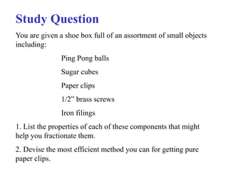
  • 1. Study Question You are given a shoe box full of an assortment of small objects including: Ping Pong balls Sugar cubes Paper clips 1/2” brass screws Iron filings 1. List the properties of each of these components that might help you fractionate them. 2. Devise the most efficient method you can for getting pure paper clips.
  • 2. Techniques of Protein Purification
  • 3. Protein isolation Selection of protein source 1. Tissues from animals 2. Microorganisms (E. coli or yeast) 3. Molecular cloning techniques Methods of solubilization 1. Osmosis lysis (with hypotonic solution) 2. Use of lysozyme (enzyme that degrades cell wall) 3. French press or sonication
  • 4. Stabilization of proteins 1. pH (think buffers!) 2. Temperature (close to 0oC) Thermal stability could be used for purification 3. Addition of protease inhibitors 4. Gentle handling (no frothing) Assay of proteins 1. If purifying an enzyme, use the reaction it catalyzes as an assay 2. If metalloprotein use the metals to follow the protein 3. Immunochemical techniques (antibodies)
  • 5. General strategy of protein purification Proteins are purified by fractionation procedures, a series of independent steps in which the properties of protein of interest are utilized to separate it from other contaminating proteins. How do we know our sample of protein is pure? We don't! The best we can do is to demonstrate by all available methods that our sample consists of only one component.
  • 6. General strategy of protein purification Characteristic Procedure Solubility 1. Salting in 2. Salting out Ionic charge: 1. Ion exchange chromatography 2. Electrophoresis 3. Isoelectric focusing Polarity: 1. Adsorption chromatography 2. Paper chromatography 3. Hydrophobic interaction chromatography Molecular size: 1. Dialysis and ultrafiltration 2. Gel electrophoresis 3. Gel filtration chromatography 4. Ultracentrifugation Binding specificity: 1. Affinity chromatography
  • 7. Solubility of a protein in aqueous solution Depends strongly on: 1. Concentrations of dissolved salts 2. pH 3. Temperature 4. Addition of water-miscible organic solvents, e.g., ethanol or acetone
  • 8. Solubility of carboxyhemoglobin at its isoelectric point as a function of ionic strength and ion type Page 131
  • 9.
  • 10. Solubility of b-lactoglobin as a function of pH at several NaCl concentrations Page 132
  • 11. Salting Out • There are hydrophobic amino acids and hydrophilic amino acids in protein molecules. After protein folding in aqueous solution, hydrophobic amino acids usually form protected hydrophobic areas while hydrophilic amino acids interact with the molecules of solvation and allow proteins to form hydrogen bonds with the surrounding water molecules. If enough of the protein surface is hydrophilic, the protein can be dissolved in water. • When the salt concentration is increased, some of the water molecules are attracted by the salt ions, which decreases the number of water molecules available to interact with the charged part of the protein. As a result of the increased demand for solvent molecules, the protein-protein interactions are stronger than the solvent-solute interactions; the protein molecules coagulate by forming hydrophobic interactions with each other. This process is known as salting out.
  • 12. Salting Out • After Proteins solubilized, they can be purified based on solubility (usually dependent on overall charge, ionic strength, polarity • Ammonium sulfate (NH4SO4) commonly used to “salt out” • Takes away water by interacting with it, makes protein less soluble because hydrophobic interactions among proteins increases • Different aliquots taken as function of salt concentration to get closer to desired protein sample of interest (30, 40, 50, 75% increments) • One fraction has protein of interest
  • 13. CENTRIFUGATION • A particle is subjected to a centrifugal force when it is rotated at a high rate of speed. The centrifugal force, F, is defined by Equation F = mω2r F = intensity of the centrifugal force m = effective mass of the sedimenting particle ω = angular velocity of rotation in rad/sec r = distance of the migrating particles from the central axis of rotation • A more common measurement of F, in terms of the earth’s gravitational force, g, is relative centrifugal force, RCF, defined by Equation RCF = (1.119 * 10-5)(rpm)2(r)
  • 14. Although the relative centrifugal force can easily be calculated, centrifugation manuals usually contain a nomograph for the convenient conversion between relative centrifugal force and speed of the centrifuge at different radii of the centrifugation spindle to a point along the centrifuge tube. A nomograph consists of three columns representing the radial distance (in mm), the relative centrifugal field and the rotor speed (in r.p.m.). For the conversion between relative centrifugal force and speed of the centrifuge spindle in r.p.m. at different radii, a straight-edge is aligned through known values in two columns, then the desired figure is read where the straight-edge intersects the third column.
  • 15. Fig. 3.1 Nomograph for the determination of the relative centrifugal field for a given rotor speed and radius. The three columns represent the radial distance (in mm), the relative centrifugal field and the rotor speed (in r.p.m.). For the conversion between relative centrifugal force and speed of the centrifuge spindle in revolutions per minute at different radii, draw a straight-edge through known values in two columns. The desired figure can then be read where the straight-edge intersects the third column. (Courtesy of Beckman- Coulter.)
  • 16. The most obvious differences between centrifuges are: •• the maximum speed at which biological specimens are subjected to increased sedimentation; •• the presence or absence of a vacuum; •• the potential for refrigeration or general manipulation of the temperature during a centrifugation run; and •• the maximum volume of samples and capacity for individual centrifugation tubes. Many different types of centrifuges are commercially available including: • large-capacity low-speed preparative centrifuges; • refrigerated high-speed preparative centrifuges; • analytical ultracentrifuges; • preparative ultracentrifuges; • large-scale clinical centrifuges; and • small-scale laboratory microfuges.
  • 17. fixed-angle rotor vertical tube rotor swinging-bucket rotor initial acceleration stage, the main centrifugal separation phase, de-acceleration and the final harvesting of separated particles in the rotor at rest.
  • 18. Differential Centrifugation • Sample is spun, after lysis, to separate unbroken cells, nuclei, other organelles and particles not soluble in buffer used • Different speeds of spin allow for particle separation
  • 19. Density-Gradient Centrifugation if the sample is centrifuged in a fluid medium that gradually increases in density from top to bottom. This technique, called density gradient centrifugation, permits the separation of multi-component mixtures of macromolecules and the measurement of sedimentation coefficients. • zonal centrifugation, in which the sample is centrifuged in a preformed gradient, A density gradient is prepared in a tube prior to centrifugation with the use of an automatic gradient mixer or manually with pipette. Both step gradient and continuous gradient systems. • Sucrose concentrations up to 60% can be used, with a density limit of 1.28g/cm3. • The various types of particles sediment as zones and remain separated from the other components. The various zones are then isolated by collecting fractions from the bottom of the tube.
  • 20. isopycnic centrifugation, in which a self-generating gradient forms during centrifugation. The sample under study is dissolved in a solution of a dense salt such as cesium chloride or cesium sulfate. The cesium salts may be used to establish gradients to an upper density limit of The solution of biological sample and cesium salt is uniformly distributed in a centrifuge tube and rotated in an ultracentrifuge. Under the influence of the centrifugal force, the cesium salt redistributes to form a continuously increasing density gradient from the top to the bottom. The macromolecules of the biological sample seek an area in the tube where the density is equal to their respective densities. That is, the macromolecules move to a region where the sum of the forces (centrifugal and frictional) is zero.
  • 21. FIGURE 4.12 A comparison of differential and density gradient measurements. A Differential centrifugation in a medium of unchanging density. B Zonal centrifugation in a prepared density gradient. C Isopycnic centrifugation; the density gradient forms during centrifugation.
  • 22. Isoelectric Points of Several Common Proteins Protein pI Pepsin 1.0 Ovalbumin (hen) 4.6 Serum albumin (human) 4.9 Tropomyosin 5.1 Insulin (bovine) 5.4 Fibrinogen (human) 5.8 g-Globulin (human) 6.6 Collagen 6.6 Myoglobin (horse) 7.0 Hemoglobin (human) 7.1 Ribonuclease A (bovine) 9.4 Cytochrome c (horse) 10.6 Histone (bovine) 10.8 Lysozyme (hen) 11.0 Salmine (salmon) 12.1
  • 23. CHROMATOGRAPHY The molecules targeted for analysis are called analytes. The mobile phase, which may be a liquid or gas, moves the sample components through a region containing the solid or liquid stationary phase, which is called the sorbent. The molecular components in the sample distribute themselves between the mobile phase and sorbent and thus have the opportunity to interact intimately with the stationary phase. If some of the sample molecules (analytes) are preferentially bound by the sorbent, they spend more time in the sorbent and are retarded in their movement through the chromatographic system. Molecules that show weak affinity for the sorbent spend more time with the mobile phase and are more easily removed or eluted from the system. The mobile phase can be collected as a function of time at the end of the chromatographic system. The mobile phase, now called the effluent, contains the purified analytes.
  • 24. When the actual adsorbing material is made into a column, it is said to be poured or packed. Application of the sample to the top of the column is loading the column. Movement of solvent through the loaded column is called developing or eluting the column. The bed volume is the total volume of solvent and adsorbing material taken up by the column. The volume taken up by the liquid phase in the column is the void volume. The elution volume is the amount of solvent required to remove a particular analyte from the column. This is analogous to values in planar chromatography.
  • 25. All components may be eluted by a single solvent or buffer. This is referred to as continual elution. In contrast, stepwise elution refers to an incremental change of solvent to aid development. The column is first eluted with a volume of one solvent and then with a second solvent. This may continue with as many solvents or solvent mixtures as desired. In general, the first solvent should be the least polar of any used in the analysis, and each additional solvent should be of greater polarity or ionic strength. Finally, adsorption columns may be developed by gradient elution brought about by a gradual change in solvent composition. The composition of the eluting solvent can be changed by the continuous mixing of two different solvents to gradually change the ratio of the two solvents. Alternatively, the concentration of a component in the solvent can be gradually increased. This is most often done by addition of a salt (KCl, NaCl, etc.). Devices are commercially available to prepare predetermined, reproducible gradients.
  • 26. Protein separation and purification by column chromatography From Lehninger Principles of Biochemistry Chromatographic separations
  • 27. Column Chromatography: Size-exclusion Gel filtration chromatography can be used to estimate molecular masses From Lehninger Principles of Biochemistry
  • 28. Theory of Gel Filtration The stationary phase consists of inert particles that contain small pores of a controlled size. Microscopic examination of a particle reveals an interior resembling a sponge. A solution containing analytes of various molecular sizes is allowed to pass through the column under the influence of continuous solvent flow. Analytes larger than the pores cannot enter the interior of the gel beads, so they are limited to the space between the beads. The volume of the column accessible to very large molecules is, therefore, greatly reduced. As a result, they are not slowed in their progress through the column and elute rapidly in a single zone. Small molecules capable of diffusing in and out of the beads have a much larger volume available to them. Therefore, they are delayed in their journey through the column bed. Molecules of intermediate size migrate through the column at a rate somewhere between those for large and small molecules.
  • 29. 1.Exclusion Limit This is defined as the molecular mass of the smallest molecule that cannot diffuse into the inner volume of the gel matrix. All molecules above this limit elute rapidly in a single zone. The exclusion limit of a typical gel, Sephadex G-50, is 30,000 daltons. All analytes having a molecular size greater than this value would pass directly through the column bed without entering the gel pores. 2. Fractionation Range Sephadex G-50 has a fractionation range of 1500 to 30,000 daltons. Analytes within this range would be separated in a somewhat linear fashion. 3. Water Regain and Bed Volume Gel chromatography media are often supplied in dehydrated form and must be swollen in a solvent, usually water, before use. The weight of water taken up by 1 g of dry gel is known as the water regain. For G-50, this value is g. This value does not include the water surrounding the gel particles, so it cannot be used as an estimate of the final volume of a packed gel column. Most commercial suppliers of gel materials provide, in addition to water regain, a bed volume value. This is the final volume taken up by 1 g of dry gel when swollen in water. For G-50, bed volume is 9 to 11 mL/g dry gel.
  • 30. 4. Gel Particle Shape and Size Ideally, gel particles should be spherical to provide a uniform bed with a high density of pores. Particle size is defined either by mesh size or bead diameter Both the degree of resolution afforded by a column and the flow rate depend on particle size. Larger particle sizes (50 to 100 mesh, 100 to 300 ) offer high flow rates but poor chromatographic separation. The opposite is true for very small particle sizes (“superfine,” 400 mesh, 10 to ). The most useful particle size, which represents a compromise between resolution and flow rate, is 100 to 200 mesh (50 to 150 ). 5. Void Volume This is the total space surrounding the gel particles in a packed column. This value is determined by measuring the volume of solvent required to elute a solute that is completely excluded from the gel matrix. Most columns can be calibrated for void volume with a dye, blue dextran, which has an average molecular mass of 2,000,000 daltons. 6. Elution Volume This is the volume of eluting buffer necessary to remove a particular analyte from a packed column.
  • 31.
  • 32.
  • 33.
  • 34. Biochemists refer to a protein's size in terms of its molecular weight, in kDa (a kilodalton, kD or kDa, is 1000 times the molecular mass of hydrogen) Each amino acid residue counts for about 110 daltons, that is, about 0.11 kDa. Sephadex G-50 1-30 kD Sephadex G-100 4-150 kD Sephadex G-200 5-600 kD Bio-Gel P-10 1.5-20 kD Bio-Gel P-30 2.4-40 kD Bio-Gel P-100 5-100 kD Bio-Gel P-300 60-400 kD Sephadex is a trademark of Pharmacia. Bio-Gel is a trademark of Bio-Rad.
  • 35. Selecting a Gel If the mixture contains macromolecules up to 120,000 in molecular weight, then Bio-Gel P-150, Sephacryl S-200HR, or Sephadex G-150 would be most appropriate. If P-100, G-100, or Sephacryl S-100HR were used, some of the highermolecular- weight proteins in the sample would elute in the void volume. On the other hand, if P-200, P-300, or G-200 were used, there would be a decrease in both resolution and flow rate. Gel Preparation and Storage The dextran and acrylamide gel products are sometimes supplied in dehydrated form and must be allowed to swell in water before use. The swelling time required differs for each gel, but the extremes are 3 to 4 hours at for highly cross-linked gels and up to 72 hours at for P-300 or G- 200. The swelling time can be shortened if a boiling-water bath is used. Agarose gels and combined polyacrylamideagarose gels are supplied in a hydrated state, so there is no need for swelling. Before a gel slurry is packed into the column, it should be defined and deaerated. Defining is necessary to remove very fine particles, which would reduce flow rates. Deaerating (removing dissolved gases) should be done on the gel slurry and all eluting buffers. Antimicrobial agents must be added to stored, hydrated gels. One of the best agents is sodium azide (0.02%).
  • 36. Column Size For fractionation purposes, it is usually not necessary to use columns greater than 100 cm in length. The ratio of bed length to width should be between 25 and 100. For group separations, columns less than 50 cm long are sufficient, and appropriate ratios of bed length to width are between 5 and 10. Eluting Buffer There are fewer restrictions on buffer choice in gel chromatography than in ion-exchange chromatography. Dextran and polyacrylamide gels are stable in the pH range 1 to 10, whereas agarose gels are limited to pH 4 to 10. Since there is such a wide range of stability of the gels, the buffer pH should be chosen on the basis of the range of stability of the macromolecules to be separated. Sample Volume If too much sample is applied to a column, resolution is decreased; if the sample size is too small, the analytes are greatly diluted. For group separations, a sample volume of 10 to 25% of the column total volume is suitable. The sample volume for fractionation procedures should be between 1 and 5% of the total volume. Column total volume is determined by measuring the volume of water in the glass column that is equivalent to the height of the packed bed. Column Flow Rate The flow rate of a gel column depends on many factors, including length of column and type and size of the gel. It is generally safe to elute a gel column at a rate slightly less than free flow. A high flow rate reduces sample diffusion or zone broadening, but may not allow complete equilibration of analyte molecules with the gel matrix. A specific flow rate cannot be recommended, since each type of gel requires a different range. The average flow rate given in literature references for small-pore-size gels is 8 to 12 mL/cm2 of cross-sectional bed area per hour (15 to 25 mL/hr). For large-pore-size gels, a value of 2 to 5mL/cm2 of cross-sectional bed area per hour (5 to 10 mL/hr) is average.
  • 37. Desalting Inorganic salts, organic solvents, and other small molecules are used extensively for the purification of macromolecules. Gel chromatography provides an inexpensive, simple, and rapid method for removal of these small molecules. One especially attractive method for desalting very small samples (0.1 mL or less) of proteins or nucleic acid solutions is to use spin columns. These are prepacked columns of polyacrylamide exclusion gels. Spin columns are used in a similar fashion to microfiltration centrifuge tubes (Chapter 3, p. 75). The sample is placed on top of the gel column and spun in a centrifuge. Large molecules are eluted from the column and collected in a reservoir. The small molecules to be removed remain in the gel. Purification of Biomolecules This is probably the most popular use of gel chromatography. Because of a gel’s ability to fractionate molecules on the basis of size, gel filtration complements other purification techniques that separate molecules on the basis of polarity and charge. Estimation of MolecularWeight The elution volume for a particular analyte is proportional to its molecular size. This indicates that it is possible to estimate the molecular weight of a molecule on the basis of its elution characteristics on a gel column. An elution curve for several standard proteins separated on Sephadex G-100 is shown in Figure 5.8. This curve, a plot of protein concentration vs. volume collected, is representative of data obtained from a gel filtration experiment. The elution volume, for each protein can be estimated as shown in the figure. A plot of log molecular mass vs. elution volume for the proteins.
  • 38. A schematic illustration of gel filtration chromatography
  • 39. FIGURE 5.8 Elution curve for a mixture of several proteins using gel-filtration chromatography. A hemoglobin; B egg albumin; C chymotrypsinogen; D myoglobin; E = cytochrome c.
  • 40. FIGURE 5.9 A plot of log molecular mass vs. elution volume for the proteins A, B, C, D, and E in Figure 5.8.
  • 42. Column Chromatography: Ion Exchange From Lehninger Principles of Biochemistry
  • 43. This form of chromatography relies on the attraction between oppositely charged stationary phase, known as an ion exchanger, and analyte. It is frequently chosen for the separation and purification of proteins, peptides, nucleic acids, polynucleotides and other charged molecules, mainly because of its high resolving power and high capacity. There are two types of ion exchanger, namely cation and anion exchangers. Cation exchangers possess negatively charged groups and these will attract positively charged cations. These exchangers are also called acidic ion exchangers because their negative charges result from the ionisation of acidic groups. Anion exchangers have positively charged groups that will attract negatively charged anions. The term basic ion exchangers is also used to describe these exchangers, as positive chargesgenerally result from the association of protons with basic groups. Matrices used include polystyrene, cellulose and agarose. Functional ionic groups include sulphonate (–SO–3) and quaternary ammonium (–N+R3), both of which are strong exchangers because they are totally ionised at all normal working pH values,and carboxylate (–COO-) and diethylammonium (–HN+(CH2CH3)2), both of which are termed weak exchangers because they are ionised over only a narrow range of pH values.
  • 44.
  • 45.
  • 46. FIGURE 5.6 The effect of pH on the net charge of a protein.
  • 47. Eluent pH The pH of the buffer selected as eluent should be at least one pH unit above or below the isoionic point of the analytes. In general, cationic buffers such as Tris, pyridine and alkylamines are used in conjunction with anion exchangers, and anionic buffers such as acetate, barbiturate and phosphate are used with cation exchangers. Elution Gradient elution is far more common than isocratic elution. Continuous or stepwise pH and ionic strength gradients may be employed but continuous gradients tend to give better resolution with less peak tailing. Generally with an anion exchanger, the pH gradient decreases and the ionic strength increases, whereas for cation exchangers both the pH and ionic gradients increase during the elution.
  • 48. A schematic diagram illustrating the separation of several proteins by ion exchange chromatography using stepwise elution
  • 49. A gradient is a change in the proportion of the two (or more) solvents that make up the mobile phase. With a pump controlling flow rates and proportioning valves on all the solvent reservoirs, it is possible to gradually or abruptly change the mobile phase composition. A Step Gradient is an abrupt change in mobile phase composition. An example of a step gradient is shown here (blue line), where the salt concentration in the mobile phase is changed from 0.1 M to 0.2 M at 10 min, then from 0.2 M to 0.3 M at 20 min.