SlideShare a Scribd company logo
1 of 59
Respiratory System: Functional Anatomy
• Major function — respiration
– O2 in tissues, CO2 out of tissues
• Respiratory and circulatory systems cooperate
• Major organs
– Nose, nasal cavity, and paranasal sinuses
– Pharynx
– Larynx
– Trachea
– Bronchi and their branches
– Lungs and alveoli
• Respiratory zone — gas exchange
– Respiratory bronchioles, alveolar ducts, and alveoli
• Conducting zone — gas transport
– Everything else; cleanses, warms, humidifies air
• Diaphragm and intercostal muscles promote ventilation
• Pulmonary ventilation — air into and out of lungs
• External respiration — gasexchange between lungs and
blood
• Transport — gases in blood
• Internal respiration — gasexchange between blood and
tissues
The Nose – Outside
• Provides an airway for respiration
• Moistens and warms the air
• Filters and cleans the air
• Resonating chamber for speech
• Olfaction
• Two regions — external nose and nasal cavity
• External nose — root, bridge, dorsum nasi, and apex
– Philtrum — shallow vertical groove inferior to apex
– Nostrils (nares) — bounded laterally by alae
The Nose – Inside
• Nasal cavity — within and posterior to external
nose
– Divided by midline nasal septum
– Posterior nasal apertures (choanae) open
into nasal pharynx
– Roof — ethmoid and sphenoid bones
– Floor — hard (bone) and soft palates
(muscle)
• Nasal vestibule — nasal cavity immediately
superior to nostrils
– Vibrissae (hairs) filter particles
• Nasal cavity lined with mucous membranes
– Olfactory mucosa
• Olfactory epithelium
• Respiratory mucosa
– Pseudostratified ciliated columnar epithelium
– Mucous and serous secretions contain lysozyme and defensins
– Cilia move contaminated mucus posteriorly to throat
– Inspired air warmed by plexuses of capillaries and veins
– Sensory nerve endings trigger sneezing
Nasal Cavity
• Nasal conchae-superior, middle, and inferior
– Protrude medially from lateral walls
– Increase mucosal area
– Enhance air turbulence
• Nasal meatus
– Groove inferior to each concha
• During inhalation, conchae and nasal mucosa
– Filter, heat, and moisten air
• During exhalation these structures
– Reclaim heat and moisture
• Paranasal sinuses
– In frontal, sphenoid, ethmoid, and maxillary
bones
– Lighten skull; secrete mucus; help to warm
and moisten air
• Rhinitis
– Inflammation of nasal mucosa
– Nasal mucosa continuous with mucosa of respiratory tract 
spreads from nose  throat  chest
– Spreads to tear ducts and paranasal sinuses causing
• Blocked sinus passageways  air absorbed  vacuum 
sinus headache
Pharynx
• Muscular tube from base of skull to C6
– Connects nasal cavity and mouth to larynx and esophagus
– Composed of skeletal muscle
• Three regions
• Nasopharynx
– Air passageway posterior to nasal cavity
– Lining - pseudostratified columnar epithelium
– Soft palate and uvula close nasopharynx during swallowing
– Pharyngeal tonsil (adenoids) on posterior wall
– Pharyngotympanic (auditory) tubes drain and equalize pressure in middle ear; open into lateral
walls
• Oropharynx
– Passageway for food and air from level of soft palate to epiglottis
– Lining of stratified squamous epithelium
– Isthmus of fauces-opening to oral cavity
– Palatine tonsils-in lateral walls of fauces
– Lingual tonsil-on posterior surface of tongue
• Laryngopharynx
– Passageway for food and air
– Extends to larynx, where continuous with esophagus
– Lined with stratified squamous epithelium
Larynx
• Attaches to hyoid bone; opens into
laryngopharynx; continuous with trachea
• Functions
– Provides patent airway
– Routes air and food into proper channels
– Voice production
• Houses vocal folds
• Superior portion–stratified squamous epithelium
• Inferior to vocal folds–pseudostratified ciliated
columnar epithelium
• Nine cartilages of larynx
– All hyaline cartilage except epiglottis
– Thyroid cartilage with laryngeal
prominence (Adam's apple)
– Ring-shaped cricoid cartilage
– Paired arytenoid, cuneiform, and
corniculate cartilages
– Epiglottis-elastic cartilage; covers laryngeal
inlet during swallowing; covered in taste
bud-containing mucosa
Vocal folds
• Vocal ligaments-deep to laryngeal mucosa
– Attach arytenoid cartilages to thyroid
cartilage
– Contain elastic fibers
– Form core of vocal folds (true vocal cords)
• Glottis-opening between vocal folds
• Folds vibrate to produce sound as air
rushes up from lungs
• Vestibular folds (false vocal cords)
– Superior to vocal folds
– No part in sound production
– Help to close glottis during swallowing
• Speech-intermittent release of expired air while
opening and closing glottis
• Pitch — length and tension of vocal cords
• Loudness — force of air
• Pharynx, oral, nasal, and sinus cavities amplify
and enhance sound
• Language — muscles of pharynx, tongue, soft
palate, and lips
• Vocal folds may act as sphincter to prevent air
passage
• Example-Valsalva's maneuver
– Glottis closes to prevent exhalation
– Abdominal muscles contract
– Intra-abdominal pressure rises
– Emptying rectum or stabilizes trunk
Trachea
• Windpipe–from larynx into mediastinum
• Wall composed of three layers
– Mucosa-ciliated pseudostratified epithelium with
goblet cells
– Submucosa-connective tissue with seromucous glands
– Adventitia-outermost layer made of connective tissue;
encases C-shaped rings of hyaline cartilage
• Trachealis muscle
– Connects posterior parts of cartilage rings
– Contracts during coughing to expel mucus
• Carina
– Spar of cartilage on last, expanded tracheal cartilage
– Point where trachea branches into two main bronchi
Bronchi and Subdivisions
• Air passages undergo 23 orders of branching  bronchial (respiratory) tree
• From tips of bronchial tree  conducting zone structures  respiratory zone structures
• Trachea → right and left main (primary) bronchi
• Each main bronchus enters hilum of one lung
– Right main bronchus wider, shorter, more vertical than left
• Each main bronchus branches into lobar (secondary) bronchi (three on right, two on left)
– Each lobar bronchus supplies one lobe
• Each lobar bronchus branches into segmental (tertiary) bronchi
– Segmental bronchi divide repeatedly
• Branches become smaller and smaller 
– Bronchioles-less than 1 mm in diameter
– Terminal bronchioles-smallest-less than
0.5 mm diameter
• From bronchi through bronchioles, structural changes occur
– Cartilage rings become irregular plates; in bronchioles elastic fibers replace cartilage
– Epithelium changes from pseudostratified columnar to cuboidal; cilia and goblet cells become sparse
– Relative amount of smooth muscle increases
• Allows constriction
Respiratory zone
• Begins as terminal bronchioles  respiratory bronchioles  alveolar ducts  alveolar sacs
– Alveolar sacs contain clusters of alveoli
• ~300 million alveoli make up most of lung volume
• Sites of gas exchange
Conducting and Respiratory Zones
Respiratory Membrane. Alveoli
• Alveolar and capillary walls and their fused basement
membranes
– ~0.5-µ m-thick; gas exchange across membrane by
simple diffusion
• Alveolar walls
– Single layer of squamous epithelium (type I alveolar
cells)
• Scattered cuboidal type II alveolar cells secrete surfactant
and antimicrobial proteins
• Surrounded by fine elastic fibers and pulmonary capillaries
• Alveolar pores connect adjacent alveoli
• Equalize air pressure throughout lung
• Alveolar macrophages keep alveolar surfaces sterile
– 2 million dead macrophages/hour carried by cilia 
throat  swallowed
Alveolar Anatomy
Lungs
• Occupy all thoracic cavity except mediastinum
• Root — site of vascular and bronchial
attachment to mediastinum
• Costal surface — anterior, lateral, and posterior
surfaces
• Composed primarily of alveoli
• Stroma — elastic connective tissue  elasticity
• Apex-superior tip; deep to clavicle
• Base-inferior surface; rests on diaphragm
• Hilum-on mediastinal surface; site for entry/exit of blood
vessels, bronchi, lymphatic vessels, and nerves
• Left lung smaller than right
– Cardiac notch-concavity for heart
– Separated into superior and inferior lobes by oblique
fissure
Lungs
• Right lung
– Superior, middle, inferior lobes separated by
oblique and horizontal fissures
• Bronchopulmonary segments (10 right, 8–10
left) separated by connective tissue septa
– If diseased can be individually removed
• Lobules-smallest subdivisions visible to naked
eye; served by bronchioles and their branches
Pleurae
• Thin, double-layered serosa
• Parietal pleura – walls of pleural
cavity
• Visceral pleura – external lung
surface
• Pleural fluid fills pleural cavity
– Lubrication and surface tension
 lungs’ expansion and recoil
• Pulmonary circulation (low pressure, high volume)
– Pulmonary arteries — systemic venous blood to lungs
• Branch and feed into pulmonary capillaries
– Pulmonary veins — oxygenated blood to the heart
• Bronchial arteries — blood to lung tissue
– From aorta; enter lungs at hilum
– Part of systemic circulation
– Supply all lung tissue except alveoli
– Bronchial veins anastomose with pulmonary veins
Mechanics of Breathing. Pressures
• Pulmonary ventilation
– Inspiration-gases flow into lungs
– Expiration-gases exit lungs
• Atmospheric pressure (Patm)
– Pressure exerted by air surrounding body
– 760 mm Hg at sea level = 1 atmosphere
• Respiratory pressures
– Negative respiratory pressure < Patm
– Positive respiratory pressure > Patm
– Zero respiratory pressure = Patm
• Intrapulmonary (intra-alveolar) pressure (Ppul)
– Pressure in alveoli
– Fluctuates with breathing
– Always eventually equalizes with Patm
• Intrapleural pressure (Pip)
– Pressure in pleural cavity
– Fluctuates with breathing
– Always a negative pressure (<Patm and <Ppul)
– Fluid level must be minimal
• If accumulates  positive Pip pressure
• Negative Pip caused by
– Inward forces → lung collapse
• Elastic recoil of lungs ↓ lung size
• Surface tension of alveolar fluid ↓
alveolar size
– Outward force → enlarge lungs
• Elastic chest wall ↑ thorax outward
Pressure Relationships
• If Pip = Ppul or Patm  lungs collapse
• (Ppul – Pip) = transpulmonary pressure
– Keeps airways open
– Greater transpulmonary pressure  larger
lungs
• Atelectasis (lung collapse) due to
– Plugged bronchioles → collapse of alveoli
– Pneumothorax – air in pleural cavity
• Rupture in parietal or visceral pleura
• Air removed with chest tubes; pleurae
heal  lung reinflates
Pulmonary Ventilation. Gas Laws
• Inspiration and expiration
• Depends on volume changes in thoracic cavity
– Volume changes → pressure changes
– Pressure changes → gases flow to equalize pressure
Boyle’s Law
•Relationship between pressure and volume of a gas
– Gases fill container; if container size reduced 
increased pressure
•Pressure (P) varies inversely with volume (V):
– P1V1 = P2V2
Inspiration • Active process
– Inspiratory muscles (diaphragm and external intercostals) contract
– Thoracic volume increases  intrapulmonary pressure drops (to −1 mm Hg)
– Lungs stretched and intrapulmonary volume increases
– Air flows into lungs, down its pressure gradient, until Ppul = Patm
• Forced inspiration
• Vigorous exercise, COPD  accessory muscles (scalenes, sternocleidomastoid,
pectoralis minor)  further increase in thoracic cage size
Expiration• Quiet expiration normally passive process
– Inspiratory muscles relax
– Thoracic cavity volume decreases
– Elastic lungs recoil and intrapulmonary volume decreases  pressure increases (Ppul rises to +1 mm
Hg) 
– Air flows out of lungs down its pressure gradient until Ppul = 0
• Note: forced expiration-active process; uses abdominal (oblique and transverse) and internal intercostal
muscles
Physical Factors Influencing Pulmonary Ventilation
• Three factors hinder air passage and pulmonary ventilation;
require energy to overcome
• Airway resistance
– Friction in airways – major nonelastic source of
resistance
– F=ΔP/R
• ∆P - pressure gradient between atmosphere and
alveoli
– Resistance usually insignificant
• Large airway diameters
• Progressive branching of airways ↑ total cross-
sectional area
• Resistance greatest in medium-sized bronchi
– Resistance disappears at terminal bronchioles where
diffusion drives gas movement
• As airway resistance rises, breathing movements become more strenuous
• Severe constriction or obstruction of bronchioles
– Can prevent life-sustaining ventilation
– Can occur during acute asthma attacks; stops ventilation
• Epinephrine dilates bronchioles, reduces air resistance
• Surfactant
– Detergent-like lipid and protein complex produced by type II alveolar cells
– ↓ surface tension of alveolar fluid → no alveolar collapse
– Insufficient quantity in premature infants causes infant respiratory distress syndrome
  alveoli collapse after each breath
Alveolar Surface Tension
• Surface tension
– Attracts liquid molecules to one
another at gas-liquid interface
– Resists any increase of the surface
area of liquid
– Water – high surface tension; coats
alveolar walls  reduces them to
smallest size
Lung Compliance
• Measure of change in lung volume
that occurs with given change in
transpulmonary pressure
• Higher lung compliance  easier to
expand lungs
• Normally high due to
– Distensibility of lung tissue
– Alveolar surface tension
• Diminished by
– Nonelastic scar tissue replacing lung tissue (fibrosis)
– Reduced production of surfactant
– Decreased flexibility of thoracic cage
• Homeostatic imbalances that reduce compliance
– Deformities of thorax
– Ossification of costal cartilage
– Paralysis of intercostal muscles
Respiratory Volumes and Capacities
• Used to assess respiratory status
– Tidal volume (TV)
– Inspiratory reserve volume (IRV)
– Expiratory reserve volume (ERV)
– Residual volume (RV)
• Combinations of respiratory volumes
– Inspiratory capacity (IC)
– Functional residual capacity (FRC)
– Vital capacity (VC)
– Total lung capacity (TLC)
Dead Space. Pulmonary Function Tests
• Anatomical dead space
– No contribution to gas exchange
– Air remaining in passageways; ~150 ml
• Alveolar dead space–non-functional alveoli due
to collapse or obstruction
• Total dead space-sum of anatomical and
alveolar dead space
• Spirometer measures respiratory volumes and
capacities
• Spirometry can distinguish between
– Obstructive pulmonary disease—increased
airway resistance (e.g., bronchitis)
• TLC, FRC, RV ↑
– Restrictive disorders — VC, TLC, FRC, RV ↓
(disease or fibrosis)
• To measure rate of gas movement
– Forced vital capacity (FVC) — gas forcibly
expelled after taking deep breath
– Forced expiratory volume (FEV) — amount
of gas expelled during specific time
intervals of FVC
Alveolar Ventilation
• Minute ventilation-total amount of gas flow into or out of respiratory tract in one minute
– Normal at rest = ~ 6 L/min
– Normal with exercise = up to 200 L/min
– Only rough estimate of respiratory efficiency
• Good indicator of effective ventilation
• Alveolar ventilation rate (AVR)-flow of gases into and out of alveoli during a particular time
• Dead space normally constant
• Rapid, shallow breathing decreases AVR
AVR = frequency X (TV – dead space)
(ml/min) (breaths/min) (ml/breath)
Nonrespiratory Air Movements
• May modify normal respiratory rhythm
• Most result from reflex action; some voluntary
• Examples include-cough, sneeze, crying, laughing, hiccups, and yawns
Gas Exchanges – Blood, Lungs, and Tissues
• External respiration – diffusion of gases in lungs
• Internal respiration – diffusion of gases at body tissues
• Both involve
– Physical properties of gases
– Composition of alveolar gas
Dalton’s Law
• For mixture of gases: Ptotal = Σ Pindividual
• Partial pressure
– Pi = k C● i
Henry’s Law
• Gas mixtures in contact with liquid
– Cwater = ks P● i (each gas dissolves in proportion to its
partial pressure)
– At equilibrium, pressure in both phases equal:
• Pwater= Pair
– Amount of each gas that will dissolve depends on
• Solubility – CO2 20 times more soluble in water
than O2; little N2 dissolves in water
• Temperature – T ↑, solubility ↓
Composition of Alveolar Gas
• Alveoli contain more CO2 and water vapor than atmospheric air
– Gas exchanges in lungs
– Humidification of air
– Mixing of alveolar gas with each breath
External Respiration
• Exchange of O2 and CO2 across respiratory membrane Influenced by
– Thickness and surface area of respiratory membrane
• 0.5 to 1 µ m thick
• Large total surface area (40 times that of skin) for gas exchange
• Thicken if lungs become waterlogged and edematous  gas exchange inadequate
• Reduced surface area in emphysema (walls of adjacent alveoli break down), tumors,
inflammation, mucus
External Respiration
• Exchange of O2 and CO2 across respiratory membrane
Influenced by
– Partial pressure gradients and gas solubilities
• O2 gradient in lungs
– Venous blood Po2 = 40 mm Hg
– Alveolar Po2 = 104 mm Hg
– Oxygen to blood
• CO2 gradient in lungs
– Venous blood Pco2 = 45 mm Hg
– Alveolar Pco2 = 40 mm Hg
– CO2 to lungs
• Though gradient not as steep, CO2 diffuses in equal
amounts with oxygen
– CO2 20 times more soluble in plasma than
oxygen
Ventilation-Perfusion Coupling
• Exchange of O2 and CO2 across respiratory membrane Influenced by
– Ventilation-perfusion coupling
• Perfusion-blood flow reaching alveoli
• Ventilation-amount of gas reaching alveoli
• Ventilation and perfusion matched (coupled) for efficient gas exchange
– Never balanced for all alveoli due to
• Regional variations due to effect of gravity on blood and air flow
• Some alveolar ducts plugged with mucus
Ventilation-Perfusion Coupling
• Perfusion
– Changes in Po2 in alveoli cause changes in
diameters of arterioles
• Where alveolar O2 is high, arterioles
dilate
• Where alveolar O2 is low, arterioles
constrict
• Directs most blood where alveolar
oxygen high
• Changes in Pco2 in alveoli cause changes in
diameters of bronchioles
– Where alveolar CO2 is high, bronchioles
dilate
– Where alveolar CO2 is low, bronchioles
constrict
– Allows elimination of CO2 more rapidly
Internal Respiration
• Capillary gas exchange in body tissues
• Partial pressures and diffusion gradients reversed
compared to external respiration
– Tissue Po2 always lower than in systemic arterial blood
 oxygen from blood to tissues
– CO2  from tissues to blood
– Venous blood Po2 40 mm Hg and Pco2
45 mm Hg
O2 Transport
• Molecular O2 carried in blood
– 1.5% dissolved in plasma
– 98.5% loosely bound to each Fe of hemoglobin (Hb) in
RBCs
• 4 O2 per Hb
• Oxyhemoglobin (HbO2)
• Deoxyhemoglobin (HHb – no O2)
• Loading and unloading of O2 → change in shape of Hb
– As O2 binds, Hb affinity for O2 increases and vice versa
– Cooperative binding
• All four heme groups carry O2 – full saturation
• Rate of loading and unloading of O2 regulated to ensure
adequate oxygen delivery to cells
– Po2
– Temperature
– Blood pH
– Pco2
– Concentration of BPG–produced by RBCs during
glycolysis; levels rise when oxygen levels chronically
low
Hemoglobin Dissociation Curve
• Oxygen-hemoglobin dissociation curve
• Hemoglobin saturation plotted against Po2 is S-
shaped curve
• Binding and release of O2 influenced by Po2
• In arterial blood
– Po2 = 100 mm Hg
– Contains 20 ml oxygen per 100 ml blood (20 vol %)
– Hb is 98% saturated
• Further increases in Po2 (e.g., breathing deeply) produce minimal increases in O2 binding
• In venous blood
– Po2 = 40 mm Hg
– Contains 15 vol % oxygen
– Hb is 75% saturated
• Venous reserve
• Oxygen remaining in venous blood
Factors Influencing Hb Saturation
• Increases in temperature, H+
, Pco2, and BPG
– Modify structure of hemoglobin; its affinity for O2 ↓
– Occur in systemic capillaries
– ↑ O2 unloading from blood
– Shift O2-Hb curve to right
• Decreases in these factors shift curve to left
– Decreases oxygen unloading from blood
• As cells metabolize glucose and use O2
– Pco2 and H+
↑ in capillary blood  blood pH ↓, Pco2 ↑
– Heat ↑  Hb affinity for O2 ↓  oxygen unloading ↑
– Bohr effect: oxygen unloading in tissues that use O2
• Hypoxia
– Inadequate O2 delivery to tissues  cyanosis
– Anemic hypoxia–too few RBCs; abnormal or too little Hb
– Ischemic hypoxia–impaired/blocked circulation
– Histotoxic hypoxia–cells unable to use O2, as in metabolic
poisons
– Hypoxemic hypoxia–abnormal ventilation; pulmonary disease
– Carbon monoxide poisoning–especially from fire; 200X
greater affinity for Hb than oxygen
CO2 Transport
• CO2 transported in blood in three forms
– 7 to 10% dissolved in plasma
– 20% bound to globin of hemoglobin (carbaminohemoglobin)
– 70% transported as bicarbonate ions (HCO3
–
) in plasma
• CO2 combines with water to form carbonic acid (H2CO3), which quickly dissociates
• Occurs primarily in RBCs, where carbonic anhydrase reversibly and rapidly catalyzes reaction
• In systemic capillaries
– HCO3
–
quickly diffuses from RBCs into plasma
• Chloride shift occurs
– Outrush of HCO3
–
from RBCs balanced as Cl–
moves into RBCs from plasma
• In pulmonary capillaries
– HCO3
–
moves into RBCs (while Cl-
move out); binds with H+
to form H2CO3
– H2CO3 split by carbonic anhydrase into CO2 and water
– CO2 diffuses into alveoli
Haldane Effect
• Amount of CO2 transported affected by Po2
– HHb forms carbaminohemoglobin and buffers H+
more
easily 
– Po2 and hemoglobin saturation ↓; CO2 in blood ↑
• ↑ CO2 exchange in tissues and lungs
• At tissues, as more CO2 enters blood
– O2 unloading ↑ (Bohr effect)
– HbO2 releases O2and readily forms
carbaminohemoglobin
• Carbonic acid–bicarbonate buffer system–resists changes in blood pH
– If H+
concentration in blood rises, excess H+
is removed by combining with HCO3
–
 H2CO3
– If H+
concentration begins to drop, H2CO3 dissociates, releasing H+
– HCO3
–
is alkaline reserve of carbonic acid-bicarbonate buffer system
• Changes in respiratory rate and depth affect blood pH
– Slow, shallow breathing  increased CO2 in blood drop in pH
– Rapid, deep breathing  decreased CO2 in blood  rise in pH
• Higher brain centers, chemoreceptors, and other reflexes
• Neural controls
– Neurons in reticular formation of medulla and pons
– Clustered neurons in medulla important
• Ventral respiratory group
• Dorsal respiratory group
• Ventral respiratory group (VRG)
– Rhythm-generating and integrative center
– Sets eupnea (12–15 breaths/minute)
• Normal respiratory rate and rhythm
– Its inspiratory neurons excite inspiratory muscles via
phrenic (diaphragm) and intercostal nerves (external
intercostals)
– Expiratory neurons inhibit inspiratory neurons
• Dorsal respiratory group (DRG)
– Near root of cranial nerve IX
– Integrates input from peripheral stretch and
chemoreceptors; sends information  VRG
Control of Respiration. Medullary Respiratory Centers
Pontine Respiratory Centers
• Influence and modify activity of VRG
• Smooth out transition between inspiration and
expiration and vice versa
• Transmit impulses to VRG  modify and fine-
tune breathing rhythms during vocalization,
sleep, exercise
Respiratory Rhythm
•Not well understood
•One hypothesis
– Pacemaker neurons with intrinsic
rhythmicity
•Most widely accepted hypothesis
– Reciprocal inhibition of two sets of
interconnected pacemaker neurons in
medulla that generate rhythm
Factors influencing Breathing Rate and Depth
• Depth determined by how actively respiratory center stimulates respiratory muscles
• Rate determined by how long inspiratory center active
• Both modified in response to changing body demands
– Most important are changing levels of CO2, O2, and H+
– Sensed by central and peripheral chemoreceptors
Chemical Factors
• Influence of Pco2 (most potent; most closely
controlled)
– blood Pco2 levels ↑ (hypercapnia), CO2
accumulates in brain 
– CO2 in brain hydrated  carbonic acid 
dissociates, releasing H+
 pH ↓
– H+
stimulates central chemoreceptors of
brain stem
– Chemoreceptors synapse with respiratory
regulatory centers  depth and rate of
breathing ↑  blood Pco2 ↓  pH ↑
• Hyperventilation –depth/rate of breathing ↑;
exceeds body's need to remove CO2
  blood CO2 levels ↓ (hypocapnia) 
cerebral vasoconstriction and cerebral
ischemia  dizziness, fainting
• Apnea – breathing cessation from abnormally
low Pco2
Chemical Factors
• Influence of Po2
– Peripheral chemoreceptors in aortic and
carotid bodies – arterial O2 level sensors
• When excited, cause respiratory
centers to increase ventilation
– ↓ Po2 – normally slight effect on ventilation
• O2 reservoir in HbO2
• Requires substantial drop in arterial
Po2 (to 60 mm Hg) to stimulate
increased ventilation
• Influence of arterial pH
– Can modify respiratory rate and rhythm
even if CO2 and O2 levels normal
– Mediated by peripheral chemoreceptors
– ↓ pH may reflect
• CO2 retention; accumulation of lactic
acid; excess ketone bodies
– Respiratory system controls ↑ pH by ↑
respiratory rate and depth
• ↑CO2 levels most powerful respiratory stimulant
• Normally blood Po2 affects breathing only
indirectly by influencing peripheral
chemoreceptor sensitivity to changes in Pco2
• When arterial Po2 falls below 60 mm Hg, it
becomes major stimulus for respiration (via
peripheral chemoreceptors)
• Changes in arterial pH resulting from CO2
retention or metabolic factors act indirectly
through peripheral chemoreceptors
Influence of Higher Brain Centers
• Hypothalamic controls act through limbic system to modify rate and depth of respiration
– Example-breath holding that occurs in anger or gasping with pain
• Rise in body temperature increases respiratory rate
• Cortical controls-direct signals from cerebral motor cortex that bypass medullary controls
– Example-voluntary breath holding
• Brain stem reinstates breathing when blood CO2 critical
Inflation Reflex
• Hering-Breuer Reflex (inflation reflex)
– Stretch receptors in pleurae and airways
stimulated by lung inflation
• Inhibitory signals to medullary
respiratory centers end inhalation and
allow expiration
• Acts as protective response more than
normal regulatory mechanism
Pulmonary Irritant Reflexes
• Receptors in bronchioles respond to irritants
– Communicate with respiratory centers via
vagal nerve afferents
• Promote reflexive constriction of air passages
• Same irritant  cough in trachea or bronchi;
sneeze in nasal cavity
Respiratory Adjustments: Exercise and High Altitude
• Adjustments geared to both intensity and
duration of exercise
• Hyperpnea
– ↑ ventilation (10 to 20 fold)
• Pco2, Po2, and pH remain during exercise≡
• Three neural factors cause increase in
ventilation as exercise begins
– Psychological stimuli
– Simultaneous cortical motor activation of
skeletal muscles and respiratory centers
– Excitatory impulses to respiratory centers
from proprioceptors
• Ventilation declines suddenly as exercise ends
because the three neural factors shut off
• Gradual decline to baseline because of decline in
CO2 flow after exercise ends
• Exercise  anaerobic respiration  lactic acid
– Not from poor respiratory function; from
insufficient cardiac output or skeletal
muscle inability to increase oxygen uptake
• Quick travel to altitudes above 2400 meters
(8000 feet) may  symptoms of acute mountain
sickness (AMS)
– Atmospheric pressure and Po2 levels ↓
– Headaches, shortness of breath, nausea,
and dizziness
– (Lethal) cerebral and pulmonary edema
• Acclimatization-respiratory and hematopoietic
adjustments to high altitude
– Chemoreceptors – more responsive to Pco2
when Po2 ↓
– ↓ in Po2 directly stimulates peripheral
chemoreceptors
– Minute ventilation ↑ and stabilizes in few
days to 2–3 L/min higher than at sea level
• Always lower-than-normal Hb saturation levels
– Less O2 available
• ↓ blood O2 → ↑ production of EPO in kidneys
• RBC ↑ slowly → long-term compensation
COPD. Emphysema
• Chronic obstructive pulmonary disease (COPD)
– Exemplified by chronic bronchitis and emphysema
– Irreversible decrease in ability to force air out of lungs
– Other common features
• History of smoking in 80% of patients
• Dyspnea - labored breathing ("air hunger")
• Coughing and frequent pulmonary infections
• Most develop respiratory failure
(hypoventilation) accompanied by respiratory
acidosis, hypoxemia
• Emphysema
– Permanent enlargement of alveoli; destruction of
alveolar walls; decreased lung elasticity 
• Accessory muscles necessary for breathing
  exhaustion from energy usage
• Hyperinflation  flattened diaphragm  reduced
ventilation efficiency
• Damaged pulmonary capillaries  enlarged right
ventricle
Homeostatic Imbalance
• Chronic bronchitis
– Inhaled irritants  chronic excessive mucus 
inflamed and fibrosed lower respiratory passageways
 obstructed airways  impaired lung ventilation and
gas exchange  frequent pulmonary infections
• COPD symptoms and treatment
– Strength of innate respiratory drive  different
symptoms in patients
• "Pink puffers"–thin; near-normal blood gases
• "Blue bloaters"–stocky, hypoxic
– Treated with bronchodilators, corticosteroids, oxygen,
sometimes surgery
• Asthma – reversible COPD
– Characterized by coughing, dyspnea, wheezing, and
chest tightness
– Active inflammation of airways precedes
bronchospasms
– Airway inflammation is immune response caused by
release of interleukins, production of IgE, and
recruitment of inflammatory cells
– Airways thickened with inflammatory exudate magnify
effect of bronchospasms
Tuberculosis. Lung Cancer.
• Tuberculosis (TB)
– Infectious disease caused by bacterium
Mycobacterium tuberculosis
– Symptoms-fever, night sweats, weight loss,
racking cough, coughing up blood
– Treatment- 12-month course of antibiotics
• Are antibiotic resistant strains
• Lung cancer
– Leading cause of cancer deaths in North
America
– 90% of all cases result of smoking
– Three most common types
• Adenocarcinoma (~40% of cases)
peripheral lung areas (bronchial
glands, alveolar cells)
• Squamous cell carcinoma (20–40% of
cases) in bronchial epithelium
• Small cell carcinoma (~20% of cases) -
lymphocyte-like cells; originate in
primary bronchi; metastasize
http://www.lung.org/
Lung Cancer
• Lung cancer
– Early detection key to survival
– Helical CT scan better than chest X ray
– Developing breath test of gold nanoparticles
– If no metastasis  surgery to remove diseased lung
tissue
– If metastasis  radiation and chemotherapy
• Potential new therapies for lung cancer
– Antibodies targeting growth factors required by tumor;
or deliver toxic agents to tumor
– Cancer vaccines to stimulate immune system
– Gene therapy to replace defective genes
http://www.aboutcancer.com/
Early Development
• Upper respiratory structures develop first
• Olfactory placodes invaginate into olfactory pits ( nasal cavities) by fourth week
• Laryngotracheal bud present by fifth week
• Mucosae of bronchi and lung alveoli present by eighth week
• By 28th week, premature baby can breathe on its own
• During fetal life, lungs filled with fluid and blood bypasses lungs
• Gas exchange takes place via placenta
Age and Respiratory System
• At birth, respiratory centers activated, alveoli inflate, and lungs begin to function
• Two weeks after birth before lungs fully inflated
• Respiratory rate highest in newborns and slows until adulthood
• Lungs continue to mature and more alveoli formed until young adulthood
• Respiratory efficiency decreases in old age
http://www.nursingconsult.com/
Cystic Fibrosis
• Cystic fibrosis
– Most common lethal genetic disease in
North America
– Abnormal, viscous mucus clogs
passageways  bacterial infections
• Affects lungs, pancreatic ducts,
reproductive ducts
– Cause–abnormal gene for Cl-
membrane
channel
http://learn.genetics.utah.edu/http://www.nhlbi.nih.gov/
• Treatments for cystic fibrosis
– Mucus-dissolving drugs; chest percussion to
loosen mucus; antibiotics
– Research into
• Introducing normal genes
• Prodding different protein  Cl-
channel
• Freeing patient's abnormal protein
from ER to  Cl-
channels
• Inhaling hypertonic saline to thin
mucus

More Related Content

What's hot

Nasal cavity
Nasal cavityNasal cavity
Nasal cavityOHupdates
 
独中高中生物Chapter 13 Part 3 Peripheral nervous syste,
独中高中生物Chapter 13 Part 3 Peripheral nervous syste,独中高中生物Chapter 13 Part 3 Peripheral nervous syste,
独中高中生物Chapter 13 Part 3 Peripheral nervous syste,Yee Sing Ong
 
Infra temporal fossa
Infra temporal fossaInfra temporal fossa
Infra temporal fossaRam Raju
 
Lesson 2 ventilation and gas exchange
Lesson 2 ventilation and gas exchange Lesson 2 ventilation and gas exchange
Lesson 2 ventilation and gas exchange Ferhad Shakir
 
Respiratory
Respiratory Respiratory
Respiratory Wendy Lin
 
Nose and nasal cavity anatomy
Nose and nasal cavity anatomyNose and nasal cavity anatomy
Nose and nasal cavity anatomyChirag Ajmera
 
The sense of smell (1)
The sense of smell (1)The sense of smell (1)
The sense of smell (1)Aftab Badshah
 
Nose and paranasal sinuses.
Nose  and  paranasal  sinuses.Nose  and  paranasal  sinuses.
Nose and paranasal sinuses.Srishti Verma
 
4@neaural control of respiration
4@neaural control of respiration4@neaural control of respiration
4@neaural control of respirationMohanad Mohanad
 
Lymphatic system ppt
Lymphatic system pptLymphatic system ppt
Lymphatic system pptRikkideogade
 
Anatomy and physiology of nose and PNS
Anatomy  and physiology of nose and PNSAnatomy  and physiology of nose and PNS
Anatomy and physiology of nose and PNSDr Krishna Koirala
 

What's hot (20)

Nasal cavity
Nasal cavityNasal cavity
Nasal cavity
 
Physiology of Nose.pptx
Physiology of Nose.pptxPhysiology of Nose.pptx
Physiology of Nose.pptx
 
Physiology of smell
Physiology of smellPhysiology of smell
Physiology of smell
 
独中高中生物Chapter 13 Part 3 Peripheral nervous syste,
独中高中生物Chapter 13 Part 3 Peripheral nervous syste,独中高中生物Chapter 13 Part 3 Peripheral nervous syste,
独中高中生物Chapter 13 Part 3 Peripheral nervous syste,
 
Infra temporal fossa
Infra temporal fossaInfra temporal fossa
Infra temporal fossa
 
Olfaction ppt
Olfaction pptOlfaction ppt
Olfaction ppt
 
Larynx
LarynxLarynx
Larynx
 
Lesson 2 ventilation and gas exchange
Lesson 2 ventilation and gas exchange Lesson 2 ventilation and gas exchange
Lesson 2 ventilation and gas exchange
 
Respiratory
Respiratory Respiratory
Respiratory
 
Respiratory system physio
Respiratory system physioRespiratory system physio
Respiratory system physio
 
Nose and nasal cavity anatomy
Nose and nasal cavity anatomyNose and nasal cavity anatomy
Nose and nasal cavity anatomy
 
Gustaation
GustaationGustaation
Gustaation
 
Anatomy of larynx
Anatomy of larynxAnatomy of larynx
Anatomy of larynx
 
Scalp[1]
Scalp[1]Scalp[1]
Scalp[1]
 
The sense of smell (1)
The sense of smell (1)The sense of smell (1)
The sense of smell (1)
 
Nose and paranasal sinuses.
Nose  and  paranasal  sinuses.Nose  and  paranasal  sinuses.
Nose and paranasal sinuses.
 
4@neaural control of respiration
4@neaural control of respiration4@neaural control of respiration
4@neaural control of respiration
 
Lymphatic system ppt
Lymphatic system pptLymphatic system ppt
Lymphatic system ppt
 
Anatomy and physiology of nose and PNS
Anatomy  and physiology of nose and PNSAnatomy  and physiology of nose and PNS
Anatomy and physiology of nose and PNS
 
nail anatomy.pptx
nail anatomy.pptxnail anatomy.pptx
nail anatomy.pptx
 

Viewers also liked

Regulatory Briefing Landowner Liability Protections and All Appropriate Inquiry
Regulatory Briefing  Landowner Liability Protections and All Appropriate InquiryRegulatory Briefing  Landowner Liability Protections and All Appropriate Inquiry
Regulatory Briefing Landowner Liability Protections and All Appropriate InquiryLoren Larson, CHMM, CSO, CEI
 
JE_Urkunde Bachelor_Universität Heidelberg
JE_Urkunde Bachelor_Universität HeidelbergJE_Urkunde Bachelor_Universität Heidelberg
JE_Urkunde Bachelor_Universität HeidelbergJohannes Erath
 
LAWS OF HAMMURABI
LAWS OF HAMMURABILAWS OF HAMMURABI
LAWS OF HAMMURABIHelao Silas
 
Phase 1 Environmental Site Assessment Buyer's Guide
Phase 1 Environmental Site Assessment Buyer's GuidePhase 1 Environmental Site Assessment Buyer's Guide
Phase 1 Environmental Site Assessment Buyer's GuideLoren Larson, CHMM, CSO, CEI
 
презентація досвіду роботи теорія
презентація досвіду роботи теоріяпрезентація досвіду роботи теорія
презентація досвіду роботи теоріяltasenko
 
University of Oxford
University of OxfordUniversity of Oxford
University of OxfordARI_223
 
Odoo / OpenERP for media industry
Odoo / OpenERP for media industryOdoo / OpenERP for media industry
Odoo / OpenERP for media industryGaurav Kumar
 
Odoo OpenERP 7 Repair Management
Odoo OpenERP 7 Repair ManagementOdoo OpenERP 7 Repair Management
Odoo OpenERP 7 Repair Managementpragmatic123
 
KirschAutopsyPresentation
KirschAutopsyPresentationKirschAutopsyPresentation
KirschAutopsyPresentationKristen Kirsch
 
تحليل الاحتياج
تحليل الاحتياجتحليل الاحتياج
تحليل الاحتياجHusain Alsalman
 
Chapter 22: Respiratory System (#3)
Chapter 22: Respiratory System (#3)Chapter 22: Respiratory System (#3)
Chapter 22: Respiratory System (#3)Caroline Tokarski
 

Viewers also liked (16)

Regulatory Briefing Landowner Liability Protections and All Appropriate Inquiry
Regulatory Briefing  Landowner Liability Protections and All Appropriate InquiryRegulatory Briefing  Landowner Liability Protections and All Appropriate Inquiry
Regulatory Briefing Landowner Liability Protections and All Appropriate Inquiry
 
Safety evaluation handbook
Safety evaluation handbookSafety evaluation handbook
Safety evaluation handbook
 
JE_Urkunde Bachelor_Universität Heidelberg
JE_Urkunde Bachelor_Universität HeidelbergJE_Urkunde Bachelor_Universität Heidelberg
JE_Urkunde Bachelor_Universität Heidelberg
 
LAWS OF HAMMURABI
LAWS OF HAMMURABILAWS OF HAMMURABI
LAWS OF HAMMURABI
 
Phase 1 Environmental Site Assessment Buyer's Guide
Phase 1 Environmental Site Assessment Buyer's GuidePhase 1 Environmental Site Assessment Buyer's Guide
Phase 1 Environmental Site Assessment Buyer's Guide
 
презентація досвіду роботи теорія
презентація досвіду роботи теоріяпрезентація досвіду роботи теорія
презентація досвіду роботи теорія
 
University of Oxford
University of OxfordUniversity of Oxford
University of Oxford
 
Energy requirments of poultry by Dr sunil kumar B
Energy requirments of poultry by Dr sunil kumar BEnergy requirments of poultry by Dr sunil kumar B
Energy requirments of poultry by Dr sunil kumar B
 
Ppts of odoo
Ppts of odooPpts of odoo
Ppts of odoo
 
Odoo / OpenERP for media industry
Odoo / OpenERP for media industryOdoo / OpenERP for media industry
Odoo / OpenERP for media industry
 
Odoo OpenERP 7 Repair Management
Odoo OpenERP 7 Repair ManagementOdoo OpenERP 7 Repair Management
Odoo OpenERP 7 Repair Management
 
KirschAutopsyPresentation
KirschAutopsyPresentationKirschAutopsyPresentation
KirschAutopsyPresentation
 
MCCC 2015 Business Awards Dinner Program Book
MCCC 2015 Business Awards Dinner Program BookMCCC 2015 Business Awards Dinner Program Book
MCCC 2015 Business Awards Dinner Program Book
 
تحليل الاحتياج
تحليل الاحتياجتحليل الاحتياج
تحليل الاحتياج
 
Chapter 22: Respiratory System (#3)
Chapter 22: Respiratory System (#3)Chapter 22: Respiratory System (#3)
Chapter 22: Respiratory System (#3)
 
Prolactina y Reproducción humana
Prolactina y Reproducción humanaProlactina y Reproducción humana
Prolactina y Reproducción humana
 

Similar to The respiratory system

3 Respiratory System.ppt
3 Respiratory System.ppt3 Respiratory System.ppt
3 Respiratory System.pptRamarGurusamy2
 
anatomy of rspiratory system, 2022.ppt
anatomy of rspiratory system, 2022.pptanatomy of rspiratory system, 2022.ppt
anatomy of rspiratory system, 2022.pptmomda
 
Respiratory system
Respiratory systemRespiratory system
Respiratory systemDAWN V TOMY
 
Respiratory Pathway.pptx
Respiratory Pathway.pptxRespiratory Pathway.pptx
Respiratory Pathway.pptxAkashAnand55
 
respiratory system and the related organs
respiratory system and the related organsrespiratory system and the related organs
respiratory system and the related organsCharutaKunjeer1
 
respiratory system and related organs anatomy physiology
respiratory system and related organs anatomy physiologyrespiratory system and related organs anatomy physiology
respiratory system and related organs anatomy physiologyCharutaKunjeer1
 
Respiratory System
Respiratory SystemRespiratory System
Respiratory SystemAsad Kamran
 
Respiratory system
Respiratory systemRespiratory system
Respiratory systemRupali Patil
 
respiratory system Sem 2 HAP.ppt
respiratory system Sem 2 HAP.pptrespiratory system Sem 2 HAP.ppt
respiratory system Sem 2 HAP.pptsachinshitole12
 
Nasal cavity (1).pptx
Nasal cavity (1).pptxNasal cavity (1).pptx
Nasal cavity (1).pptxsomal6
 
Anatomy of respiratory system
Anatomy of respiratory systemAnatomy of respiratory system
Anatomy of respiratory systemSELINA SRAVANTHI
 
L12__Respiratory_system_anatomy[1].pptx
L12__Respiratory_system_anatomy[1].pptxL12__Respiratory_system_anatomy[1].pptx
L12__Respiratory_system_anatomy[1].pptxssuser31c469
 
anatomyandphysiologyoftherespiratorysystem-180129061553.pdf
anatomyandphysiologyoftherespiratorysystem-180129061553.pdfanatomyandphysiologyoftherespiratorysystem-180129061553.pdf
anatomyandphysiologyoftherespiratorysystem-180129061553.pdfsafuraqazi
 
Anatomy and physiology of the respiratory system
Anatomy and physiology of the respiratory systemAnatomy and physiology of the respiratory system
Anatomy and physiology of the respiratory systemDipali Harkhani
 

Similar to The respiratory system (20)

Respiratory system full
Respiratory system fullRespiratory system full
Respiratory system full
 
2304 respiratory
2304 respiratory2304 respiratory
2304 respiratory
 
3 Respiratory System.ppt
3 Respiratory System.ppt3 Respiratory System.ppt
3 Respiratory System.ppt
 
anatomy of rspiratory system, 2022.ppt
anatomy of rspiratory system, 2022.pptanatomy of rspiratory system, 2022.ppt
anatomy of rspiratory system, 2022.ppt
 
Respiratory system
Respiratory systemRespiratory system
Respiratory system
 
akee ass.pptx
akee ass.pptxakee ass.pptx
akee ass.pptx
 
Respiratory Pathway.pptx
Respiratory Pathway.pptxRespiratory Pathway.pptx
Respiratory Pathway.pptx
 
Human respiration
Human respirationHuman respiration
Human respiration
 
respiratory system and the related organs
respiratory system and the related organsrespiratory system and the related organs
respiratory system and the related organs
 
respiratory system and related organs anatomy physiology
respiratory system and related organs anatomy physiologyrespiratory system and related organs anatomy physiology
respiratory system and related organs anatomy physiology
 
Respiratory System
Respiratory SystemRespiratory System
Respiratory System
 
Respiratory system
Respiratory systemRespiratory system
Respiratory system
 
Respiratory System Notes
Respiratory System NotesRespiratory System Notes
Respiratory System Notes
 
respiratory system Sem 2 HAP.ppt
respiratory system Sem 2 HAP.pptrespiratory system Sem 2 HAP.ppt
respiratory system Sem 2 HAP.ppt
 
Nasal cavity (1).pptx
Nasal cavity (1).pptxNasal cavity (1).pptx
Nasal cavity (1).pptx
 
Anatomy of respiratory system
Anatomy of respiratory systemAnatomy of respiratory system
Anatomy of respiratory system
 
L12__Respiratory_system_anatomy[1].pptx
L12__Respiratory_system_anatomy[1].pptxL12__Respiratory_system_anatomy[1].pptx
L12__Respiratory_system_anatomy[1].pptx
 
Respiratory system
Respiratory systemRespiratory system
Respiratory system
 
anatomyandphysiologyoftherespiratorysystem-180129061553.pdf
anatomyandphysiologyoftherespiratorysystem-180129061553.pdfanatomyandphysiologyoftherespiratorysystem-180129061553.pdf
anatomyandphysiologyoftherespiratorysystem-180129061553.pdf
 
Anatomy and physiology of the respiratory system
Anatomy and physiology of the respiratory systemAnatomy and physiology of the respiratory system
Anatomy and physiology of the respiratory system
 

Recently uploaded

Software Engineering Methodologies (overview)
Software Engineering Methodologies (overview)Software Engineering Methodologies (overview)
Software Engineering Methodologies (overview)eniolaolutunde
 
microwave assisted reaction. General introduction
microwave assisted reaction. General introductionmicrowave assisted reaction. General introduction
microwave assisted reaction. General introductionMaksud Ahmed
 
Z Score,T Score, Percential Rank and Box Plot Graph
Z Score,T Score, Percential Rank and Box Plot GraphZ Score,T Score, Percential Rank and Box Plot Graph
Z Score,T Score, Percential Rank and Box Plot GraphThiyagu K
 
The byproduct of sericulture in different industries.pptx
The byproduct of sericulture in different industries.pptxThe byproduct of sericulture in different industries.pptx
The byproduct of sericulture in different industries.pptxShobhayan Kirtania
 
Russian Call Girls in Andheri Airport Mumbai WhatsApp 9167673311 💞 Full Nigh...
Russian Call Girls in Andheri Airport Mumbai WhatsApp  9167673311 💞 Full Nigh...Russian Call Girls in Andheri Airport Mumbai WhatsApp  9167673311 💞 Full Nigh...
Russian Call Girls in Andheri Airport Mumbai WhatsApp 9167673311 💞 Full Nigh...Pooja Nehwal
 
Accessible design: Minimum effort, maximum impact
Accessible design: Minimum effort, maximum impactAccessible design: Minimum effort, maximum impact
Accessible design: Minimum effort, maximum impactdawncurless
 
Ecosystem Interactions Class Discussion Presentation in Blue Green Lined Styl...
Ecosystem Interactions Class Discussion Presentation in Blue Green Lined Styl...Ecosystem Interactions Class Discussion Presentation in Blue Green Lined Styl...
Ecosystem Interactions Class Discussion Presentation in Blue Green Lined Styl...fonyou31
 
1029 - Danh muc Sach Giao Khoa 10 . pdf
1029 -  Danh muc Sach Giao Khoa 10 . pdf1029 -  Danh muc Sach Giao Khoa 10 . pdf
1029 - Danh muc Sach Giao Khoa 10 . pdfQucHHunhnh
 
Disha NEET Physics Guide for classes 11 and 12.pdf
Disha NEET Physics Guide for classes 11 and 12.pdfDisha NEET Physics Guide for classes 11 and 12.pdf
Disha NEET Physics Guide for classes 11 and 12.pdfchloefrazer622
 
JAPAN: ORGANISATION OF PMDA, PHARMACEUTICAL LAWS & REGULATIONS, TYPES OF REGI...
JAPAN: ORGANISATION OF PMDA, PHARMACEUTICAL LAWS & REGULATIONS, TYPES OF REGI...JAPAN: ORGANISATION OF PMDA, PHARMACEUTICAL LAWS & REGULATIONS, TYPES OF REGI...
JAPAN: ORGANISATION OF PMDA, PHARMACEUTICAL LAWS & REGULATIONS, TYPES OF REGI...anjaliyadav012327
 
mini mental status format.docx
mini    mental       status     format.docxmini    mental       status     format.docx
mini mental status format.docxPoojaSen20
 
Interactive Powerpoint_How to Master effective communication
Interactive Powerpoint_How to Master effective communicationInteractive Powerpoint_How to Master effective communication
Interactive Powerpoint_How to Master effective communicationnomboosow
 
A Critique of the Proposed National Education Policy Reform
A Critique of the Proposed National Education Policy ReformA Critique of the Proposed National Education Policy Reform
A Critique of the Proposed National Education Policy ReformChameera Dedduwage
 
Kisan Call Centre - To harness potential of ICT in Agriculture by answer farm...
Kisan Call Centre - To harness potential of ICT in Agriculture by answer farm...Kisan Call Centre - To harness potential of ICT in Agriculture by answer farm...
Kisan Call Centre - To harness potential of ICT in Agriculture by answer farm...Krashi Coaching
 
Beyond the EU: DORA and NIS 2 Directive's Global Impact
Beyond the EU: DORA and NIS 2 Directive's Global ImpactBeyond the EU: DORA and NIS 2 Directive's Global Impact
Beyond the EU: DORA and NIS 2 Directive's Global ImpactPECB
 
Introduction to Nonprofit Accounting: The Basics
Introduction to Nonprofit Accounting: The BasicsIntroduction to Nonprofit Accounting: The Basics
Introduction to Nonprofit Accounting: The BasicsTechSoup
 
Mastering the Unannounced Regulatory Inspection
Mastering the Unannounced Regulatory InspectionMastering the Unannounced Regulatory Inspection
Mastering the Unannounced Regulatory InspectionSafetyChain Software
 
Activity 01 - Artificial Culture (1).pdf
Activity 01 - Artificial Culture (1).pdfActivity 01 - Artificial Culture (1).pdf
Activity 01 - Artificial Culture (1).pdfciinovamais
 

Recently uploaded (20)

Software Engineering Methodologies (overview)
Software Engineering Methodologies (overview)Software Engineering Methodologies (overview)
Software Engineering Methodologies (overview)
 
microwave assisted reaction. General introduction
microwave assisted reaction. General introductionmicrowave assisted reaction. General introduction
microwave assisted reaction. General introduction
 
Z Score,T Score, Percential Rank and Box Plot Graph
Z Score,T Score, Percential Rank and Box Plot GraphZ Score,T Score, Percential Rank and Box Plot Graph
Z Score,T Score, Percential Rank and Box Plot Graph
 
The byproduct of sericulture in different industries.pptx
The byproduct of sericulture in different industries.pptxThe byproduct of sericulture in different industries.pptx
The byproduct of sericulture in different industries.pptx
 
Russian Call Girls in Andheri Airport Mumbai WhatsApp 9167673311 💞 Full Nigh...
Russian Call Girls in Andheri Airport Mumbai WhatsApp  9167673311 💞 Full Nigh...Russian Call Girls in Andheri Airport Mumbai WhatsApp  9167673311 💞 Full Nigh...
Russian Call Girls in Andheri Airport Mumbai WhatsApp 9167673311 💞 Full Nigh...
 
Mattingly "AI & Prompt Design: Structured Data, Assistants, & RAG"
Mattingly "AI & Prompt Design: Structured Data, Assistants, & RAG"Mattingly "AI & Prompt Design: Structured Data, Assistants, & RAG"
Mattingly "AI & Prompt Design: Structured Data, Assistants, & RAG"
 
Accessible design: Minimum effort, maximum impact
Accessible design: Minimum effort, maximum impactAccessible design: Minimum effort, maximum impact
Accessible design: Minimum effort, maximum impact
 
Ecosystem Interactions Class Discussion Presentation in Blue Green Lined Styl...
Ecosystem Interactions Class Discussion Presentation in Blue Green Lined Styl...Ecosystem Interactions Class Discussion Presentation in Blue Green Lined Styl...
Ecosystem Interactions Class Discussion Presentation in Blue Green Lined Styl...
 
1029 - Danh muc Sach Giao Khoa 10 . pdf
1029 -  Danh muc Sach Giao Khoa 10 . pdf1029 -  Danh muc Sach Giao Khoa 10 . pdf
1029 - Danh muc Sach Giao Khoa 10 . pdf
 
INDIA QUIZ 2024 RLAC DELHI UNIVERSITY.pptx
INDIA QUIZ 2024 RLAC DELHI UNIVERSITY.pptxINDIA QUIZ 2024 RLAC DELHI UNIVERSITY.pptx
INDIA QUIZ 2024 RLAC DELHI UNIVERSITY.pptx
 
Disha NEET Physics Guide for classes 11 and 12.pdf
Disha NEET Physics Guide for classes 11 and 12.pdfDisha NEET Physics Guide for classes 11 and 12.pdf
Disha NEET Physics Guide for classes 11 and 12.pdf
 
JAPAN: ORGANISATION OF PMDA, PHARMACEUTICAL LAWS & REGULATIONS, TYPES OF REGI...
JAPAN: ORGANISATION OF PMDA, PHARMACEUTICAL LAWS & REGULATIONS, TYPES OF REGI...JAPAN: ORGANISATION OF PMDA, PHARMACEUTICAL LAWS & REGULATIONS, TYPES OF REGI...
JAPAN: ORGANISATION OF PMDA, PHARMACEUTICAL LAWS & REGULATIONS, TYPES OF REGI...
 
mini mental status format.docx
mini    mental       status     format.docxmini    mental       status     format.docx
mini mental status format.docx
 
Interactive Powerpoint_How to Master effective communication
Interactive Powerpoint_How to Master effective communicationInteractive Powerpoint_How to Master effective communication
Interactive Powerpoint_How to Master effective communication
 
A Critique of the Proposed National Education Policy Reform
A Critique of the Proposed National Education Policy ReformA Critique of the Proposed National Education Policy Reform
A Critique of the Proposed National Education Policy Reform
 
Kisan Call Centre - To harness potential of ICT in Agriculture by answer farm...
Kisan Call Centre - To harness potential of ICT in Agriculture by answer farm...Kisan Call Centre - To harness potential of ICT in Agriculture by answer farm...
Kisan Call Centre - To harness potential of ICT in Agriculture by answer farm...
 
Beyond the EU: DORA and NIS 2 Directive's Global Impact
Beyond the EU: DORA and NIS 2 Directive's Global ImpactBeyond the EU: DORA and NIS 2 Directive's Global Impact
Beyond the EU: DORA and NIS 2 Directive's Global Impact
 
Introduction to Nonprofit Accounting: The Basics
Introduction to Nonprofit Accounting: The BasicsIntroduction to Nonprofit Accounting: The Basics
Introduction to Nonprofit Accounting: The Basics
 
Mastering the Unannounced Regulatory Inspection
Mastering the Unannounced Regulatory InspectionMastering the Unannounced Regulatory Inspection
Mastering the Unannounced Regulatory Inspection
 
Activity 01 - Artificial Culture (1).pdf
Activity 01 - Artificial Culture (1).pdfActivity 01 - Artificial Culture (1).pdf
Activity 01 - Artificial Culture (1).pdf
 

The respiratory system

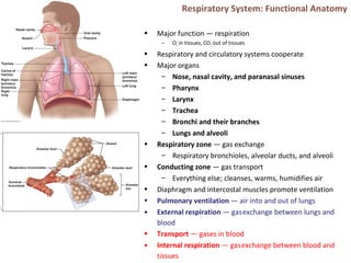
  • 1. Respiratory System: Functional Anatomy • Major function — respiration – O2 in tissues, CO2 out of tissues • Respiratory and circulatory systems cooperate • Major organs – Nose, nasal cavity, and paranasal sinuses – Pharynx – Larynx – Trachea – Bronchi and their branches – Lungs and alveoli • Respiratory zone — gas exchange – Respiratory bronchioles, alveolar ducts, and alveoli • Conducting zone — gas transport – Everything else; cleanses, warms, humidifies air • Diaphragm and intercostal muscles promote ventilation • Pulmonary ventilation — air into and out of lungs • External respiration — gasexchange between lungs and blood • Transport — gases in blood • Internal respiration — gasexchange between blood and tissues
  • 2. The Nose – Outside • Provides an airway for respiration • Moistens and warms the air • Filters and cleans the air • Resonating chamber for speech • Olfaction • Two regions — external nose and nasal cavity • External nose — root, bridge, dorsum nasi, and apex – Philtrum — shallow vertical groove inferior to apex – Nostrils (nares) — bounded laterally by alae
  • 3. The Nose – Inside • Nasal cavity — within and posterior to external nose – Divided by midline nasal septum – Posterior nasal apertures (choanae) open into nasal pharynx – Roof — ethmoid and sphenoid bones – Floor — hard (bone) and soft palates (muscle) • Nasal vestibule — nasal cavity immediately superior to nostrils – Vibrissae (hairs) filter particles • Nasal cavity lined with mucous membranes – Olfactory mucosa • Olfactory epithelium • Respiratory mucosa – Pseudostratified ciliated columnar epithelium – Mucous and serous secretions contain lysozyme and defensins – Cilia move contaminated mucus posteriorly to throat – Inspired air warmed by plexuses of capillaries and veins – Sensory nerve endings trigger sneezing
  • 4. Nasal Cavity • Nasal conchae-superior, middle, and inferior – Protrude medially from lateral walls – Increase mucosal area – Enhance air turbulence • Nasal meatus – Groove inferior to each concha • During inhalation, conchae and nasal mucosa – Filter, heat, and moisten air • During exhalation these structures – Reclaim heat and moisture • Paranasal sinuses – In frontal, sphenoid, ethmoid, and maxillary bones – Lighten skull; secrete mucus; help to warm and moisten air • Rhinitis – Inflammation of nasal mucosa – Nasal mucosa continuous with mucosa of respiratory tract  spreads from nose  throat  chest – Spreads to tear ducts and paranasal sinuses causing • Blocked sinus passageways  air absorbed  vacuum  sinus headache
  • 5. Pharynx • Muscular tube from base of skull to C6 – Connects nasal cavity and mouth to larynx and esophagus – Composed of skeletal muscle • Three regions • Nasopharynx – Air passageway posterior to nasal cavity – Lining - pseudostratified columnar epithelium – Soft palate and uvula close nasopharynx during swallowing – Pharyngeal tonsil (adenoids) on posterior wall – Pharyngotympanic (auditory) tubes drain and equalize pressure in middle ear; open into lateral walls • Oropharynx – Passageway for food and air from level of soft palate to epiglottis – Lining of stratified squamous epithelium – Isthmus of fauces-opening to oral cavity – Palatine tonsils-in lateral walls of fauces – Lingual tonsil-on posterior surface of tongue • Laryngopharynx – Passageway for food and air – Extends to larynx, where continuous with esophagus – Lined with stratified squamous epithelium
  • 6. Larynx • Attaches to hyoid bone; opens into laryngopharynx; continuous with trachea • Functions – Provides patent airway – Routes air and food into proper channels – Voice production • Houses vocal folds • Superior portion–stratified squamous epithelium • Inferior to vocal folds–pseudostratified ciliated columnar epithelium • Nine cartilages of larynx – All hyaline cartilage except epiglottis – Thyroid cartilage with laryngeal prominence (Adam's apple) – Ring-shaped cricoid cartilage – Paired arytenoid, cuneiform, and corniculate cartilages – Epiglottis-elastic cartilage; covers laryngeal inlet during swallowing; covered in taste bud-containing mucosa
  • 7. Vocal folds • Vocal ligaments-deep to laryngeal mucosa – Attach arytenoid cartilages to thyroid cartilage – Contain elastic fibers – Form core of vocal folds (true vocal cords) • Glottis-opening between vocal folds • Folds vibrate to produce sound as air rushes up from lungs • Vestibular folds (false vocal cords) – Superior to vocal folds – No part in sound production – Help to close glottis during swallowing • Speech-intermittent release of expired air while opening and closing glottis • Pitch — length and tension of vocal cords • Loudness — force of air • Pharynx, oral, nasal, and sinus cavities amplify and enhance sound • Language — muscles of pharynx, tongue, soft palate, and lips • Vocal folds may act as sphincter to prevent air passage • Example-Valsalva's maneuver – Glottis closes to prevent exhalation – Abdominal muscles contract – Intra-abdominal pressure rises – Emptying rectum or stabilizes trunk
  • 8. Trachea • Windpipe–from larynx into mediastinum • Wall composed of three layers – Mucosa-ciliated pseudostratified epithelium with goblet cells – Submucosa-connective tissue with seromucous glands – Adventitia-outermost layer made of connective tissue; encases C-shaped rings of hyaline cartilage • Trachealis muscle – Connects posterior parts of cartilage rings – Contracts during coughing to expel mucus • Carina – Spar of cartilage on last, expanded tracheal cartilage – Point where trachea branches into two main bronchi
  • 9. Bronchi and Subdivisions • Air passages undergo 23 orders of branching  bronchial (respiratory) tree • From tips of bronchial tree  conducting zone structures  respiratory zone structures • Trachea → right and left main (primary) bronchi • Each main bronchus enters hilum of one lung – Right main bronchus wider, shorter, more vertical than left • Each main bronchus branches into lobar (secondary) bronchi (three on right, two on left) – Each lobar bronchus supplies one lobe • Each lobar bronchus branches into segmental (tertiary) bronchi – Segmental bronchi divide repeatedly • Branches become smaller and smaller  – Bronchioles-less than 1 mm in diameter – Terminal bronchioles-smallest-less than 0.5 mm diameter
  • 10. • From bronchi through bronchioles, structural changes occur – Cartilage rings become irregular plates; in bronchioles elastic fibers replace cartilage – Epithelium changes from pseudostratified columnar to cuboidal; cilia and goblet cells become sparse – Relative amount of smooth muscle increases • Allows constriction Respiratory zone • Begins as terminal bronchioles  respiratory bronchioles  alveolar ducts  alveolar sacs – Alveolar sacs contain clusters of alveoli • ~300 million alveoli make up most of lung volume • Sites of gas exchange Conducting and Respiratory Zones
  • 11. Respiratory Membrane. Alveoli • Alveolar and capillary walls and their fused basement membranes – ~0.5-µ m-thick; gas exchange across membrane by simple diffusion • Alveolar walls – Single layer of squamous epithelium (type I alveolar cells) • Scattered cuboidal type II alveolar cells secrete surfactant and antimicrobial proteins • Surrounded by fine elastic fibers and pulmonary capillaries • Alveolar pores connect adjacent alveoli • Equalize air pressure throughout lung • Alveolar macrophages keep alveolar surfaces sterile – 2 million dead macrophages/hour carried by cilia  throat  swallowed
  • 13. Lungs • Occupy all thoracic cavity except mediastinum • Root — site of vascular and bronchial attachment to mediastinum • Costal surface — anterior, lateral, and posterior surfaces • Composed primarily of alveoli • Stroma — elastic connective tissue  elasticity • Apex-superior tip; deep to clavicle • Base-inferior surface; rests on diaphragm • Hilum-on mediastinal surface; site for entry/exit of blood vessels, bronchi, lymphatic vessels, and nerves • Left lung smaller than right – Cardiac notch-concavity for heart – Separated into superior and inferior lobes by oblique fissure
  • 14. Lungs • Right lung – Superior, middle, inferior lobes separated by oblique and horizontal fissures • Bronchopulmonary segments (10 right, 8–10 left) separated by connective tissue septa – If diseased can be individually removed • Lobules-smallest subdivisions visible to naked eye; served by bronchioles and their branches
  • 15. Pleurae • Thin, double-layered serosa • Parietal pleura – walls of pleural cavity • Visceral pleura – external lung surface • Pleural fluid fills pleural cavity – Lubrication and surface tension  lungs’ expansion and recoil • Pulmonary circulation (low pressure, high volume) – Pulmonary arteries — systemic venous blood to lungs • Branch and feed into pulmonary capillaries – Pulmonary veins — oxygenated blood to the heart • Bronchial arteries — blood to lung tissue – From aorta; enter lungs at hilum – Part of systemic circulation – Supply all lung tissue except alveoli – Bronchial veins anastomose with pulmonary veins
  • 16. Mechanics of Breathing. Pressures • Pulmonary ventilation – Inspiration-gases flow into lungs – Expiration-gases exit lungs • Atmospheric pressure (Patm) – Pressure exerted by air surrounding body – 760 mm Hg at sea level = 1 atmosphere • Respiratory pressures – Negative respiratory pressure < Patm – Positive respiratory pressure > Patm – Zero respiratory pressure = Patm • Intrapulmonary (intra-alveolar) pressure (Ppul) – Pressure in alveoli – Fluctuates with breathing – Always eventually equalizes with Patm • Intrapleural pressure (Pip) – Pressure in pleural cavity – Fluctuates with breathing – Always a negative pressure (<Patm and <Ppul) – Fluid level must be minimal • If accumulates  positive Pip pressure • Negative Pip caused by – Inward forces → lung collapse • Elastic recoil of lungs ↓ lung size • Surface tension of alveolar fluid ↓ alveolar size – Outward force → enlarge lungs • Elastic chest wall ↑ thorax outward
  • 17. Pressure Relationships • If Pip = Ppul or Patm  lungs collapse • (Ppul – Pip) = transpulmonary pressure – Keeps airways open – Greater transpulmonary pressure  larger lungs • Atelectasis (lung collapse) due to – Plugged bronchioles → collapse of alveoli – Pneumothorax – air in pleural cavity • Rupture in parietal or visceral pleura • Air removed with chest tubes; pleurae heal  lung reinflates
  • 18. Pulmonary Ventilation. Gas Laws • Inspiration and expiration • Depends on volume changes in thoracic cavity – Volume changes → pressure changes – Pressure changes → gases flow to equalize pressure Boyle’s Law •Relationship between pressure and volume of a gas – Gases fill container; if container size reduced  increased pressure •Pressure (P) varies inversely with volume (V): – P1V1 = P2V2
  • 19. Inspiration • Active process – Inspiratory muscles (diaphragm and external intercostals) contract – Thoracic volume increases  intrapulmonary pressure drops (to −1 mm Hg) – Lungs stretched and intrapulmonary volume increases – Air flows into lungs, down its pressure gradient, until Ppul = Patm • Forced inspiration • Vigorous exercise, COPD  accessory muscles (scalenes, sternocleidomastoid, pectoralis minor)  further increase in thoracic cage size
  • 20. Expiration• Quiet expiration normally passive process – Inspiratory muscles relax – Thoracic cavity volume decreases – Elastic lungs recoil and intrapulmonary volume decreases  pressure increases (Ppul rises to +1 mm Hg)  – Air flows out of lungs down its pressure gradient until Ppul = 0 • Note: forced expiration-active process; uses abdominal (oblique and transverse) and internal intercostal muscles
  • 21.
  • 22. Physical Factors Influencing Pulmonary Ventilation • Three factors hinder air passage and pulmonary ventilation; require energy to overcome • Airway resistance – Friction in airways – major nonelastic source of resistance – F=ΔP/R • ∆P - pressure gradient between atmosphere and alveoli – Resistance usually insignificant • Large airway diameters • Progressive branching of airways ↑ total cross- sectional area • Resistance greatest in medium-sized bronchi – Resistance disappears at terminal bronchioles where diffusion drives gas movement • As airway resistance rises, breathing movements become more strenuous • Severe constriction or obstruction of bronchioles – Can prevent life-sustaining ventilation – Can occur during acute asthma attacks; stops ventilation • Epinephrine dilates bronchioles, reduces air resistance
  • 23. • Surfactant – Detergent-like lipid and protein complex produced by type II alveolar cells – ↓ surface tension of alveolar fluid → no alveolar collapse – Insufficient quantity in premature infants causes infant respiratory distress syndrome   alveoli collapse after each breath Alveolar Surface Tension • Surface tension – Attracts liquid molecules to one another at gas-liquid interface – Resists any increase of the surface area of liquid – Water – high surface tension; coats alveolar walls  reduces them to smallest size
  • 24. Lung Compliance • Measure of change in lung volume that occurs with given change in transpulmonary pressure • Higher lung compliance  easier to expand lungs • Normally high due to – Distensibility of lung tissue – Alveolar surface tension • Diminished by – Nonelastic scar tissue replacing lung tissue (fibrosis) – Reduced production of surfactant – Decreased flexibility of thoracic cage • Homeostatic imbalances that reduce compliance – Deformities of thorax – Ossification of costal cartilage – Paralysis of intercostal muscles
  • 25. Respiratory Volumes and Capacities • Used to assess respiratory status – Tidal volume (TV) – Inspiratory reserve volume (IRV) – Expiratory reserve volume (ERV) – Residual volume (RV) • Combinations of respiratory volumes – Inspiratory capacity (IC) – Functional residual capacity (FRC) – Vital capacity (VC) – Total lung capacity (TLC)
  • 26. Dead Space. Pulmonary Function Tests • Anatomical dead space – No contribution to gas exchange – Air remaining in passageways; ~150 ml • Alveolar dead space–non-functional alveoli due to collapse or obstruction • Total dead space-sum of anatomical and alveolar dead space • Spirometer measures respiratory volumes and capacities • Spirometry can distinguish between – Obstructive pulmonary disease—increased airway resistance (e.g., bronchitis) • TLC, FRC, RV ↑ – Restrictive disorders — VC, TLC, FRC, RV ↓ (disease or fibrosis) • To measure rate of gas movement – Forced vital capacity (FVC) — gas forcibly expelled after taking deep breath – Forced expiratory volume (FEV) — amount of gas expelled during specific time intervals of FVC
  • 27. Alveolar Ventilation • Minute ventilation-total amount of gas flow into or out of respiratory tract in one minute – Normal at rest = ~ 6 L/min – Normal with exercise = up to 200 L/min – Only rough estimate of respiratory efficiency • Good indicator of effective ventilation • Alveolar ventilation rate (AVR)-flow of gases into and out of alveoli during a particular time • Dead space normally constant • Rapid, shallow breathing decreases AVR AVR = frequency X (TV – dead space) (ml/min) (breaths/min) (ml/breath)
  • 28. Nonrespiratory Air Movements • May modify normal respiratory rhythm • Most result from reflex action; some voluntary • Examples include-cough, sneeze, crying, laughing, hiccups, and yawns
  • 29. Gas Exchanges – Blood, Lungs, and Tissues • External respiration – diffusion of gases in lungs • Internal respiration – diffusion of gases at body tissues • Both involve – Physical properties of gases – Composition of alveolar gas Dalton’s Law • For mixture of gases: Ptotal = Σ Pindividual • Partial pressure – Pi = k C● i Henry’s Law • Gas mixtures in contact with liquid – Cwater = ks P● i (each gas dissolves in proportion to its partial pressure) – At equilibrium, pressure in both phases equal: • Pwater= Pair – Amount of each gas that will dissolve depends on • Solubility – CO2 20 times more soluble in water than O2; little N2 dissolves in water • Temperature – T ↑, solubility ↓
  • 30. Composition of Alveolar Gas • Alveoli contain more CO2 and water vapor than atmospheric air – Gas exchanges in lungs – Humidification of air – Mixing of alveolar gas with each breath
  • 31. External Respiration • Exchange of O2 and CO2 across respiratory membrane Influenced by – Thickness and surface area of respiratory membrane • 0.5 to 1 µ m thick • Large total surface area (40 times that of skin) for gas exchange • Thicken if lungs become waterlogged and edematous  gas exchange inadequate • Reduced surface area in emphysema (walls of adjacent alveoli break down), tumors, inflammation, mucus
  • 32. External Respiration • Exchange of O2 and CO2 across respiratory membrane Influenced by – Partial pressure gradients and gas solubilities • O2 gradient in lungs – Venous blood Po2 = 40 mm Hg – Alveolar Po2 = 104 mm Hg – Oxygen to blood • CO2 gradient in lungs – Venous blood Pco2 = 45 mm Hg – Alveolar Pco2 = 40 mm Hg – CO2 to lungs • Though gradient not as steep, CO2 diffuses in equal amounts with oxygen – CO2 20 times more soluble in plasma than oxygen
  • 33. Ventilation-Perfusion Coupling • Exchange of O2 and CO2 across respiratory membrane Influenced by – Ventilation-perfusion coupling • Perfusion-blood flow reaching alveoli • Ventilation-amount of gas reaching alveoli • Ventilation and perfusion matched (coupled) for efficient gas exchange – Never balanced for all alveoli due to • Regional variations due to effect of gravity on blood and air flow • Some alveolar ducts plugged with mucus
  • 34. Ventilation-Perfusion Coupling • Perfusion – Changes in Po2 in alveoli cause changes in diameters of arterioles • Where alveolar O2 is high, arterioles dilate • Where alveolar O2 is low, arterioles constrict • Directs most blood where alveolar oxygen high • Changes in Pco2 in alveoli cause changes in diameters of bronchioles – Where alveolar CO2 is high, bronchioles dilate – Where alveolar CO2 is low, bronchioles constrict – Allows elimination of CO2 more rapidly
  • 35. Internal Respiration • Capillary gas exchange in body tissues • Partial pressures and diffusion gradients reversed compared to external respiration – Tissue Po2 always lower than in systemic arterial blood  oxygen from blood to tissues – CO2  from tissues to blood – Venous blood Po2 40 mm Hg and Pco2 45 mm Hg
  • 36. O2 Transport • Molecular O2 carried in blood – 1.5% dissolved in plasma – 98.5% loosely bound to each Fe of hemoglobin (Hb) in RBCs • 4 O2 per Hb • Oxyhemoglobin (HbO2) • Deoxyhemoglobin (HHb – no O2) • Loading and unloading of O2 → change in shape of Hb – As O2 binds, Hb affinity for O2 increases and vice versa – Cooperative binding • All four heme groups carry O2 – full saturation • Rate of loading and unloading of O2 regulated to ensure adequate oxygen delivery to cells – Po2 – Temperature – Blood pH – Pco2 – Concentration of BPG–produced by RBCs during glycolysis; levels rise when oxygen levels chronically low
  • 37. Hemoglobin Dissociation Curve • Oxygen-hemoglobin dissociation curve • Hemoglobin saturation plotted against Po2 is S- shaped curve • Binding and release of O2 influenced by Po2
  • 38. • In arterial blood – Po2 = 100 mm Hg – Contains 20 ml oxygen per 100 ml blood (20 vol %) – Hb is 98% saturated • Further increases in Po2 (e.g., breathing deeply) produce minimal increases in O2 binding
  • 39. • In venous blood – Po2 = 40 mm Hg – Contains 15 vol % oxygen – Hb is 75% saturated • Venous reserve • Oxygen remaining in venous blood
  • 40. Factors Influencing Hb Saturation • Increases in temperature, H+ , Pco2, and BPG – Modify structure of hemoglobin; its affinity for O2 ↓ – Occur in systemic capillaries – ↑ O2 unloading from blood – Shift O2-Hb curve to right • Decreases in these factors shift curve to left – Decreases oxygen unloading from blood • As cells metabolize glucose and use O2 – Pco2 and H+ ↑ in capillary blood  blood pH ↓, Pco2 ↑ – Heat ↑  Hb affinity for O2 ↓  oxygen unloading ↑ – Bohr effect: oxygen unloading in tissues that use O2 • Hypoxia – Inadequate O2 delivery to tissues  cyanosis – Anemic hypoxia–too few RBCs; abnormal or too little Hb – Ischemic hypoxia–impaired/blocked circulation – Histotoxic hypoxia–cells unable to use O2, as in metabolic poisons – Hypoxemic hypoxia–abnormal ventilation; pulmonary disease – Carbon monoxide poisoning–especially from fire; 200X greater affinity for Hb than oxygen
  • 41. CO2 Transport • CO2 transported in blood in three forms – 7 to 10% dissolved in plasma – 20% bound to globin of hemoglobin (carbaminohemoglobin) – 70% transported as bicarbonate ions (HCO3 – ) in plasma • CO2 combines with water to form carbonic acid (H2CO3), which quickly dissociates • Occurs primarily in RBCs, where carbonic anhydrase reversibly and rapidly catalyzes reaction
  • 42. • In systemic capillaries – HCO3 – quickly diffuses from RBCs into plasma • Chloride shift occurs – Outrush of HCO3 – from RBCs balanced as Cl– moves into RBCs from plasma
  • 43. • In pulmonary capillaries – HCO3 – moves into RBCs (while Cl- move out); binds with H+ to form H2CO3 – H2CO3 split by carbonic anhydrase into CO2 and water – CO2 diffuses into alveoli
  • 44. Haldane Effect • Amount of CO2 transported affected by Po2 – HHb forms carbaminohemoglobin and buffers H+ more easily  – Po2 and hemoglobin saturation ↓; CO2 in blood ↑ • ↑ CO2 exchange in tissues and lungs • At tissues, as more CO2 enters blood – O2 unloading ↑ (Bohr effect) – HbO2 releases O2and readily forms carbaminohemoglobin • Carbonic acid–bicarbonate buffer system–resists changes in blood pH – If H+ concentration in blood rises, excess H+ is removed by combining with HCO3 –  H2CO3 – If H+ concentration begins to drop, H2CO3 dissociates, releasing H+ – HCO3 – is alkaline reserve of carbonic acid-bicarbonate buffer system • Changes in respiratory rate and depth affect blood pH – Slow, shallow breathing  increased CO2 in blood drop in pH – Rapid, deep breathing  decreased CO2 in blood  rise in pH
  • 45. • Higher brain centers, chemoreceptors, and other reflexes • Neural controls – Neurons in reticular formation of medulla and pons – Clustered neurons in medulla important • Ventral respiratory group • Dorsal respiratory group • Ventral respiratory group (VRG) – Rhythm-generating and integrative center – Sets eupnea (12–15 breaths/minute) • Normal respiratory rate and rhythm – Its inspiratory neurons excite inspiratory muscles via phrenic (diaphragm) and intercostal nerves (external intercostals) – Expiratory neurons inhibit inspiratory neurons • Dorsal respiratory group (DRG) – Near root of cranial nerve IX – Integrates input from peripheral stretch and chemoreceptors; sends information  VRG Control of Respiration. Medullary Respiratory Centers
  • 46. Pontine Respiratory Centers • Influence and modify activity of VRG • Smooth out transition between inspiration and expiration and vice versa • Transmit impulses to VRG  modify and fine- tune breathing rhythms during vocalization, sleep, exercise Respiratory Rhythm •Not well understood •One hypothesis – Pacemaker neurons with intrinsic rhythmicity •Most widely accepted hypothesis – Reciprocal inhibition of two sets of interconnected pacemaker neurons in medulla that generate rhythm
  • 47. Factors influencing Breathing Rate and Depth • Depth determined by how actively respiratory center stimulates respiratory muscles • Rate determined by how long inspiratory center active • Both modified in response to changing body demands – Most important are changing levels of CO2, O2, and H+ – Sensed by central and peripheral chemoreceptors
  • 48. Chemical Factors • Influence of Pco2 (most potent; most closely controlled) – blood Pco2 levels ↑ (hypercapnia), CO2 accumulates in brain  – CO2 in brain hydrated  carbonic acid  dissociates, releasing H+  pH ↓ – H+ stimulates central chemoreceptors of brain stem – Chemoreceptors synapse with respiratory regulatory centers  depth and rate of breathing ↑  blood Pco2 ↓  pH ↑ • Hyperventilation –depth/rate of breathing ↑; exceeds body's need to remove CO2   blood CO2 levels ↓ (hypocapnia)  cerebral vasoconstriction and cerebral ischemia  dizziness, fainting • Apnea – breathing cessation from abnormally low Pco2
  • 49. Chemical Factors • Influence of Po2 – Peripheral chemoreceptors in aortic and carotid bodies – arterial O2 level sensors • When excited, cause respiratory centers to increase ventilation – ↓ Po2 – normally slight effect on ventilation • O2 reservoir in HbO2 • Requires substantial drop in arterial Po2 (to 60 mm Hg) to stimulate increased ventilation • Influence of arterial pH – Can modify respiratory rate and rhythm even if CO2 and O2 levels normal – Mediated by peripheral chemoreceptors – ↓ pH may reflect • CO2 retention; accumulation of lactic acid; excess ketone bodies – Respiratory system controls ↑ pH by ↑ respiratory rate and depth • ↑CO2 levels most powerful respiratory stimulant • Normally blood Po2 affects breathing only indirectly by influencing peripheral chemoreceptor sensitivity to changes in Pco2 • When arterial Po2 falls below 60 mm Hg, it becomes major stimulus for respiration (via peripheral chemoreceptors) • Changes in arterial pH resulting from CO2 retention or metabolic factors act indirectly through peripheral chemoreceptors
  • 50. Influence of Higher Brain Centers • Hypothalamic controls act through limbic system to modify rate and depth of respiration – Example-breath holding that occurs in anger or gasping with pain • Rise in body temperature increases respiratory rate • Cortical controls-direct signals from cerebral motor cortex that bypass medullary controls – Example-voluntary breath holding • Brain stem reinstates breathing when blood CO2 critical
  • 51. Inflation Reflex • Hering-Breuer Reflex (inflation reflex) – Stretch receptors in pleurae and airways stimulated by lung inflation • Inhibitory signals to medullary respiratory centers end inhalation and allow expiration • Acts as protective response more than normal regulatory mechanism Pulmonary Irritant Reflexes • Receptors in bronchioles respond to irritants – Communicate with respiratory centers via vagal nerve afferents • Promote reflexive constriction of air passages • Same irritant  cough in trachea or bronchi; sneeze in nasal cavity
  • 52. Respiratory Adjustments: Exercise and High Altitude • Adjustments geared to both intensity and duration of exercise • Hyperpnea – ↑ ventilation (10 to 20 fold) • Pco2, Po2, and pH remain during exercise≡ • Three neural factors cause increase in ventilation as exercise begins – Psychological stimuli – Simultaneous cortical motor activation of skeletal muscles and respiratory centers – Excitatory impulses to respiratory centers from proprioceptors • Ventilation declines suddenly as exercise ends because the three neural factors shut off • Gradual decline to baseline because of decline in CO2 flow after exercise ends • Exercise  anaerobic respiration  lactic acid – Not from poor respiratory function; from insufficient cardiac output or skeletal muscle inability to increase oxygen uptake • Quick travel to altitudes above 2400 meters (8000 feet) may  symptoms of acute mountain sickness (AMS) – Atmospheric pressure and Po2 levels ↓ – Headaches, shortness of breath, nausea, and dizziness – (Lethal) cerebral and pulmonary edema • Acclimatization-respiratory and hematopoietic adjustments to high altitude – Chemoreceptors – more responsive to Pco2 when Po2 ↓ – ↓ in Po2 directly stimulates peripheral chemoreceptors – Minute ventilation ↑ and stabilizes in few days to 2–3 L/min higher than at sea level • Always lower-than-normal Hb saturation levels – Less O2 available • ↓ blood O2 → ↑ production of EPO in kidneys • RBC ↑ slowly → long-term compensation
  • 53. COPD. Emphysema • Chronic obstructive pulmonary disease (COPD) – Exemplified by chronic bronchitis and emphysema – Irreversible decrease in ability to force air out of lungs – Other common features • History of smoking in 80% of patients • Dyspnea - labored breathing ("air hunger") • Coughing and frequent pulmonary infections • Most develop respiratory failure (hypoventilation) accompanied by respiratory acidosis, hypoxemia • Emphysema – Permanent enlargement of alveoli; destruction of alveolar walls; decreased lung elasticity  • Accessory muscles necessary for breathing   exhaustion from energy usage • Hyperinflation  flattened diaphragm  reduced ventilation efficiency • Damaged pulmonary capillaries  enlarged right ventricle
  • 54. Homeostatic Imbalance • Chronic bronchitis – Inhaled irritants  chronic excessive mucus  inflamed and fibrosed lower respiratory passageways  obstructed airways  impaired lung ventilation and gas exchange  frequent pulmonary infections • COPD symptoms and treatment – Strength of innate respiratory drive  different symptoms in patients • "Pink puffers"–thin; near-normal blood gases • "Blue bloaters"–stocky, hypoxic – Treated with bronchodilators, corticosteroids, oxygen, sometimes surgery • Asthma – reversible COPD – Characterized by coughing, dyspnea, wheezing, and chest tightness – Active inflammation of airways precedes bronchospasms – Airway inflammation is immune response caused by release of interleukins, production of IgE, and recruitment of inflammatory cells – Airways thickened with inflammatory exudate magnify effect of bronchospasms
  • 55. Tuberculosis. Lung Cancer. • Tuberculosis (TB) – Infectious disease caused by bacterium Mycobacterium tuberculosis – Symptoms-fever, night sweats, weight loss, racking cough, coughing up blood – Treatment- 12-month course of antibiotics • Are antibiotic resistant strains • Lung cancer – Leading cause of cancer deaths in North America – 90% of all cases result of smoking – Three most common types • Adenocarcinoma (~40% of cases) peripheral lung areas (bronchial glands, alveolar cells) • Squamous cell carcinoma (20–40% of cases) in bronchial epithelium • Small cell carcinoma (~20% of cases) - lymphocyte-like cells; originate in primary bronchi; metastasize http://www.lung.org/
  • 56. Lung Cancer • Lung cancer – Early detection key to survival – Helical CT scan better than chest X ray – Developing breath test of gold nanoparticles – If no metastasis  surgery to remove diseased lung tissue – If metastasis  radiation and chemotherapy • Potential new therapies for lung cancer – Antibodies targeting growth factors required by tumor; or deliver toxic agents to tumor – Cancer vaccines to stimulate immune system – Gene therapy to replace defective genes http://www.aboutcancer.com/
  • 57. Early Development • Upper respiratory structures develop first • Olfactory placodes invaginate into olfactory pits ( nasal cavities) by fourth week • Laryngotracheal bud present by fifth week • Mucosae of bronchi and lung alveoli present by eighth week • By 28th week, premature baby can breathe on its own • During fetal life, lungs filled with fluid and blood bypasses lungs • Gas exchange takes place via placenta
  • 58. Age and Respiratory System • At birth, respiratory centers activated, alveoli inflate, and lungs begin to function • Two weeks after birth before lungs fully inflated • Respiratory rate highest in newborns and slows until adulthood • Lungs continue to mature and more alveoli formed until young adulthood • Respiratory efficiency decreases in old age http://www.nursingconsult.com/
  • 59. Cystic Fibrosis • Cystic fibrosis – Most common lethal genetic disease in North America – Abnormal, viscous mucus clogs passageways  bacterial infections • Affects lungs, pancreatic ducts, reproductive ducts – Cause–abnormal gene for Cl- membrane channel http://learn.genetics.utah.edu/http://www.nhlbi.nih.gov/ • Treatments for cystic fibrosis – Mucus-dissolving drugs; chest percussion to loosen mucus; antibiotics – Research into • Introducing normal genes • Prodding different protein  Cl- channel • Freeing patient's abnormal protein from ER to  Cl- channels • Inhaling hypertonic saline to thin mucus