Best VIP Call Girls Noida Sector 75 Call Me: 8448380779
Embryology of nose 1440 copy
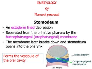
1. • An ectoderm lined depression
• Separated from the primitive pharynx by the
buccopharyngeal (oropharyngeal) membrane
• The membrane later breaks down and stomodeum
opens into the pharynx
Forms the vestibule of
the oral cavity
EMBRYOLOGY
Of
Nose and paranasal
Stomodeum
2. • By the end of 4th week,
bilateral oval-shaped
ectodermal thickenings
called ‘nasal placodes’
appear on each side of
the lower part of the
frontonasal prominence
• Nasal placodes are
primordia of the nose
and nasal cavities.
Frontonasal
prominence
3.
4. • By the end of 5th week Mesenchymal cells
proliferate at the margin of the placodes
and produce horse-shoe shaped swellings
around these.
• The sides of these swellings are called
‘medial’ and ‘lateral’ nasal prominences
• The placodes now lie in the floor of a
depression called ‘nasal pits’
Each lateral nasal prominence is separated from the maxillary swelling by nasolacrimal groove
5th weeks
6th weeks
5. 6 weeksBy the end of 6th week, nasal pits deepen and
form nasal sacs
Later medial process joins the maxillary process
forming closed maxillary arch.
Lateral nasal swelling also join maxillary process
and gives nasolacrimal duct at their junction.
frontonasal prominence gives rise to inferior
mesodermic projection-form the nasal septum
dividing the nose into two cavities.
by the 6th week,medial nasal
prominence merge to give rise to
median portion of the nose,
middle portion of upper lip,philtrum
The medial nasal swellings enlarge, grow medially and
merge with each other in the midline to form the
intermaxillary segment
The lateral nasal prominences
form the alae of the nose
6. • Initially the nasal
sacs are separated
from the oral cavity
by oronasal
membrane.
• The oronasal
membrane ruptures
by the 7th week,
communicating the
primitive nasal
cavities with the oral
cavity
7. • These communications
are called the primitive
choanae and are
located posterior to the
primary palate
• After the development
of the secondary palate,
the choanae change
their position and
become located at the
junction of nasal cavity
and the pharynx
8. The nasal septum develops as a downgrowth
from internal parts of the merged medial
nasal prominences. The fusion between the
nasal septum and the palatal processes
begins anteriorly during the ninth week and
is completed posteriorly by the 12th week,
superior to the primordium of the hard
palate
Fuses with the palatine process in 9-12
weeks, superior to the hard palate
primordium
9.
10. • The superior, middle and inferior
conchae develop on the lateral wall of
each nasal cavity
• The ectodermal epithelium in the roof
of each nasal cavity becomes
specialized as the olfactory epithelium
Some epithelial cells
differentiate into
olfactory receptor cells
(neurons). The axons of
these cells constitute
the olfactory nerves,
which grow into the
olfactory bulbs of the
brain
11. • Develops from a rod-like thickening of the ectoderm in the
floor of the nasolacrimal groove
• This solid cord of cells separates from the surface ectoderm
and lies in the underlying mesenchyme
• The cord gets canalized to form the nasolacrimal duct
• The cranial end of the duct expands to form the lacrimal sac
• The caudal end opens into the inferior meatus of the nasal
cavity
• The duct is usually becomes completely patent only after
birth
• Failure of complete canalization of the duct leads to atresia
of the duct (seen in about 6% of newborn infants)
Nasolacrimal duct
12. Maxillary sinus - first to be developed and
aerated at birth.
Sphenoidal sinus is undevoleped and non-
aereated at birth.
Aeration begins at age 3years and then
progresses posteriorly.
Ethmoid air cells-develop during puberty and
develop slowly until approximately 17-18 years
of age.
Frontal sinus is last sinus to develop ,as a direct continuation or by upward
migration of anterior ethmoidal air cells.
Remains as a small blind sac within the frontal bone till 2 years of age,from 2 to 9
years secondary pneumatization of frontal bone proceeds.
DEVELOPMENT OF PARANASAL SINUSES
At about 25 – 28 weeks of gestation, three medially
directed projections arise from the lateral wall of the
nose.
Sinuses begin developing as small sacculations of
the mucosa of the nasal meati and recesses
As the pouches or sacs develop and grow they will
invade the respective bones to form air sinuses and
cells
13. CONGENITAL ANOMALIES OF
THE NOSE
ARHINIA
Absence of the external
nose, nasal cavities, and
olfactory apparatus
due to bilateral absence of
nasal placodes
HALF NOSE
due to unilateral
absence of nasal
placode
PROBOSCIS LATERALIS
Rudimentary nasal
structure or appendage
due to imperfect fusion
between the maxillary
process and the lateral
nasal process.
14. POLYRRHINIA
Due to duplication of medial
nasal processes.
NASAL CLEFTS :
failure of the frontal
nasal process to
develop appropriately
results into two
separated halves of the
nose.
SUPERNUMERARY NOSTRIL
MIDLINE NASAL
SINUS:
incomplete fusion of
the right and left
medial nasal
prominence
15. Median cleft face syndrome (or frontonasal
dysplasia) is a rare, sporadic condition. It
results from embryonic failure of fusion of
the median nasal processes.
Congenital malformations
Median nasal cleft. Note
pronounced separation
of the nostrils.