28. Mucous Membrane
• Stratified squamous epith.:
over vocal cords and upper
part of vestibule of larynx
• Ciliated columnar epith.:
remainder of the cavity
When calcified, it can be mistaken as a foreign bidy in soft tissue Xray films
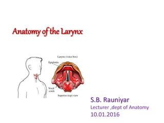
Divided into 3 parts by 2 folds of mucous membrane, namely the true and false cords.
Vestibule – lies bet. Inlet and edges of false cords
Ventricle (Morgagni) - Deep, spindle-shaped recess bet. True and false cords, lined by a mucous membrane that is covered externally by thyroarytenoid muscle
Subglottic space – lies bet. True VC and lower border of cricoid cartilage
False cords – upper set of two horizontal folds on each side of the laryngeal cavity.
TVC – covering epith. Is closely bound down to underlying vocal ligament, blood supplu here is poor hence the pearly white appearance of the vocal cords in life.
Margins of aryepiglottic folds – none on he free edges of the vocal cords
Reinke’s layer of connective tissue – lies immediately under the epith. Of larynx and superficial to elastic layer. NO GLANDS BENEATH AND NO LYMPH VESSELS IN IT.
Level of LNs
Closure of glottis – helps to increase intrathoracic and intraabdominal pressure and aids in lifting, digging, defecation, vomiting, urination or childbirth