Downloaded 82 times


![ HH6
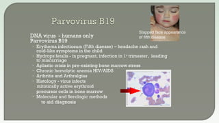
• Roseola [sixth disease]
• 6m-2yr high fever & rash
HH8
• (1) Kaposi’s sarcoma
• (2) Castleman disease
• (3) Primary effusion
lymphoma
Onion skin pattern
of Castleman
disease
1
1](https://image.slidesharecdn.com/margiemorgan-webpagevirology2019-190331040540/85/Virology-Review-2019-18-320.jpg)

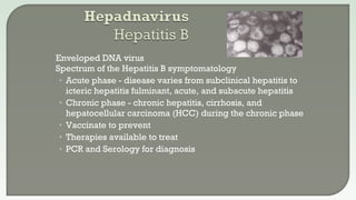
![• JC virus [John Cunningham]
Progressive multifocal leukoencephalopathy -
PML -Encephalitis of immune suppressed
Destroys oligodendrocytes in brain
• BK virus
Causes latent virus infection in kidney
Progression due to immune suppression
Hemorrhagic cystitis
• Histology
• PCR to aid diagnosis
Giant Glial Cells of JCV](https://image.slidesharecdn.com/margiemorgan-webpagevirology2019-190331040540/85/Virology-Review-2019-23-320.jpg)


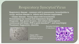
![ Disease: fever, malaise …. Death from respiratory complications or
secondary bacterial infection
Yearly H and N types dominate, most recently H1N1 and H3N2
Diagnosis
• Cell culture obsolete [RMK]
• Enzyme immunoassay (EIA) lateral flow membrane can be used in point of
care testing
• Amplification (PCR) gold standard for detection
Treatment: Amantadine and Tamiflu (Oseltamivir)
• Seasonal variation in susceptibility but Tamiflu sensitive currently
Influenza B
• Milder form of Influenza like illness
• Usually <=10% of cases /year
Vaccinate – Trivalent vaccine -2 A viruses/1 B virus](https://image.slidesharecdn.com/margiemorgan-webpagevirology2019-190331040540/85/Virology-Review-2019-39-320.jpg)
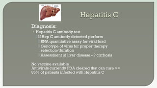
![Disease
• Fever, Rash, Dry Cough, Runny Nose, Sore throat,
inflamed eyes (photosensitive)
Can invade lung
• Respiratory spread - very contagious
• Koplik’s spot – bluish discoloration inner
lining of the cheek is pathognomonic
• Subacute sclerosing panencephalitis [SSPE]
Rare chronic degenerative neurological disease
Persistent infection with a mutated measles virus,
due to mutated virus there is total lack of an
immune response
Diagnosis: Clinical symptoms, PCR nasal or throat is best,
IgM serology can have false positive reactions
Vaccinate – MMR (Measles, Mumps, Rubella) vaccine
Treatment: Not specific, Immune globulin, vitamin A
H and E stain/ lung](https://image.slidesharecdn.com/margiemorgan-webpagevirology2019-190331040540/85/Virology-Review-2019-40-320.jpg)


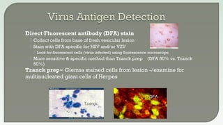
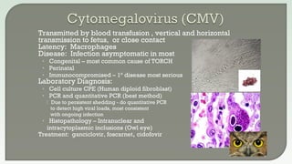
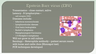
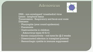
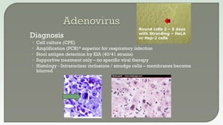
This document discusses various methods for diagnosing viral infections, including direct fluorescent antibody staining, enzyme immunoassays, viral cell culture, and molecular amplification techniques. It provides details on specific tests for different viruses, such as direct fluorescent antibody staining of lesions for HSV and VZV, enzyme immunoassays for influenza and RSV detection, and viral cell culture using various cell lines. Molecular amplification methods like PCR are described as sensitive tests for numerous viruses.


