Herpes Viruses. General properties, Laboratory diagnostics.
•Download as DOC, PDF•
3 likes•1,099 views

The document discusses herpesviruses, including their classification, properties, and laboratory diagnosis of associated human diseases. It describes the three subfamilies of herpesviruses - Alphaherpesvirinae, Betaherpesvirinae, and Gammaherpesvirinae - and provides examples such as herpes simplex virus types 1 and 2, varicella-zoster virus, cytomegalovirus, and Epstein-Barr virus. Methods for laboratory diagnosis of infections caused by these viruses are outlined, including virus isolation, serological techniques, and molecular methods. Rapid diagnosis may involve microscopy to detect intranuclear inclusion bodies. The document also covers epidemiology, pathogenesis, treatment and prophylaxis of major her

Recommended
More Related Content
What's hot
What's hot (20)
Similar to Herpes Viruses. General properties, Laboratory diagnostics.
Similar to Herpes Viruses. General properties, Laboratory diagnostics. (20)
More from Eneutron
More from Eneutron (20)
Recently uploaded
Recently uploaded (20)
Herpes Viruses. General properties, Laboratory diagnostics.
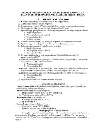
- 1. THEME: HERPESVIRUSES. GENERAL PROPERTIES. LABORATORY DIAGNOSTICS OF HUMAN DISEASES CAUSED BY HERPES VIRUSES. I. THEORETICAL QUESTIONS 1. General characteristic and classification of the herpesviruses. 2. Alpha-herpes viruses: general properties. 3. Herpes simplex virus (HSV), types, morphology, antigen structure and resistance. 4. Methods of the HSV cultivation, indication and identification. 5. Epidemiology, pathogenesis and laboratory diagnostics of the herpes simplex infection. a. Rapid diagnostics; b. Virological (cultural) method; c. Serological method d. Modern methods 6. Varicella-zoster virus (VZV), biological properties, cultivation and indication. 7. Epidemiology and pathogenesis of chickenpox and zoster infection. 8. Laboratory diagnostics of varicella-zoster infection: a. Rapid diagnostics; b. Virological (cultural) method; c. Serological method 9. General characteristic of beta-herpes viruses. Biological characteristics of cytomegalovirus (CMV). 10. Laboratory diagnostics of cytomegalic inclusion disease (congenital CMV infection) and generalized CMV infection in adults. a. Microscopic method b. Serological method 11. General characteristic of gamma-herpes viruses. Biological characteristics of Epstein- Barr virus (EBV) and sarcoma Kaposhi’s associated herpes virus (HHV 8). 12. Epidemiology, pathogenesis and laboratory diagnostics of infectious mononucleosis. a. Serological method b. Blood assay 13. Specific prophylaxis and therapy of herpes infections. Herpes viruses. Classification Family Herpesviridae is divided onto three subfamily based on the type of host cell most often infected and the site of latency. Alphaherpesvirinae includes next species: 1. Herpes simplex viruses 1 and 2 (HSV1 and HSV2) 2. Varicella-zoster virus (VZV or HHV 3) Betaherpesvirinae contains species: 1. Cytomegalovirus (CMV or HHV 5) 2. HHV6 and HHV7 Gammaherpesvirinae includes species: 1. Epstein-Barr virus (EBV or HHV4) 2. Kaposi`s sarcoma associated virus (HHV8) General properties of herpes viruses 1. Alpha-herpes viruses infect epithelial cells primarily and cause latent infections in sensory ganglia. They have relatively short replicative cycle (12-18 hrs). They are readily cultivated onto the CAM of chicken embryo and into the cell of continuous cell cultures (HeLa, Hep-2 )
- 2. 2. Beta-herpes viruses can cause infection of salivary glands and other inner organs. They replicate slowly (more than 24 hrs). They are cultivated into the human fibroblast and induce enlargement of the infected cell 3. Gamma-herpes viruses infect lymphoid cell and can be cultivated into the limphoblastoid cells. They have the highest oncogenic properties within the family Morphology of herpesviruses They enveloped DNA-including viruses with double-stranded DNA. The herpes virus capsid is icosahedral. The envelope is derived from nuclear membrane of the host cell during budding of the virus. There is additional structure named the tegument between capsid and envelope. The envelope carries virus spikes (receptors) The diameter of virion is ranged from 120 to 230 nm. General biological properties 1. Herpes viruses replicate into the nucleus of the host cell. They can cause intranuclear (Lipschutz) inclusion body. 2. They do not show any antigenic cross reaction. Any herpes virus has surface type specific and inner group specific antigens 3. They are sensitive to lipid solvents like alcohol, ether, chloroform and others. They are heat labile, but stable to freeze and lyophilization. 4. They have narrow host range and cause human infection 5. They can cause both latent and productive types of infection into the different host cells 6. The most part of the herpes viruses possess oncogenic properties Herpes simplex virus There are two types of the herpes simplex virus (HSV): 1. HSV type 1 causes herpes labialis and isolated from lesions in and around the mouth (“lesions above the waist”); 2. HSV type 2 is isolated from genital tract lesions (“lesions below the waist”) and causes herpes genitalis Cultivation of the HSV HSV has typical morphology and may be cultivated into the next alive systems: 1. Onto the chick embryo CAM (it produces small white shiny pocks) 2. Into the cell cultures (primary and continuous): it forms CPE with intranuclear inclusions and multinucleated giant cells 3. It can be propagated into conjunctiva cells of rabbits (experimental keratoconjunctivitis) Epidemiology and pathogenesis The source of infection is ill person with typical lesions. Infection is transmitted by close direct contact (labial herpes or cold sore, fever blister) or sexual intercourse (genital herpes). Viruses are present in abundant number in the skin lesions, saliva and secretions (respiratory, vaginal, etc.). Virus enters through small defects into the skin or mucous membranes and replicates locally, causing typical vesicular lesions. Then it is transported intra-axonally to the sensory ganglia (trigeminal ones at labial herpes and sacral ones at genital herpes where it causes latent infection). Sometimes virus can be reactivated into the ganglia, transported to the skin or mucous membrane and result in reccurent herpes Clinical features: HSV causes thin walled vesicles which heal without scarring. Lesions may be localized onto the face (cutaneous herpes), mucous membranes (herpetic gingivostomatitis), onto the cornea (keratoconjunctivitis, branching dendritic ulcers of cornea), onto the external sexual organs (genital herpes). The severest form is generalized herpes and congenital herpes (transplacental herpes) that have multi-organ involvement.
- 3. Laboratory diagnostics of herpes simplex infection Clinical specimens: fluid from lesions, CSF, saliva 1. Microscopy: Preparation the Tzanck smear from lesions and detection typical Tzanck cells (multinucleated giant cells with faceted nuclei and homogeneously stained chromatin); In the smear stained with Giemsa methods intranuclear incusions may be revealed (Lipschutz inclusion bodies) Other methods of rapid diagnostics: immune electron microscopy, immunofluorescence 2. Virus isolation: Cultivation onto the chick embryo CAM or into the cell cultures. Indication of virus with pock formation on the CAM or with CPE into the infected cells in the cell cultures. Identification of virus with serological test (neutralization test) 3. Serology: to detect primary infection IgM is revealed into the patient serum with ELISA; Others tests : CFT, neutralization test Treatment and prophylaxis 1. Acyclovir (Zovirax), valacyclovir (Valtrex), penciclovir inhibit viral DNA- polymerase 2. Idoxirudine, trifluridine (Viroptic) are used for eye infection Prevention is possible by avoiding of direct contact with lesions. Congenital and perinatal herpes infection is prevented by cesarean section Varicella-zoster virus (VZV) It has the typical morphology, but it is not cultivated onto the CAM of chick embryo and it is not pathogenic for laboratory animal. Virus may be cultivated into the human cell cultures (fibroblast or amnion cells) and into the HeLa cells. It does not show antigenic variation and present as single antigenic variant. It causes chickenpox after primary infection (disease of childhood), while herpes zoster arises after reactivation of the latent virus in immunocompomised patients (endogenous infection). Immunity after chickenpox is strong, long-lasting (life-long) Epidemiology and pathogenesis The source of infection is person with chickenpox or more rarely with herpes zoster. Infection is transmitted with air droplets (chickenpox) or with direct contact with lesions. The portal of entry is respiratory tract where virus is replicated, enters the bloodstream and spread with blood to skin. Typical skin lesions appear on the trunk after incubation period (1-3 weeks) and demonstrate following rash evolution: macule-papule-vesicle-pustule-scab. Virus can infect sensory ganglia of the spinal cord and remains latent many years. After activation it is spread to skin intraaxonally and causes typical lesions along the nerves on the trunk or chest Laboratory diagnostics of chickenpox and zoster infection Diagnosis is usually made on clinical findings, but some methods may confirm diagnosis at atypical duration: 1. Microscopy: Tzanck smear: in the smear stained with Giemsa methods intranuclear incusions may be revealed (Lipschutz inclusion bodies) Other methods: immune electron microscopy, immunofluorescence 2. Virus isolation is possible by infection of the cell culture. virus is indicated with CPE and identified with NT, immunofluorescence 3. Serology (detection of the antibody rising titer in the paired sera) with CFT Treatment and prophylaxis No antiviral therapy is necessary for chickenpox/varicella. Prevention is possible by active immunization with live, attenuated VZV (OKA strain). For contact person varicella-zoster immunoglobulin (VZIG) is used to prevent disease. Acyclovir is used to prevent severe infection in immunocompromised persons Cytomegalovirus (CMV) It is the largest human herpes virus (150-230 nm in diameter). It may be cultivated into the human fibroblast cell culture only. It causes specific CPE: enlargement of the infected cell
- 4. and prominent intranuclear inclusions (“owl-eyed” appearance). In persons with adequate immunity CMV causes subclinical or unapparent infection. In persons with waned immunity CMV can provoke generalized infection. Congenital CMV-infection often is very severe, associated with hepatosplenomegaly, jandice,hemolytic anemia, and microcephaly, chorioretinitis Epidemiology of CMV infection CMV is transmitted by different ways: 1. It is transmitted across the placenta (congenital CMV infection) 2. It can be transmitted by direct contact during birth 3. Virus can replicate in saliva glands, so it can be transmitted with saliva droplets 4. In adults it can be transmitted sexually 5. It also can enter human body with blood during transfusion or with transplants (grafts) Pathogenesis Infection of the fetus can cause cytomegalic inclusion disease that lead to severe pathology of the central nervous system, especially when mother was primary infected during first trimester. Infection of children and adults may be either latent or it appears as mononucleosis-like disease. Virus may be latent into leucocytes, kidney tissue and saliva glands for years. Manifested CMV infection is possible in immuno-compromised persons (HIV-patients and persons with transplants). Immunity is humoral and cell-mediated (the last is more important). Laboratory diagnostics of the cytomegalic inclusion disease and CMV infection Laboratory diagnosis is based on some methods: 1. Rapid diagnostics: demonstration into the patient urine or saliva virus-infected cells (cytomegalic cells with intranuclear inclusions) with immunofluorescence test or with Romanovsky-Giemsa staining 2. Virus isolation: Virus is isolated from urine, saliva, CSF and cultivated onto the human fibroblast cell culture. Virus indication: CPE . Virus identification: CFT, NT, ELISA, etc. 3. Serological method: demonstration in the paired patient sera four-fold rising of antibody titer (ELISA, CFT, etc.) Epstein-Barr virus (EBV) It belongs to gamma-herpes viruses and has oncogenic properties. Biological characteristics is typical for subfamily. It is widespread and can cause different diseases from latent infection in children, infectious mononucleosis in adulthood to EBV-associated malignancies such as Burkitt`s lymphoma and nasopharingeal carcinoma. Virus can be cultivated only into blast transformated human lymphocytes. Epidemiology and pathogenesis Infectious mononucleosis is transmitted with saliva of the persons with acute infection. The incubation period is about 4-8 weeks. At first virus replicates into the oropharynx cells and then spread to the blood where it infects B-lymphocytes. Clinical findings are sore throat, fever, lymphoadenopathy, splenomegaly and hepatitis B and T lymphocytes undergo blast transformation during infection; their number increase and may be up to 15%. Immunity after disease is strong, long-life. Laboratory diagnostics of infectious mononucleosis It is based on blood examination and serological tests: 1. Blood assay demonstrates abundant number of the abnormal mononuclear cells with kidney shaped nucleus 2. Serological tests are used to reveal: a. Heterophile antibodies with Paul-Bunnell test (it is based on ability of heterophile antibody to agglutinate sheep erythrocytes); b. Specific anti-EBV antibodies with ELISA and immunofluorescence. Other tests are not widely used, because virus is not readily cultivated, and it do not cause CPE in the infected transformed B-cells
- 5. II. Students Practical activities: 1. Microscopy the smear prepared from cell culture infected by herpes virus. Detect the intranuclear inclusion bodies and draw the image. 2. Estimate the complement-fixing antibody titer in paired patient’s sera with presumptive diagnosis “herpes genitalis”. Determine the rising of antibody titer and make a conclusion. 3. To make a laboratory diagnosis of CMV-infection based on results of ELISA test with sera collected from HIV-infected persons.
- 6. II. Students Practical activities: 1. Microscopy the smear prepared from cell culture infected by herpes virus. Detect the intranuclear inclusion bodies and draw the image. 2. Estimate the complement-fixing antibody titer in paired patient’s sera with presumptive diagnosis “herpes genitalis”. Determine the rising of antibody titer and make a conclusion. 3. To make a laboratory diagnosis of CMV-infection based on results of ELISA test with sera collected from HIV-infected persons.