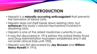
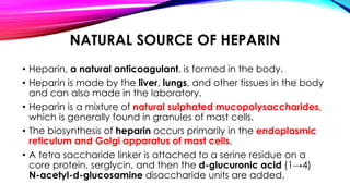
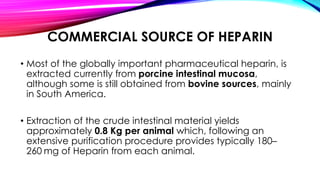
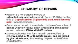
HEPARIN
Presented by: Megh.P.Vithalkar
M.Pharm Semester-1 (2020)
Department of Pharmacology.
Goa college of Pharmacy, Panaji-Goa.
INTRODUCTION
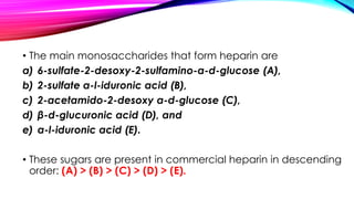
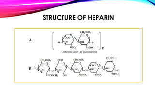
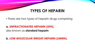
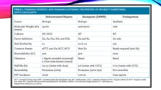
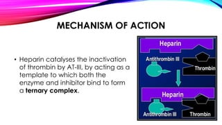
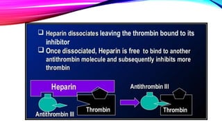
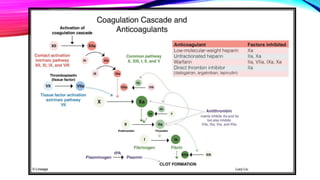
• Heparin is a naturally occurring anticoagulant that prevents
the formation of blood clots.
• Heparin does not itself break down existing clots, but
enhances the body’s natural mechanisms involved in
dissolving clots.
• Heparin is one of the oldest medicines currently in use.
• It was first discovered in 1916 before the United States Food
and Drug Administration had been established and it did
not enter clinical trials until 1935.
• Heparin was first discovered by Jay McLean and William
Henry Howell in 1916.
NATURAL SOURCE OF HEPARIN
• Heparin, a natural anticoagulant, is formed in the body.
• Heparin is made by the liver, lungs, and other tissues in the body
and can also made in the laboratory.
• Heparin is a mixture of natural sulphated mucopolysaccharides,
which is generally found in granules of mast cells.
• The biosynthesis of heparin occurs primarily in the endoplasmic
reticulum and Golgi apparatus of mast cells.
• A tetra saccharide linker is attached to a serine residue on a
core protein, serglycin, and then the d-glucuronic acid (1→4)
N-acetyl-d-glucosamine disaccharide units are added.
COMMERCIAL SOURCE OF HEPARIN
• Most of the globally important pharmaceutical heparin, is
extracted currently from porcine intestinal mucosa,
although some is still obtained from bovine sources, mainly
in South America.
• Extraction of the crude intestinal material yields
approximately 0.8 Kg per animal which, following an
extensive purification procedure provides typically 180–
260 mg of Heparin from each animal.
CHEMISTRY OF HEPARIN
• Heparin is a heterogenic mixture of
sulfonated polysaccharides made from a 15-100 repeating
units of D-glucosamine, D-glucuronic acid, and L-iduronic
acid. (monosaccharides)
• Commercial heparin is essentially a mixture of a number of
compounds with various chain lengths and of molecular
masses between 5000 and 30,000 daltons.
• Monosaccharides that form heparin are modified by
either N-acetyl, or N- or O-sulfate groups, and are joined
by glucoside bonds, thus forming polymers with different
chain lengths.
• The main monosaccharides that form heparin are
a) 6-sulfate-2-desoxy-2-sulfamino-α-d-glucose (A),
b) 2-sulfate α-l-iduronic acid (B),
c) 2-acetamido-2-desoxy α-d-glucose (C),
d) β-d-glucuronic acid (D), and
e) α-l-iduronic acid (E).
• These sugars are present in commercial heparin in descending
order: (A) > (B) > (C) > (D) > (E).
• Because of the presence of sulfonate and carboxyl
groups in the molecules, heparin is a strongly acidic
compound that is partially neutralized in the body by
substituting acidic hydrogen atoms in sulphate groups
with sodium ions.
• One unit of heparin (the "Howell unit") is an amount
approximately equivalent to 0.002 mg of pure heparin,
which is the quantity required to keep 1 ml of cat's blood
fluid for 24 hours at 0 °C.
STRUCTURE OF HEPARIN
TYPES OF HEPARIN
• There are two types of heparin drugs comprising
a. UNFRACTIONATED HEPARIN (UFH),
also known as standard heparin
b. LOW MOLECULAR WEIGHT HEPARIN (LMWH).
MECHANISM OF ACTION
• Heparin catalyses the inactivation
of thrombin by AT-III, by acting as a
template to which both the
enzyme and inhibitor bind to form
a ternary complex.
PHYSIOLOGICAL FUNCTION
• Heparin's normal role in the body is unclear.
• Heparin is usually stored within the secretory granules
of mast cells and released only into the vasculature at sites
of tissue injury.
• It has been proposed that, rather than anticoagulation, the
main purpose of heparin is defence at such sites against
invading bacteria and other foreign materials.
MEDICAL USE
• Heparin is generally used for anticoagulation for the following
conditions:
1. Acute coronary syndrome, e.g., NSTEMI
2. Atrial fibrillation
3. Deep-vein thrombosis and pulmonary embolism
4. Cardiopulmonary bypass for heart surgery
5. ECMO circuit for extracorporeal life support
6. Hemofiltration
• Heparin and its low-molecular-weight derivatives
(e.g., enoxaparin, dalteparin, tinzaparin) are effective in preventing
deep vein thrombosis and pulmonary emboli in people at high risk.
REFERENCES
• https://www.sciencedirect.com/science/article/abs/pii/S0065231808600670
• https://www.slideshare.net/venkateshkolla73/heparin-42670554
• https://www.news-medical.net/health/Heparin-History.aspx
• https://www.nature.com/articles/s41598-019-39093-
6#:~:text=Introduction,sources%2C%20mainly%20in%20South%20America.
• https://journal.chestnet.org/article/S0012-3692(15)41331-5/fulltext
• https://www.slideshare.net/IbrahimOsman5/anticoagulant-therapy-
78457821
• https://en.wikipedia.org/wiki/Heparin#Heparin_structure
• https://www.sciencedirect.com/topics/chemistry/heparin#:~:text=Heparin%
20is%20a%20heterogenic%20mixture,masses%20between%205000%20and%2
030%2C000
THANK YOU

Heparin: Source, Structure, Chemistry, MOA and Uses

  • 1.
    HEPARIN Presented by: Megh.P.Vithalkar M.PharmSemester-1 (2020) Department of Pharmacology. Goa college of Pharmacy, Panaji-Goa.
  • 2.
    INTRODUCTION • Heparin isa naturally occurring anticoagulant that prevents the formation of blood clots. • Heparin does not itself break down existing clots, but enhances the body’s natural mechanisms involved in dissolving clots. • Heparin is one of the oldest medicines currently in use. • It was first discovered in 1916 before the United States Food and Drug Administration had been established and it did not enter clinical trials until 1935. • Heparin was first discovered by Jay McLean and William Henry Howell in 1916.
  • 3.
    NATURAL SOURCE OFHEPARIN • Heparin, a natural anticoagulant, is formed in the body. • Heparin is made by the liver, lungs, and other tissues in the body and can also made in the laboratory. • Heparin is a mixture of natural sulphated mucopolysaccharides, which is generally found in granules of mast cells. • The biosynthesis of heparin occurs primarily in the endoplasmic reticulum and Golgi apparatus of mast cells. • A tetra saccharide linker is attached to a serine residue on a core protein, serglycin, and then the d-glucuronic acid (1→4) N-acetyl-d-glucosamine disaccharide units are added.
  • 4.
    COMMERCIAL SOURCE OFHEPARIN • Most of the globally important pharmaceutical heparin, is extracted currently from porcine intestinal mucosa, although some is still obtained from bovine sources, mainly in South America. • Extraction of the crude intestinal material yields approximately 0.8 Kg per animal which, following an extensive purification procedure provides typically 180– 260 mg of Heparin from each animal.
  • 5.
    CHEMISTRY OF HEPARIN •Heparin is a heterogenic mixture of sulfonated polysaccharides made from a 15-100 repeating units of D-glucosamine, D-glucuronic acid, and L-iduronic acid. (monosaccharides) • Commercial heparin is essentially a mixture of a number of compounds with various chain lengths and of molecular masses between 5000 and 30,000 daltons. • Monosaccharides that form heparin are modified by either N-acetyl, or N- or O-sulfate groups, and are joined by glucoside bonds, thus forming polymers with different chain lengths.
  • 6.
    • The mainmonosaccharides that form heparin are a) 6-sulfate-2-desoxy-2-sulfamino-α-d-glucose (A), b) 2-sulfate α-l-iduronic acid (B), c) 2-acetamido-2-desoxy α-d-glucose (C), d) β-d-glucuronic acid (D), and e) α-l-iduronic acid (E). • These sugars are present in commercial heparin in descending order: (A) > (B) > (C) > (D) > (E).
  • 7.
    • Because ofthe presence of sulfonate and carboxyl groups in the molecules, heparin is a strongly acidic compound that is partially neutralized in the body by substituting acidic hydrogen atoms in sulphate groups with sodium ions. • One unit of heparin (the "Howell unit") is an amount approximately equivalent to 0.002 mg of pure heparin, which is the quantity required to keep 1 ml of cat's blood fluid for 24 hours at 0 °C.
  • 8.
  • 9.
    TYPES OF HEPARIN •There are two types of heparin drugs comprising a. UNFRACTIONATED HEPARIN (UFH), also known as standard heparin b. LOW MOLECULAR WEIGHT HEPARIN (LMWH).
  • 11.
    MECHANISM OF ACTION •Heparin catalyses the inactivation of thrombin by AT-III, by acting as a template to which both the enzyme and inhibitor bind to form a ternary complex.
  • 14.
    PHYSIOLOGICAL FUNCTION • Heparin'snormal role in the body is unclear. • Heparin is usually stored within the secretory granules of mast cells and released only into the vasculature at sites of tissue injury. • It has been proposed that, rather than anticoagulation, the main purpose of heparin is defence at such sites against invading bacteria and other foreign materials.
  • 15.
    MEDICAL USE • Heparinis generally used for anticoagulation for the following conditions: 1. Acute coronary syndrome, e.g., NSTEMI 2. Atrial fibrillation 3. Deep-vein thrombosis and pulmonary embolism 4. Cardiopulmonary bypass for heart surgery 5. ECMO circuit for extracorporeal life support 6. Hemofiltration • Heparin and its low-molecular-weight derivatives (e.g., enoxaparin, dalteparin, tinzaparin) are effective in preventing deep vein thrombosis and pulmonary emboli in people at high risk.
  • 16.
    REFERENCES • https://www.sciencedirect.com/science/article/abs/pii/S0065231808600670 • https://www.slideshare.net/venkateshkolla73/heparin-42670554 •https://www.news-medical.net/health/Heparin-History.aspx • https://www.nature.com/articles/s41598-019-39093- 6#:~:text=Introduction,sources%2C%20mainly%20in%20South%20America. • https://journal.chestnet.org/article/S0012-3692(15)41331-5/fulltext • https://www.slideshare.net/IbrahimOsman5/anticoagulant-therapy- 78457821 • https://en.wikipedia.org/wiki/Heparin#Heparin_structure • https://www.sciencedirect.com/topics/chemistry/heparin#:~:text=Heparin% 20is%20a%20heterogenic%20mixture,masses%20between%205000%20and%2 030%2C000
  • 17.