Personnel and Equipment - Code and Rapid Response Workshop
Cystitis
1. A 71-year-old woman was brought to the emergency room
because of confusion and coffee ground vomitus. Physical
examination was unremarkable. The abdomen was soft
and non-tender. Laboratory tests showed normochro-
mic normocytic anemia, elevated inflammatory param-
eters and elevated kidney function tests. Urine analysis
showed numerous red blood cells, white blood cells and
bacteriae. Abdominal X-ray showed a curvilinear area of
radiolucency delineating the bladder wall (Figure 1a).
Non-contrast enhanced computed tomography (CT) of
the abdomen revealed multiple gas pockets within the
bladder wall (Figure 1b and 1c). A diagnosis of emphy-
sematous cystitis was made and the patient was started
empirically on intravenous piperacillin/tazobactom.
Urine culture grew E. Coli and treatment was switched
to intravenous levofloxacin. There was gradual clinical
improvement of the patient. Control ultrasonography
performed the week after admission showed a normal
bladder wall.
Emphysematous cystitis is an uncommon but severe
lower urinary tract infection characterized by the accu-
mulation of gas in and around the bladder wall produced
by bacterial or fungal fermentation. The most common
causative organisms are Escherichia Coli, Enterobacter
Aerogenes and Klebsiella Pneumonia. The majority of
patients are elderly, female and have type 2 diabetes mel-
litus. The clinical presentation of emphysematous cystitis
is nonspecific and varied, ranging from asymptomatic uri-
nary tract infection to severe peritonitis or septic shock.
Emphysematous cystitis requires aggressive treatment
Dekeyzer, S and Houthoofd, B. Emphysematous Cystitis.
Journal of the Belgian Society of Radiology. 2018;
102(1): 66, 1–2. DOI: https://doi.org/10.5334/jbsr.1192
* Universitair Ziekenhuis Antwerpen (UZA), BE
†
Sint-Andriesziekenhuis, Tielt, BE
Corresponding author: Brecht Houthoofd
(brecht.houthoofd@gmail.com)
IMAGES IN CLINICAL RADIOLOGY
Emphysematous Cystitis
Sven Dekeyzer*
and Brecht Houthoofd†
Keywords: emhysematous cystitis; bladder; urinary tract; infection
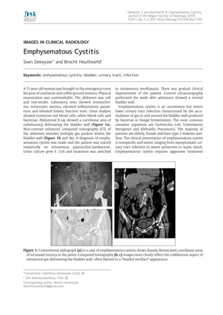
Figure 1: Conventional radiograph (a) in a case of emphysematous cystitis shows sharply demarcated, curvilinear areas
of increased lucency in the pelvis. Computed tomography (b, c) images more clearly reflect the cobblestone aspect of
intramural gas delineating the bladder wall, often likened to a “beaded necklace” appearance.
![Dekeyzer and Houthoofd: Emphysematous CystitisArt. 66, page 2 of 2
How to cite this article: Dekeyzer, S and Houthoofd, B. Emphysematous Cystitis. Journal of the Belgian Society of Radiology.
2018; 102(1): 66, 1–2. DOI: https://doi.org/10.5334/jbsr.1192
Submitted: 02 September 2016 Accepted: 08 September 2016 Published: 12 October 2018
Copyright: © 2018 The Author(s). This is an open-access article distributed under the terms of the Creative Commons
Attribution 4.0 International License (CC-BY 4.0), which permits unrestricted use, distribution, and reproduction in any medium,
provided the original author and source are credited. See http://creativecommons.org/licenses/by/4.0/.
OPEN ACCESS
Journal of the Belgian Society of Radiology is a peer-reviewed open access journal
published by Ubiquity Press.
with parenteral antibiotics, bladder drainage and control
of sugar level and the overall average mortality rate is
approximately 7% [1].
Because of the nonspecific presentation, imaging plays
an important role in the diagnosis. Plain abdominal X-rays
can show a rim of gas lucency outlining the bladder wall
and/or air fluid levels within the bladder. Computed
tomography (CT) can more accurately define the extent
and severity of the disease, can detect cases that are
not apparent on plain radiography and can help in dif-
ferentiating emphysematous cystitis from colovesical
fistula, intra-abdominal abcesses, neoplastic disease or
emphysematous pyelonephritis.
Competing Interests
The authors have no competing interests to declare.
Reference
1. Amano, M and Shimizu, T. Emphysematous
cystitis: A review of the literature. Intern Med.
2014; 53: 79–82. DOI: https://doi.org/10.2169/
internalmedicine.53.1121](data:image/gif;base64,R0lGODlhAQABAIAAAAAAAP///yH5BAEAAAAALAAAAAABAAEAAAIBRAA7)