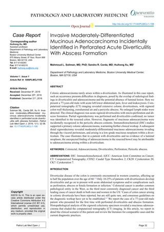
2. PATHOLOGY AND LABORATORY MEDICINE
Open Journal
http://dx.doi.org/10.17140/PLMOJ-1-106
Pathol Lab Med Open J Page 33
CASE PRESENTATION
A 77-year-old male patient was referred to our medical center
from an outside hospital after presenting for the first time with
acute onset of abdominal pain localized to the left lower quad-
rant, nausea, vomiting, fever, chills, malaise, anorexia and di-
arrhea. The laboratory results were found to be unremarkable
except for a white blood cell count of 16,300 /µL and creatinine
level of 1.7 mg/dL. Electrocardiogram was unremarkable and
arterial oxygen saturation was 95%. A CT scan with intravenous
contrast revealed extensive colonic diverticulosis, thickening of
the wall of the sigmoid colon, consistent with sigmoid diverticu-
litis and extraluminal air compatible with a micro-perforation.
In addition, a 4.9×4.6 cm abscess was identified adjacent to the
colon. No bowel obstruction or enlarged (abdominal or pelvic)
lymph nodes were recognized by.
The patient’s past medical history included bladder
cancer with cystectomy and neobladder reconstruction, appen-
dectomy, and prostatectomy 15 years earlier. The histopatholog-
ical type and staging of the bladder cancer is not known. More-
over, the patient also had a history of coronary artery disease
with coronary angioplasty and stent placement, atrial fibrillation
with placement of an inferior vena cava filter, chronic obstruc-
tive pulmonary disease, diabetes mellitus, hypertension and
deep venous thrombosis. He was also a heavy smoker for 24
years who quit at the age of 40. There was no history of alcohol
use or known allergies.
Due to the persistence of abdominal pain and severe
anorexia, the patient underwent exploratory laparotomy, during
which diverticulitis was confirmed. A partial (sigmoid) colec-
tomy was performed. The main resection specimen consisted of
a 9.5 cm long segment of sigmoid colon with attached perico-
lic fat, as well as pelvic abscess contents, containing gelatinous
material admixed with firm pink tan tissue. Upon opening the
bowel, the congested mucosa showed many diverticula ranging
in depth from 0.5 to 1.5 cm associated with necrotic tissues (Fig-
ure 1A). Histopathological examination revealed diverticulitis
(Figure 1B) and pericolonic adhesions, but no tumor. The pelvic
abscess contents showed mucinous adenocarcinoma with posi-
tive staining for CDX2 and CK20 and negative staining for CK7
consistent with a primary colonic adenocarcinoma (Figure 2).
Figure 1: Panel A shows a gross picture of a cross section of the sigmoid colon with an arrow indicating a di-
verticulum and an asterisk indicating the site of perforation. Panel B shows 40x magnification of an H&E stained
section showing a diverticulum with surrounding acute and chronic inflammatory infiltrate.
Figure 2: Panel A is H&E at 40x showing mucinous adenocarcinoma. The immunohistochemical profile
shows the tumor cells with negative staining for CK7 (B) and positive staining for CK20 (C) and CDX2 (D).
3. PATHOLOGY AND LABORATORY MEDICINE
Open Journal
http://dx.doi.org/10.17140/PLMOJ-1-106
Pathol Lab Med Open J Page 34
On post-operative day 4, due to worsening clinical
symptoms of perforated sigmoid diverticulitis and newly de-
veloped peritonitis, a subsequent distal sigmoid colectomy was
performed. Upon opening the bowel, the hemorrhagic colonic
mucosa showed multiple perforated diverticula surrounded with
necrotic tissue, but no visible masses were identified. Histopath-
ological examination revealed a moderately-differentiated mu-
cinous adenocarcinoma (Figure 3) measuring 0.9 cm in greatest
dimension associated with the perforation site in the distal sig-
moid colon. The tumor had moderate intramural and peritumor
lymphocytic responses. Immunohistochemical analysis again
showed positive immunostaining of the tumor cells for CK20
and CDX2 and negative immunostaining for CK7, supporting
the diagnosis of a primary colonic adenocarcinoma. The tumor
was found to invade through the muscularis propria into perico-
lonic soft tissue and visceral peritoneum. The proximal, distal
and radial margins were not involved and there was no perineu-
ral invasion, lymph-vascular invasion, or regional lymph node
metastases. In addition, a low grade mucinous neoplasm was
seen in the vicinity of the mucinous adenocarcinoma (Figure
4). Additional pathological findings included ischemic necrosis,
multiple diverticula, acute and chronic inflammation, abscess
formation and acute organizing serositis. As such, the tumor
was classified according to the American Joint Committee on
Cancer (AJCC) Pathological Staging 7th
edition as (pT4a, pN0).
Additionally, the specimen was submitted for KRAS mutation
analysis, revealing that KRAS codon 12 mutation (c.35G>T)
was present. Unfortunately, the patient died within six days of
presentation to our hospital from septicemia as a complication
of perforated diverticulitis.
DISCUSSION AND LITERATURE REVIEW
The incidence of diverticulosis and diverticulitis has been rising
in the western world due to dietary habits and prolonged life ex-
pectancy such that diverticulosis now affects 48% of the devel-
oped world population over the age of 50.1,2
Diverticulitis typi-
cally presents as “acute abdomen” with acute abdominal pain,
fever, diarrhea or constipation, nausea and vomiting.1,8,9
Aging
and western diet are also risk factors for increased incidence of
colorectal cancer which is the third most common cancer type
in the US. The presentation of colorectal cancers can vary con-
siderably, from being entirely asymptomatic to bleeding per rec-
tum, melena or constipation up to frank intestinal obstruction.
Colorectal cancers arising in colonic diverticulitis have been
reported but remain very rare with few reported cases.4-7,10-17
This co-occurrence is proposed to represent a diagnostically and
therapeutically significant correlation8
rather than a causal rela-
tionship18
with only poorly defined guidelines existing to guide
diagnosis and eventual management of the coexisting condi-
tions. Interestingly, benign polyps are also reported to arise in
association with colonic diverticula.19,20
From a prognostic point of view, colorectal carcinoma
arising from diverticula may have a higher likelihood of extra-
mural spread to neighboring organs due to the deficiency of the
muscular layer in diverticula. In support of this, Yagi et al15
re-
ported a case of a sigmoid colon adenocarcinoma arising from
a colonic diverticulum and invading the urinary blabber via a
colovesical fistula. Importantly, attempts at endoscopic mucosal
resection can be complicated by iatrogenic perforation requiring
subsequent hemicolectomy.13
Rendering the proper diagnosis is one of the chal-
lenges for concomitant diverticulitis and adenocarcinoma given
the different diagnostic techniques for each clinical entity. The
diagnostic modality of choice for colorectal adenocarcinoma is
colonoscopy which may fail in identifying submucosal lesions
especially in the absence of frank mucosal involvement. On
the other hand, CT scan is the standard diagnostic modality of
choice for diverticulitis, but its findings can be non-specific. In-
deed, thickening of the intestinal wall, localized inflammatory
reaction, enlarged pericolic lymph nodes, are all radiological
findings commonly found in colorectal carcinomas as well.21
Figure 3: H&E at 100x of mucinous adenocarcinoma arising within a
diverticulum.
Figure 4: H&E at 40x, low grade mucinous neoplasm near the diver-
ticulum.
4. PATHOLOGY AND LABORATORY MEDICINE
Open Journal
http://dx.doi.org/10.17140/PLMOJ-1-106
Pathol Lab Med Open J Page 35
Likewise, barium enema, positron emission tomography-com-
puted tomography and magnetic resonance imaging may fail in
distinguishing inflammatory from malignant conditions. In ad-
dition to radiological ambiguity, some diverticulitis cases have
been reported to present typically as reduced stool caliber due to
considerable stenosis rather than the conventional acute abdo-
men presentation.22
Therefore, distinguishing diverticulitis from
carcinoma-induced stricture can be challenging without histo-
pathological examination.
Studies show that the incidence of colorectal cancer
at the site of diverticulitis one year after the episode is 44-fold
higher compared to the age-matched general population.23
Simi-
larly, adenomas are reported to have a significantly higher in-
cidence in association with diverticular disease.24
Therefore,
practice guidelines recommend performing a colonoscopy after
the resolution of an episode of diverticulitis regardless of the
clinical or radiological presentation, in order to detect unsus-
pected etiologies, particularly neoplastic conditions.25
However,
because the incidence of carcinoma is significantly lower fol-
lowing an uncomplicated diverticulitis (0.3%) than following a
complicated one (7.6%), it has been proposed that endoscopic
evaluation may be skipped in patients with uncomplicated diver-
ticulitis.26
We propose that in a patient with a long history of di-
verticulitis, multiple colorectal cancer high risk factors, and any
discrepancy between radiology, clinical and pathological find-
ings, a thorough investigation is warranted.
CONFLICTS OF INTEREST
The authors declare that they have no conflicts of interest.
REFERENCES
1. Brian West A. The pathology of diverticulosis: Classical
concepts and mucosal changes in diverticula. J Clin Gastro-
enterol. 2006; 40(3): S126-S131. doi: 10.1097/01.mcg.00002
25508.90417.07
2. Hughes LE. Postmortem survey of diverticular disease of the
colon. I. Diverticulosis and diverticulitis. Gut. 1969; 10(5): 336-
344. Web site. http://gut.bmj.com/content/10/5/336.long. Ac-
cessed December 5, 2016.
3. Marley AR, Nan H. Epidemiology of colorectal cancer. Int J
Mol Epidemiol Genet. 2016; 7(3): 105-114.
4. Cohn KH, Weimar JA, Fani K, DeSoto-LaPaix F. Adenocar-
cinoma arising within a colonic diverticulum: Report of two
cases and review of the literature. Surgery. 1993; 113(2): 223-
226. Web site. http://europepmc.org/abstract/med/8430371. Ac-
cessed December 5, 2016.
5. Hines JR, Gordon RT. Adenocarcinoma arising in a diverticu-
lar abscess of the colon: Report of a case. Dis Colon Rectum.
1975; 18(1): 49-51. doi: 10.1007/BF02587240
6. Bellows CF, Haque S.Adenocarcinoma within a diverticulum:
A common tumor arising in an uncommon location. Dig Dis Sci.
2002; 47(12): 2758-2759. doi: 10.1023/A:1021013423752
7. McCraw RC, Wilson SM, Brown FM, Gardner WA. Ad-
enocarcinoma arising in a sigmoid diverticulum: Report of a
case. Dis Colon Rectum. 1976; 19(6): 553-556. doi: 10.1007/
BF02590952
8. Localio SA, Stahl WM. Diverticular disease of the alimentary
tract. I. The colon. Curr Probl Surg. 1967; 1-78.
9. Simpson J. Perception and the origin of symptoms in diverticu-
lar disease. Dig Dis. 2012; 30(1): 75-79. doi: 10.1159/000335723
10. Drut R. Adenoacanthoma arising in a diverticulum of the co-
lon: Report of a case. Dis Colon Rectum. 1974; 17(2): 258-261.
doi: 10.1007/BF02588113
11. Kajiwara H, Umemura S, Mukai M, Sadahiro S, Tsutsumi Y.
Adenocarcinoma arising within a colonic diverticulum. Pathol
Int. 1996; 46(7): 538-539.
12. Kikuchi T, Kotanagi H, Kon H, Koyama K, Ito S, Otaka M.
Mucosal carcinoma within a colonic diverticulum. J Gastroen-
terol. 1999; 34(5): 622-625. doi: 10.1007/s005350050383
13. Fu KI, Hamahata Y, Tsujinaka Y. Early colon cancer within a
diverticulum treated by magnifying chromoendoscopy and lapa-
roscopy. World J Gastroenterol. 2010; 16(12): 1545-1547. doi:
10.3748/WJG.v16.i12.1545
14. Nomi M, Umemoto S, Kikutake T, et al. A case of carcinoma
arising in a diverticulum of the transverse colon. Gan To Kagaku
Ryoho. 2014; 41(12): 1680-1682. Web site. http://europepmc.
org/abstract/med/25731294. Accessed December 5, 2016.
15. Yagi Y, Shoji Y, Sasaki S, et al. Sigmoid colon cancer arising
in a diverticulum of the colon with involvement of the urinary
bladder: A case report and review of the literature. BMC Gastro-
enterol. 2014; 14: 90. doi: 10.1186/1471-230X-14-90
16. Kobayashi N, Hirabayashi K, Matsui T, et al. Depressed-
type colon cancer in a patient with diverticulosis. Endoscopy.
2008; 40(Suppl 2): E44. doi: 10.1055/s-2007-966854
17. Imai K, Hotta K, Ono H. Unusual colonic mucosal cancer
extending into a diverticulum. Dig Endosc. 2014; 26(6): 752.
doi: 10.1111/den.12336
18. McCallum A, Eastwood MA, Smith AN, Fulton PM. Co-
lonic diverticulosis in patients with colorectal cancer and in
controls. Scand J Gastroenterol. 1988; 23(3): 284-286. doi:
5. PATHOLOGY AND LABORATORY MEDICINE
Open Journal
http://dx.doi.org/10.17140/PLMOJ-1-106
Pathol Lab Med Open J Page 36
10.3109/00365528809093866
19. Martich V, Kutashy M, Gasparaitis A. Polyp arising in a co-
lonic diverticulum. AJR Am J Roentgenol. 1992; 159(6): 1348.
doi: 10.2214/ajr.159.6.1442418
20. Barr YR, Brazowski E, Leider-Trejo L. Villous adenoma in
a perforated colonic diverticulum. Int J Colorectal Dis. 2006;
21(3): 282-284. doi: 10.1007/s00384-004-0694-1
21. Balthazar EJ, Megibow A, Schinella RA, Gordon R. Limi-
tations in the CT diagnosis of acute diverticulitis: Comparison
of CT, contrast enema, and pathologic findings in 16 patients.
AJR Am J Roentgenol. 1990; 154(2): 281-285. doi: 10.2214/
ajr.154.2.2105015
22. Nishiyama N, Mori H, Kobara H, et al. Difficulty in dif-
ferentiating two cases of sigmoid stenosis by diverticulitis from
cancer. World J Gastroenterol. 2012; 18(27): 3623-3626. doi:
10.3748/wjg.v18.i27.3623
23. Meyer J, Thomopoulos T, Usel M, et al. The incidence of
colon cancer among patients diagnosed with left colonic or sig-
moid acute diverticulitis is higher than in the general population.
Surg Endosc. 2015; 29(11): 3331-3337. doi: 10.1007/s00464-
015-4093-1
24. Morini S, Hassan C, Zullo A, et al. Diverticular disease as
a risk factor for sigmoid colon adenomas. Dig Liver Dis. 2002;
34(9): 635-639. doi: 10.1016/S1590-8658(02)80206-7
25. Feingold D, Steele SR, Lee S, et al. Practice parameters for
the treatment of sigmoid diverticulitis. Dis Colon Rectum. 2014;
57(3): 284-294. doi: 10.1097/DCR.0000000000000075
26. de Vries HS, Boerma D, Timmer R, et al. Routine colonos-
copy is not required in uncomplicated diverticulitis: A system-
atic review. Surg Endosc. 2014; 28(7): 2039-2047. doi: 10.1007/
s00464-014-3447-4