![United Prime Publications. LLC., clinandmedimages.com 2
Volume 7 Issue 5-2023 Case Report
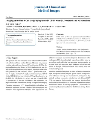
Figure 1: Contrast-enhanced CT images in portal phase. A. Multiple well-delimited hypovascular hepatic nodules. Some nodules are associated with
intrahepatic bile duct dilatation; B and C. Multiple bilateral hypovascular renal nodules. Infiltrative soft-tissue pancreatic mass with main pancreatic
duct dilatation; D. Infiltrative nodules in the interventricular septum e left ventricular myocardial wall.
Figure 2: Two-dimensional transthoracic echocardiogram on longitudinal parasternal view depicting multiple hypoechoic nodules in the latero-inferior
wall and in the interventricular septum causing an increase in the left ventricular walls thickness.
2. Discussion
The spectrum of opportunistic diseases in advanced HIV is exten-
sive, and there is an increased risk of neoplasms. Given the pa-
tient’s clinical presentation and our local epidemiological context,
the primary differential diagnoses included tuberculosis, dissemi-
nated histoplasmosis, cryptococcosis, and non-Hodgkin lympho-
ma.
Our patient’s CT showed a disseminated disease, with multiple
well-defined solid lesions affecting liver, kidneys, pancreas and
heart. Also, the TTE showed a cystic pattern in myocardium. It
was difficult to formulate a strong hypothesis, even more because
the patient’s blood tests were almost normal. High level of fer-
ritin is non-specific, but hyperuricemia highlights neoplasm. Al-
though lymphoma became our main hypothesis, the presentation
was atypical, with no lesions in spleen nor lymph nodes enlarge-
ment. Searching for similar images led us to several differential
diagnoses. A case of angiosarcoma presented similar low-attenua-
tion lesions throughout the liver, but also a large spleen lesion [1].
Eshiwe at al. described a case of disseminated tuberculosis in an
immunocompetent woman with multiple hepatic lesions sugges-
tive of abscesses, in addition to other lesions in lungs and pericar-
dial effusion [2]. A review of concurrent hepatic and spleen lesions
described cases of melanoma, disseminated candidiasis, sarcoido-
sis and lymphoma, not like our patient [3]. Another review of pe-
riportal pathologies showed a very similar hepatic lesions from a
woman with lymphoma, but it was limited to describing only liver
lesions [4]. Kidney spread of lymphoma is common and the image
finding in most cases is multiple solid parenchymal masses [5].
Kidney abscesses, pyelonephritis and IgG4 disease can present
similar pattern on CT, but the clinical history and physical exam
usually help in the differentialdiagnosis.
Particularly, we do not recall a myocardium involvement like this
case. On CT the image was poorly delimited, suggestive of an in-
filtrative disease, but on TTE the lesions were well delimited with
cystic aspect. Looking exclusively at the TTE we thought about
hydatidosis, but the abdominal lesions were not consistent with](data:image/gif;base64,R0lGODlhAQABAIAAAAAAAP///yH5BAEAAAAALAAAAAABAAEAAAIBRAA7)
Recommended
Recommended
More Related Content
Similar to Imaging of Diffuse B Cell Large Lymphoma in Liver, Kidneys, Pancreas and Myocardium in a Case Report
Similar to Imaging of Diffuse B Cell Large Lymphoma in Liver, Kidneys, Pancreas and Myocardium in a Case Report (20)
More from semualkaira
More from semualkaira (20)
Recently uploaded
Recently uploaded (20)
Imaging of Diffuse B Cell Large Lymphoma in Liver, Kidneys, Pancreas and Myocardium in a Case Report
- 1. Case Report ISSN: 2640-9615 Volume 7 Imaging of Diffuse B Cell Large Lymphoma in Liver, Kidneys, Pancreas and Myocardium in a Case Report * Corresponding author: Andréa Varon, Infectious Diseases National Institute / Fiocruz, Bonsucesso Federal Hospital, Rio de Janeiro, Brazil Received: 28 Sep 2023 Accepted: 01 Nov 2023 Published: 10 Nov 2023 J Short Name: JCMI Copyright: ©2023 Varon A, This is an open access article distributed under the terms of the Creative Commons Attribution Li- cense, which permits unrestricted use, distribution, and build upon your work non-commercially. Citation: Varon A, Imaging of Diffuse B Cell Large Lymphoma in Liver, Kidneys, Pancreas and Myocardium in a Case Report. J Clin Med Img. 2023; V7(5): 1-3 Journal of Clinical and Medical Images United Prime Publications. LLC., clinandmedimages.com 1 Varon A¹, ²* , Saraiva RM¹, Netto TAL¹, Zeferino TCA², Astacio GSM¹ and Theodoro PHN¹ 1 Infectious Diseases National Institute / Fiocruz, Rio de Janeiro, Brazil 2 Bonsucesso Federal Hospital, Rio de Janeiro, Brazil 1. Case Report A 41-year-old man was transferred to our Infectious Diseases Unit with a history of three weeks of fever, abdominal pain, nausea, vomiting and weight loss. He was HIV positive since 2013 but dis- continued antiretroviral therapy in 2019. At admission he was afe- brile, with haemoglobin level of 12.8 g/dL, leucocytes 4,340 cells/ mm³, platelets 237,000 cells/mm³, reative C protein 14.1 mg/dL, urea 48 mg/dL, creatinin 0.98 mg/dL, normal electrolytes, AST 90 U/mL, ALT 126 U/mL, total bilirrubin 0.75 mg/dL, albumin 3.5 g/ dL, DHL 344 U/L, ferritin 914 ng/mL, uric acid 8.5 mg/dL, CD4 74 cells/mm³ (11%) and HIV viral load 111,000 copies/mm³. His physical exam was unremarkable, except for hepatosplenomegaly. Computer tomography (CT) showed multiple well-defined hy- povascular nodules in liver and kidneys, a large hypovascular and infiltrative mass in pancreas and sparse small hypovascular and infiltrative nodules in myocardium (Figure 1). Transthoracic echo- cardiogram (TTE) showed multiple hypoechoic nodules in the la- tero-inferior wall and in the interventricular septum, causing an increase in the left ventricular walls thickness, and an isoechoic nodule in the right ventricular outflow tract, causing mild flow obstruction (Figure 2). Infectious diseases investigation included cryptococcal serum an- tigen, histoplasma urinary antigen, sputum culture for mycobac- teria, hidatidosis serology and blood cultures, all negatives. He- patitis B serology was positive only for total anti-HBc, and HBV PCR was negative. Antirretroviral was restarted with tenofovir, lamivudine and dolutegravir. An ultrasound guided hepatic biopsy demonstrated diffuse B cell large lymphoma. Unfortunately, his condition rapidly deteriorated and he died before starting chemo- therapy.
- 2. United Prime Publications. LLC., clinandmedimages.com 2 Volume 7 Issue 5-2023 Case Report Figure 1: Contrast-enhanced CT images in portal phase. A. Multiple well-delimited hypovascular hepatic nodules. Some nodules are associated with intrahepatic bile duct dilatation; B and C. Multiple bilateral hypovascular renal nodules. Infiltrative soft-tissue pancreatic mass with main pancreatic duct dilatation; D. Infiltrative nodules in the interventricular septum e left ventricular myocardial wall. Figure 2: Two-dimensional transthoracic echocardiogram on longitudinal parasternal view depicting multiple hypoechoic nodules in the latero-inferior wall and in the interventricular septum causing an increase in the left ventricular walls thickness. 2. Discussion The spectrum of opportunistic diseases in advanced HIV is exten- sive, and there is an increased risk of neoplasms. Given the pa- tient’s clinical presentation and our local epidemiological context, the primary differential diagnoses included tuberculosis, dissemi- nated histoplasmosis, cryptococcosis, and non-Hodgkin lympho- ma. Our patient’s CT showed a disseminated disease, with multiple well-defined solid lesions affecting liver, kidneys, pancreas and heart. Also, the TTE showed a cystic pattern in myocardium. It was difficult to formulate a strong hypothesis, even more because the patient’s blood tests were almost normal. High level of fer- ritin is non-specific, but hyperuricemia highlights neoplasm. Al- though lymphoma became our main hypothesis, the presentation was atypical, with no lesions in spleen nor lymph nodes enlarge- ment. Searching for similar images led us to several differential diagnoses. A case of angiosarcoma presented similar low-attenua- tion lesions throughout the liver, but also a large spleen lesion [1]. Eshiwe at al. described a case of disseminated tuberculosis in an immunocompetent woman with multiple hepatic lesions sugges- tive of abscesses, in addition to other lesions in lungs and pericar- dial effusion [2]. A review of concurrent hepatic and spleen lesions described cases of melanoma, disseminated candidiasis, sarcoido- sis and lymphoma, not like our patient [3]. Another review of pe- riportal pathologies showed a very similar hepatic lesions from a woman with lymphoma, but it was limited to describing only liver lesions [4]. Kidney spread of lymphoma is common and the image finding in most cases is multiple solid parenchymal masses [5]. Kidney abscesses, pyelonephritis and IgG4 disease can present similar pattern on CT, but the clinical history and physical exam usually help in the differentialdiagnosis. Particularly, we do not recall a myocardium involvement like this case. On CT the image was poorly delimited, suggestive of an in- filtrative disease, but on TTE the lesions were well delimited with cystic aspect. Looking exclusively at the TTE we thought about hydatidosis, but the abdominal lesions were not consistent with
- 3. United Prime Publications. LLC., clinandmedimages.com 3 Volume 7 Issue 5-2023 Case Report this diagnosis. After confirmation of lymphoma, we postulated that this pattern of hypoechogenic nodules could be lymphoma- tous granulomas. Ideally, we should have performed a necropsy for better elucidation if we had the resource. 3. Conclusion HIV patients with multiple abdominal solid lesions should always have the suspicion of lymphoma, even in the absence of lymph nodes involvement. Multiple hypoechogenic myocardial lesions should rise the suspicion of a granulomatous disease.