More Related Content Similar to BioSight targets it rightly - structural homology and beyond in e.coli dhodh (20) 1. Dihydroorotate dehydrogenase (DHODH) is a mitochondrial
enzyme involved in the de novo biosynthesis of Uridine Mono-
Phosphate (UMP) as it catalyzes the oxidation of dihydroorotate
to orotate (ORO). Besides, DHODH has been extensively
studied for therapeutic purposes and is currently known as the
therapeutic target of leflunomide, a treatment for rheumatoid
and psoriatic arthitis1
. The present study challenges BioBind
, an application for the detection and ranking of similar sites
of biological macromolecules, on the active site of E. coli
DHODH. Researches on DHODH active site revealed that
the binding of ORO is strongly dependent on a flexible loop
bearing a serine, anchor point for the binding.
METHODOLOGY
BioSight embeddes BioBind, a surface similarity based al-
gorithm using models borrowed to the alpha-shape theory.
The algorithm takes a given surface pattern as query, detects
motifs at the surface of target macromolecules, seeks for the
best superpositions and ranks them by similarity using a sco-
ring function2
: best results have the highest scores and lowest
ranks. The query pattern can be automatically defined using
atoms nearby a given ligand. Here, the E. coli DHODH
(PYRD_ECOLI, PDB ID 1F763
) crystallized with its endoge-
nous ligand ORO was used. To build the query pattern,
atoms were chosen within a radius of about 6.5 Å around
the ligand (chain A). The analysis was performed on all
structures from the PDB database.
RESULTS
Asexpected,thetoprankedstructuresretrievedbyBioBind (highlighted
in red in Fig.2) correspond to the E. coli DHODH chains A,
B, D, E. Human DHODH shares 41% sequence identity and a
highly similar folding pattern with E. coli DHODH. For 44 out of
the 46 human DHODH structures (PYRD_HUMAN) listed in the
PDB database, BioBind attributed an excellent similarity score
(see orange bars in Fig.2) and accurately spotted the similar
surface corresponding to the counterpart site of ORO. For other
DHODH structures (yellow bars in Fig. 2), 129 out of the 260
chains corresponding to 17 unique homologs are in the leading
pack. In the other structures, either the critical loop is in a different
conformation, or it has not been resolved (respectively in yellow
and cyan Fig. 1B).
INTRODUCTION
/CASE STUDY
BioSight targets it rightly - structural
homology and beyond in e.coli dhodh
SUMMARY
BioSight* is a BIONEXT’s web platform dedicated to the retrieval and assessment of local surface similarities.
Here, BioSight was challenged on a structure of the protein E. coli dihydroorotate dehydrogenase (E. coli
DHODH) complexed with orotate. Among the first hits retrieved were legitimate DHODH homologs and
also the similarly folded dihydropyrimidine dehydrogenase. Surprisingly, the molybdenum storage protein
was also significantly highly ranked by BioSight despite its structural differences with the DHODH query.
Overall, in a couple of hours, BioSight was not only able to retrieve all known homologous DHODH
proteins but also to retrieve proteins with a different fold, nonetheless relevant.
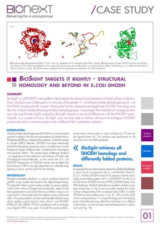
A.Dihydroorotate dehydrogenase (PDB ID 1F763
, chain A) complexed with its substrate orotate (ORO, spheres) - B.Superposition of three DHODH’s binding site. Binding
site residues (1F76 in blue) are displayed in lines, atoms defining the query site in balls & sticks. In some structures (in yellow) a flexible loop is in a different conformation, in
others (in cyan) it is missing. - C. MoSto protein complexed with two phosphate ions (spheres) in the UMP binding site.
Fig.1
A. B. C.
BioSight retrieves all
DHODH homologs and
differently folded proteins.
«
© BIONEXT 2017. Technology developed in Strasbourg, France. Bionext may be registered trademarks or service marks of Bionext registered in many jurisdictions worldwide. This document is
current as of the initial date of publication and may be changed by Bionext at any time.
https://biosight.bionext.com
01
Surface
similarity
DHODH
Structural
analysis
2. The lower BioBind ’s scores is then inevitable since seven atoms
of the loop are used by BioBind to define the query site
(see Fig. 1B). Moreover, among the best ranked structures,
BioBind further identified dihydropyrimidine dehydrogenase
[NADP(+)] (DPYD_PIG, in magenta in Fig. 2), whose fold
around the binding site is similar to the DHODH’s fold despite
different function and amino acid sequence (21.6% identity,
35.9% similarity). BioBind also retrieved the molybdenum
storage (MoSto) protein (MOSB_AZOVD, in green in Fig.
2) involved in the pyrimidine nucleotide biosynthetic process,
known to display an uridylate kinase function and thus an
UMP binding site. Despite its broadly different fold compared
to the query (see Fig.1, A and C), BioBind spotted the UMP
binding site, relevant concidering that UMP and ORO ligands
share the uracil substructure (see Fig. 3, green substructure).
CONCLUSION
Based on ORO binding site of the E. coli DHODH and
within a couple of hours, BioBind appears strongly efficient
to retrieve structures of all DHODH homologs provided
that the critical loop of the binding site was complete
enough and in a similar conformation. Besides, BioBind
further retrieved poorly related as well as a non related
yet relevant proteins. This analysis brings further evidence
of the potential of BioBind for the analysis of proteins of
therapeutic interest. Our fully secured platform can be ac-
cessed and challenged from https://biosight.bionext.com
REFERENCES
1. Leban, J., & Vitt, D. (2011). Human dihydroorotate dehydrogenase inhibitors, a
novel approach for the treatment of autoimmune and inflammatory diseases. Arznei-
mittelforschung, 61(01), 66-72.
2. Bionext submitted publication to BioInformatics. Available on demand for private
communication.
3. Nørager, S. et al. (2002). E. coli dihydroorotate dehydrogenase reveals
structural and functional distinctions between different classes of dihydroorotate
dehydrogenases. Structure, 10(9), 1211-1223.
4. Björnberg, O. et al. (1999). The activity of Escherichia coli dihydroorotate
dehydrogenase is dependent on a conserved loop identified by sequence homology,
mutagenesis, and limited proteolysis. Biochemistry, 38(10), 2899-2908.
*https://biosight.bionext.com
Fig.2: Biobind’s score distribution for all PDB structures. BioBind’s results are scored between 0 and 1. A score of 1 corresponds to structural identity.
Fig.3: 2D structures of ORO and UMP. The common uracil
substructure is displayed in green.
© BIONEXT 2017. Technology developed in Strasbourg, France. Bionext may be registered trademarks or service marks of Bionext registered in many jurisdictions worldwide. This document is
current as of the initial date of publication and may be changed by Bionext at any time.
https://biosight.bionext.com
ABOUT BIONEXT
BIONEXTisaFrenchbioinformaticscompanyfounded
in 2009 and dedicated to the better understanding
and treatment of various diseases. As such, BIONEXT
focuses on pharmaceutical problematics with a ma-
jor interest in guiding the prediction of biochemical
compounds for pharmaceuticals targets. BIONEXT
aims to accelerate and improve the effectiveness
of drug development and biological research by
developing programs and software-based tools to
tackle problematics of prime importance in the phar-
maceutical field, such as drug profile assessment or
suggestion of drug repurposing. Our first application
BioBind is based on the now accepted principle
that similar receptors bind similar ligands.
www.bionext.com
Surface
similarity
DHODH
Structural
analysis
02
![The lower BioBind ’s scores is then inevitable since seven atoms
of the loop are used by BioBind to define the query site
(see Fig. 1B). Moreover, among the best ranked structures,
BioBind further identified dihydropyrimidine dehydrogenase
[NADP(+)] (DPYD_PIG, in magenta in Fig. 2), whose fold
around the binding site is similar to the DHODH’s fold despite
different function and amino acid sequence (21.6% identity,
35.9% similarity). BioBind also retrieved the molybdenum
storage (MoSto) protein (MOSB_AZOVD, in green in Fig.
2) involved in the pyrimidine nucleotide biosynthetic process,
known to display an uridylate kinase function and thus an
UMP binding site. Despite its broadly different fold compared
to the query (see Fig.1, A and C), BioBind spotted the UMP
binding site, relevant concidering that UMP and ORO ligands
share the uracil substructure (see Fig. 3, green substructure).
CONCLUSION
Based on ORO binding site of the E. coli DHODH and
within a couple of hours, BioBind appears strongly efficient
to retrieve structures of all DHODH homologs provided
that the critical loop of the binding site was complete
enough and in a similar conformation. Besides, BioBind
further retrieved poorly related as well as a non related
yet relevant proteins. This analysis brings further evidence
of the potential of BioBind for the analysis of proteins of
therapeutic interest. Our fully secured platform can be ac-
cessed and challenged from https://biosight.bionext.com
REFERENCES
1. Leban, J., & Vitt, D. (2011). Human dihydroorotate dehydrogenase inhibitors, a
novel approach for the treatment of autoimmune and inflammatory diseases. Arznei-
mittelforschung, 61(01), 66-72.
2. Bionext submitted publication to BioInformatics. Available on demand for private
communication.
3. Nørager, S. et al. (2002). E. coli dihydroorotate dehydrogenase reveals
structural and functional distinctions between different classes of dihydroorotate
dehydrogenases. Structure, 10(9), 1211-1223.
4. Björnberg, O. et al. (1999). The activity of Escherichia coli dihydroorotate
dehydrogenase is dependent on a conserved loop identified by sequence homology,
mutagenesis, and limited proteolysis. Biochemistry, 38(10), 2899-2908.
*https://biosight.bionext.com
Fig.2: Biobind’s score distribution for all PDB structures. BioBind’s results are scored between 0 and 1. A score of 1 corresponds to structural identity.
Fig.3: 2D structures of ORO and UMP. The common uracil
substructure is displayed in green.
© BIONEXT 2017. Technology developed in Strasbourg, France. Bionext may be registered trademarks or service marks of Bionext registered in many jurisdictions worldwide. This document is
current as of the initial date of publication and may be changed by Bionext at any time.
https://biosight.bionext.com
ABOUT BIONEXT
BIONEXTisaFrenchbioinformaticscompanyfounded
in 2009 and dedicated to the better understanding
and treatment of various diseases. As such, BIONEXT
focuses on pharmaceutical problematics with a ma-
jor interest in guiding the prediction of biochemical
compounds for pharmaceuticals targets. BIONEXT
aims to accelerate and improve the effectiveness
of drug development and biological research by
developing programs and software-based tools to
tackle problematics of prime importance in the phar-
maceutical field, such as drug profile assessment or
suggestion of drug repurposing. Our first application
BioBind is based on the now accepted principle
that similar receptors bind similar ligands.
www.bionext.com
Surface
similarity
DHODH
Structural
analysis
02](data:image/gif;base64,R0lGODlhAQABAIAAAAAAAP///yH5BAEAAAAALAAAAAABAAEAAAIBRAA7)