3D bioprinting
•
5 likes•1,105 views

Bioprinting Techniques: inkjet, extrusion, laser-assisted

Recommended
More Related Content
What's hot
What's hot (20)
Similar to 3D bioprinting
Similar to 3D bioprinting (20)
More from Margarida Rodrigues
More from Margarida Rodrigues (20)
Recently uploaded
Recently uploaded (20)
3D bioprinting
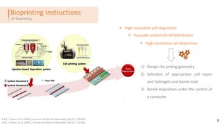
- 1. Bioprinting Instructions 2211 3D Bioprinting XA.B.C. Fulano, et al. (ANO), Journal com Nome Abreviado, 94(12): 123-456. A.B.C. Fulano, et al. (ANO), Journal com Nome Abreviado, 94(12): 123-456 Accurate control of cell distribution High-resolution cell deposition High-resolution cell deposition 1) Design the priting geometry 2) Selection of appropriate cell types and hydrogels and bioink load 3) Bioink deposition under the control of a computer
- 2. Bioprinting Techniques 2211 3D Bioprinting XA.B.C. Fulano, et al. (ANO), Journal com Nome Abreviado, 94(12): 123-456. A.B.C. Fulano, et al. (ANO), Journal com Nome Abreviado, 94(12): 123-456 InkJet Extrusion Laser-Assisted
- 3. Inkjet Bioprinting ‘’Drop-on-Drop’’ 2211 3D Bioprinting XA.B.C. Fulano, et al. (ANO), Journal com Nome Abreviado, 94(12): 123-456. A.B.C. Fulano, et al. (ANO), Journal com Nome Abreviado, 94(12): 123-456 Hydrogel with encapsulated cells form the bioink ; Thermal or acoustic forces to eject drops; System drop-to drop The material needs to be in the liquid state to form drops and after form a solid 3D organized structure Crosslinking strategies Regeneration of lesions in situ Controlled volumes of liquid delivered High resolution, precision and speed High cell viability (80%-90%) Droplet size and deposition rate are controlled electronically • Microelectromechanical system causes: • Mechanical stress • Non uniform droplet size • Frequent clogging of the nozzle • Cannot be used with: • High viscosity materials • High cell density • Cells settle and clog the bioprinter head Advantages Disadvantages
- 4. Extrusion Bioprinting 2211 3D Bioprinting XA.B.C. Fulano, et al. (ANO), Journal com Nome Abreviado, 94(12): 123-456. A.B.C. Fulano, et al. (ANO), Journal com Nome Abreviado, 94(12): 123-456 Wide variety of materials printed Deposition of very high cell densities High resolution, speed and spatial control Allows deposition of spheroids multicellular complex : self-assembling strategy • Exposure to large mechanical stress that can reduce cell viability • Pneumatic nozzles can not precisely control the deposited mass • Cell survival rates in the range of 40-86 % Advantages Disadvantages Movement along x,y,z axes To dispense bioink it is used air-force pump or a mechanical screw plunger Prints uninterrupted cylindrical lines Can print aggregates with high cell density and diverse viscosities Generation of tissues and organs
- 5. Laser-Assisted Bioprinting 2211 3D Bioprinting XA.B.C. Fulano, et al. (ANO), Journal com Nome Abreviado, 94(12): 123-456. A.B.C. Fulano, et al. (ANO), Journal com Nome Abreviado, 94(12): 123-456 No contact of the dispenser with the bioink ‘’Nozzle-free’’ avoids the clogging problem Does not cause mechanical stress to the cells Prints diverse materials and highly viscous High cell viability (>95%) • Cellular exposure to laser • High-cost method • Does not comprises droplet size and quality • Does not accurately target cell position deposition • Metallic residues are formed owing to the vaporization of the metallic laser-absorbing layer Advantages Disadvantages Donor layer containing an energy-absorbing layer on the top and a layer of bioink solution suspended on the bottom. The laser pulse creates a high-pressure bubble at the interface of the donor layer propelling the suspended bioink.
- 6. Case study: Cell Viability 2211 3D Bioprinting XA.B.C. Fulano, et al. (ANO), Journal com Nome Abreviado, 94(12): 123-456. A.B.C. Fulano, et al. (ANO), Journal com Nome Abreviado, 94(12): 123-456 Cell viability can be controlled by varying the parameters like pressure and nozzle diameter Aim Understand cell state: whether If they were live, injured or necrotic Different nozzle diameter : 150,250 and 400 µ Dispensed pressures: 5, 10, 20 and 40 psi Percentage of live cells Percentage of injured cells Percentage of dead cells It can be seen that the effect of pressure is significantly larger than the effect of the nozzle diameter.
- 7. Case study: Generation of Cell Cocultures through ATS an ADE 2211 3D Bioprinting XA.B.C. Fulano, et al. (ANO), Journal com Nome Abreviado, 94(12): 123-456. A.B.C. Fulano, et al. (ANO), Journal com Nome Abreviado, 94(12): 123-456 Patterning cocultures of cells are indispensable to understand the interaction between multiple cells Techniques to dispense cells fail on dispense high-viability cell; through long-time and large areas Aqueous two-phase system Acoustic droplet ejection I. Contact-free II. Nozzle-less Aqueous two-phase system
- 8. Case study: Generation of Cell Cocultures through ATS an ADE 2211 3D Bioprinting XA.B.C. Fulano, et al. (ANO), Journal com Nome Abreviado, 94(12): 123-456. A.B.C. Fulano, et al. (ANO), Journal com Nome Abreviado, 94(12): 123-456 Patterning cocultures of cells are indispensable to understand the interaction between multiple cells Techniques to dispense cells fail on dispense high-viability cell; through long-time and large areas Aqueous two-phase system Acoustic droplet ejection I. Contact-free II. Nozzle-less Source plate Fixed distance between the transducer and the source plate First, multiple wells were punched in the PDMS slab of the source plate to house each different cell type
- 9. Case study: Generation of Cell Cocultures through ATS an ADE 2211 3D Bioprinting XA.B.C. Fulano, et al. (ANO), Journal com Nome Abreviado, 94(12): 123-456. A.B.C. Fulano, et al. (ANO), Journal com Nome Abreviado, 94(12): 123-456 The maximum height of travel for an ejected droplet increased linearly with increasing acoustic pressure amplitude and pulse duration of the applied ultrasound Droplet volume increased linearly with increasing acoustic pressure and pulse duration, following a similar trend to what was observed for ejection height Two simple pattern types, circle arrays and lines, were created to demonstrate the principal of spatial patterning.
- 10. Case study: Skin replacement therapy 2211 3D Bioprinting XA.B.C. Fulano, et al. (ANO), Journal com Nome Abreviado, 94(12): 123-456. A.B.C. Fulano, et al. (ANO), Journal com Nome Abreviado, 94(12): 123-456 It was used a free-form fabrication 3D printing to engineer a human plasma-derived bilayer skin using human fibroblasts(hFBs) and keratinocytes (hKCs) Bioprinter-Extrusion Method: a)Human Plasma b)CaCl2 ; c)hFBS; d)hKCs; C : heated bed; D:program system
- 11. The first 3 syringes were extruded into a P100 tissue plate and incubated for 30 minutes for polymerization hKCs were printed–> incubation during night Printed skin equivalents were transplanted on to the backs of mice Also, skin equivalents were printed on transwell inserts and hKCs were differentiated at the air-liquid surface Case study: Skin replacement therapy 3D Bioprinting In vivo: The substitute skin was placed on the wounds and covered by skin previously removed (8 weeks). In vitro: differentiation at the air-liquid interface for 17 days at 37 ºC.
- 12. Case study: Skin replacement therapy 3D Bioprinting InvitruInvivo Printed equivalents have a similar structure to normal human skin; Also expression of keratin K10; Similar structure with normal strata characteristics; Green stain: Keratin K5 Green junction: collagen VII Red stain: Keratin K10 Arrows: capillary (dermal and epidermal markers) Immunofluoresce using skin markers Normal Human skin
- 13. Case study: Skin replacement therapy 3D Bioprinting Surpassing known methods that require high number of cellular and acellular layers to form dermis and epidermis. This bioprinting method and bioinks allows the production of human bilayer skin, using human plasma and hFBs and hKCs 3D bioprinting is a suitable technology to generate bioengineered skin for therapeutical and industrial applications Conclusions
Editor's Notes
- Thermal inkjet printers function by electrically heating the print head to produce pulses of pressure that force droplets from the nozzle. Several studies have demonstrated that this localized heating, which can range from 200 °C to 300 °C, does not have a substantial impact either on the stability of biological molecules, such as DNA52,53, or on the viability or post-printing function of mammalian cells. It has been demonstrated that the short duration of the heating (~2 μs) results in an overall temperature rise of only 4–10 °C in the printer head55. The advantages of thermal inkjet printers include high print speed, low cost and wide availability. However, the risk of exposing cells and materials to thermal and mechanical stress, low droplet directionality, nonuniform droplet size, frequent clogging of the nozzle and unreliable cell encapsulation pose considerable disadvantages for the use of these printers in 3D bioprinting. Many inkjet printers contain a piezoelectric crystal that creates an acoustic wave inside the print head to break the liquid into droplets at regular intervals. Applying a voltage to a piezoelectric material induces a rapid change in shape, which in turn generates the pressure needed to eject droplets from the nozzle56. Other However, because current printer heads are based on microelectromechanical system (MEMS) devices, there is a relatively small deformation generated by either thermal or piezoelectric actuation at the nozzle opening. As a result, MEMS-based printer heads cannot squeeze out high viscosity materials (N15 mPa/s) and do not work well with bioinks with high cell density Our group64 and others65 have shown that this limitation could be addressed by using materials that can be crosslinked after deposition by printing using chemical, pH or ultraviolet mechanisms. However, the requirement for crosslinking often slows the bioprinting process and involves chemical modification of naturally occurring ECM materials, which changes both their chemical and material properties. Additionally, some crosslinking mechanisms require products or conditions that are toxic to cells, which results in decreased viability and functionality66. Another limitation encountered by users of inkjet-based bioprinting technology is the difficulty in achieving biologically relevant cell densities. Often, low cell concentrations (fewer than 10 million cells/ml)42 are used to facilitate droplet formation, avoid nozzle clogging and reduce shear stress60. Higher cell concentrations The inkjet approach facilitated the deposition of either primary cells or stem cell types with uniform density throughout the volume of the lesion, and maintained high cell viability and function after printing. These studies demonstrate the potential of inkjet-based bioprinting to regenerate functional structures
- cell survival rates are in the range of 40–86%, with the rate decreasing with increasing extrusion pressure and increasing nozzle gauge76,80. The decreased viability of cells deposited by microextrusion is likely to result from the shear stresses inflicted on cells in viscous fluids. Dispensing pressure may have a more substantial effect on cell viability than the nozzle diameter90. Although cell viability can be maintained using low pressures and large nozzle sizes, the drawback may be a major loss of resolution and print speed. Maintaining high viability is essential for achieving tissue functionality. Additionally, improvements in nozzle, syringe or motor-control systems might reduce print times as well as allow deposition of multiple diverse materials simultaneously82 Additionally, improvements in nozzle, syringe or motor-control systems might reduce print times as well as allow deposition of multiple diverse materials simultaneously82. Microextrusion bioprinters have been used to fabricate multiple tissue types, including aortic valves93, branched vascular trees94 and in vitro pharmokinetic95 as well as tumor models96 Depending on the viscoelastic properties of the building blocks, the apposed cell aggregates fuse with each other, forming a cohesive macroscopic construct. One advantage of the self-assembling spheroid strategy is potentially accelerated tissue organization and the ability to direct the formation of complex structures.
- Micropatterning strategies fall broadly into two categories: (i) those that rely on substrate features, such as biochemical or topographic patterns to position cells,11–13 and (ii) those that actively dispense cells.14,1 To generate a scratch-free cell migration assay, we utilize an aqueous two-phase system (ATPS) consisting of polyethy lene glycol (PEG) and dextran (DEX) as the phase-forming polymers. [18] In this method, a submicroliter droplet of the DEX phase is spotted and dried on the fl oor of standard microwells. When the PEG phase containing suspended cells is added to these wells, the dried DEX spot rehydrates to form an immiscible droplet. The PEG-DEX biphasic system interfacial tension excludes cells from adhering to the part of the substrate covered by the rehydrating drop. This generates a well-defi ned circular cell-excluded area within a cell monolayer without the need for any special equipment or procedures other than standard pipetting equipment accessible at high throughput screening facilities. After washing away the ATPS medium, the rate and degree to which cells fi ll up the available space provides a measure of cell migration in a manner similar to wound healing assays. Pulsed ultrasound exposures were used to generate dextran droplets that were collected on the destination plate HIFU: high intensity focused ultrasound
- Micropatterning strategies fall broadly into two categories: (i) those that rely on substrate features, such as biochemical or topographic patterns to position cells,11–13 and (ii) those that actively dispense cells.14,1 To generate a scratch-free cell migration assay, we utilize an aqueous two-phase system (ATPS) consisting of polyethy lene glycol (PEG) and dextran (DEX) as the phase-forming polymers. [18] In this method, a submicroliter droplet of the DEX phase is spotted and dried on the fl oor of standard microwells. When the PEG phase containing suspended cells is added to these wells, the dried DEX spot rehydrates to form an immiscible droplet. The PEG-DEX biphasic system interfacial tension excludes cells from adhering to the part of the substrate covered by the rehydrating drop. This generates a well-defi ned circular cell-excluded area within a cell monolayer without the need for any special equipment or procedures other than standard pipetting equipment accessible at high throughput screening facilities. After washing away the ATPS medium, the rate and degree to which cells fi ll up the available space provides a measure of cell migration in a manner similar to wound healing assays. Pulsed ultrasound exposures were used to generate dextran droplets that were collected on the destination plate HIFU: high intensity focused ultrasound
- Micropatterning strategies fall broadly into two categories: (i) those that rely on substrate features, such as biochemical or topographic patterns to position cells,11–13 and (ii) those that actively dispense cells.14,1 To generate a scratch-free cell migration assay, we utilize an aqueous two-phase system (ATPS) consisting of polyethy lene glycol (PEG) and dextran (DEX) as the phase-forming polymers. [18] In this method, a submicroliter droplet of the DEX phase is spotted and dried on the fl oor of standard microwells. When the PEG phase containing suspended cells is added to these wells, the dried DEX spot rehydrates to form an immiscible droplet. The PEG-DEX biphasic system interfacial tension excludes cells from adhering to the part of the substrate covered by the rehydrating drop. This generates a well-defi ned circular cell-excluded area within a cell monolayer without the need for any special equipment or procedures other than standard pipetting equipment accessible at high throughput screening facilities. After washing away the ATPS medium, the rate and degree to which cells fi ll up the available space provides a measure of cell migration in a manner similar to wound healing assays. Pulsed ultrasound exposures were used to generate dextran droplets that were collected on the destination plate HIFU: high intensity focused ultrasound
- The function of CaCl2 is to induce the coagulation of the plasma fibrinogen into a fibrin hydrogel. Transwell permeable supports are backed by extensive citations, protocols, and scientific support—all to help create a cell culture that more closely mimics an in vivo environment The printing process was designed to produce a fibroblast-containing fibrin hydrogel covered with a monolayer of hKCs fibrin-based dermal matrix previously developed by our group for the production of large skin surfaces, useful in the treatment of severe and extensive burns, wounds with loss of substance and skin fragility diseases
- In vivo and in vitro assays were performed to analyse the viability of these constructs and their capacity to generate a terminally differentiated skin.
- In vivo and in vitro assays were performed to analyse the viability of these constructs and their capacity to generate a terminally differentiated skin. The correct formation of the dermoepidermal junction of the skin was confirmed by labelling with an antibody against human collagen VII (figure 4(B) green staining), the protein forming the anchoring fibrils that bind together epidermis and dermis. From the immunohistochemical point of view, K14 is a well-established marker of epidermal basal cells as shown *Este metodo é mais fácil em vez de imprimir um grande numero cellular e acellular em camadas, o que temos é depossição simultaneal de fibroblastos, plasma humano e cacl2 e por fim uma mono layer de hKCs staining with dermal and epidermal markers) The results showed that the generated skin had correct architecture Secondly, the structure and functionality of the printed human skin was further analysed on skinhumanized Mice and several differentiation markers such as keratin 5 (a marker of proliferative basal keratinocytes), human vimentin (a marker of hFBs), humancollagen type VII (a marker of dermoepidermal basal membrane), keratin 10 (a marker of suprabasal differentiated keratinocytes) and human-filaggrin (a marker of keratinocyte terminal differentiation) Sobre os capilares This finding represents additional evidence of the functionality of our skin regenerated from human bioprinted equivalents.
- In vivo and in vitro assays were performed to analyse the viability of these constructs and their capacity to generate a terminally differentiated skin. The correct formation of the dermoepidermal junction of the skin was confirmed by labelling with an antibody against human collagen VII (figure 4(B) green staining), the protein forming the anchoring fibrils that bind together epidermis and dermis. From the immunohistochemical point of view, K14 is a well-established marker of epidermal basal cells as shown *Este metodo é mais fácil em vez de imprimir um grande numero cellular e acellular em camadas, o que temos é depossição simultaneal de fibroblastos, plasma humano e cacl2 e por fim uma mono layer de hKCs staining with dermal and epidermal markers) The results showed that the generated skin had correct architecture Secondly, the structure and functionality of the printed human skin was further analysed on skinhumanized Mice and several differentiation markers such as keratin 5 (a marker of proliferative basal keratinocytes), human vimentin (a marker of hFBs), humancollagen type VII (a marker of dermoepidermal basal membrane), keratin 10 (a marker of suprabasal differentiated keratinocytes) and human-filaggrin (a marker of keratinocyte terminal differentiation) Sobre os capilares This finding represents additional evidence of the functionality of our skin regenerated from human bioprinted equivalents.