
Recommended
More Related Content
What's hot
What's hot (20)
Similar to 3D Bioprinting
Similar to 3D Bioprinting (20)
Recently uploaded
Recently uploaded (20)
3D Bioprinting
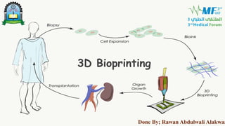
- 1. FR Add a footer 1 3D Bioprinting Done By; Rawan Abdulwali Alakwaa
- 2. FR IN THIS LECTURE: • Justification • 3D Bioprinting concept. • 3D Bioprinting process: • Generating printing paths. • Selecting Bioinks. • Start Bioprinting. • Current advances in 3D Bioprinting.
- 3. Add a footer 3
- 4. FR JUSTIFICATION • Organ failure is drastically increasing. Often, clinical treatments are limited by a paucity of available donors and immune rejection of donated tissue. In the search for alternatives for the repair or replacement of malfunctioning human tissues and organs, tissue 3D Bioprinting is being explored as a promising solution.
- 5. 3D BIOPRINTING • Bioprinting is the layer-by-layer deposition of cells to create 3D tissue that closely resembles native tissue in form and function from basic biological building blocks. Add a footer 5
- 6. FR BIOPRINTING PROCESS • To successfully create bioprinted tissues, it is necessary to: • Generate the printing paths • Select appropriate bioinks • Control the bioprinter and perform quality control after printing. Add a footer 6
- 7. FR GENERATING THE PRINTING PATHS Add a footer 7
- 8. 1. CREATING THE 3D MODEL Add a footer 8 The correct anatomical shape of the desired tissue construct is achieved by: representing clinical imaging (CT or MRI) data as a computer 3D model STL file format as an intermediate between model and print path generation.
- 9. 2. SLICING In a process analogous to histological sectioning, printing paths are created by “slicing” these STL model into layers and creating bioprinter tool paths that trace out the perimeter and interior features of each slice.
- 10. FR SELECTING THE APPROPRIATE BIOINK Add a footer 10
- 11. BIOINK • The Bioink is the bioprintable material consisting of living cells and biomaterials that resemble the ECM to support cells. • An ideal bioink should possess proper mechanical, rheological, and biological properties of the target tissues.
- 12. FR BIOINK CLASSIFICATION OF BIOINKS Scaffold Based Bioinks • Consist of cells dispersed within biomaterials that help to create a conducive environment for cell proliferation as well as providing structural support. Scaffold Free Bioinks • Cells are bioprinted without a supporting hydrogel and therefore cells are loaded in higher concentrations which then deposit their own ECM. Add a footer 12
- 13. FR Add a footer 13 SCAFFOLD BASED BIOINKS •Hydrogel: • Natural: including collagen, fibrin, chitosan, and alginate. • Synthetic: including methacrylated gelatin, Pluronic, and polyethylene glycol. • Hybrid. •Decellularized matrix components obtained by chopping tissue into small fragments, lysing the cells, and extracting the remaining ECM. TYPES OF BIOINKS USED
- 14. SCAFFOLD BASED BIOINKS •Microcarriers have recently been used in bioprinting to increase the cell density in bioinks. They are porous particles designed to promote cell attachment, survival, and expansion. Add a footer 14
- 16. CELL SOURCE • The cell component of the bioink should be autologous and patient specific. Add a footer 16
- 17. STEM CELLS Add a footer 17
- 18. FR STEM CELLS IN 3D BIOPRINTING • Stem cells have the properties of self-renewal and potency, representing an unlimited cell source for 3D bioprinting and regenerative medicine. However, autologous adult stem cells have to be obtained invasively through bone marrow harvesting, adipose tissue extraction by liposuction or least invasively using blood apheresis.
- 19. FR STEM CELLS IN 3D BIOPRINTING • In 2006, with the discovery of human induced pluripotent stem cells (iPSCs) , patient-specific stem cell lines can now be created from mature cell types, such as peripheral blood mononuclear cells by venipuncture, or dermal skin fibroblasts by skin biopsy. This ability to reprogram patient-specific cells also allows for the creation of stem cells containing heritable mutations, which can then be bioprinted and studied.
- 20. FR STEM CELLS IN 3D BIOPRINTING • This has the potential to improve current understanding of disease mechanisms and phenotypic variability, while minimizing rejection when transplanted back into the host for the purposes of tissue regeneration.
- 22. FR Add a footer 22
- 23. FR START TO BIOPRINT Add a footer 23
- 25. INKJET 3D BIOPRINTING • Bioink is stored in the ink cartridge which is connected to a printer head and acts as the source during the electronically controlled printing process. During printing, the printer heads are squeezed by a thermal or piezoelectric actuator to generate droplets of a controllable size. Add a footer 25
- 26. FR • (1) low cost due to similar structure with commercial printers • (2) high printing speed • (3) relatively high cell viability (usually from 80% to 90%). The advantages of inkjet printing include: INKJET 3D BIOPRINTING Add a footer 26
- 27. EXTRUSION BASED BIOPRINTING • Extrusion printing is a modification of inkjet printing. In order to print the viscous materials inkjet printers cannot deposit, extrusion printing uses either an air-force pump, piston or a mechanical screw plunger to dispense bioinks. Add a footer 27
- 28. LASER ASSISTED BIOPRINTING • A focused laser pulse is applied to stimulate a small area of the absorbing layer. This laser pulse vaporizes a portion of the donor layer, creating a high-pressure bubble at the interface of the bioink layer and propelling the suspended bioink. Add a footer 28
- 30. CARDIOVASCULAR TISSUE CURRENT ADVANCES Add a footer 30 • Creating 3D cardiovascular tissue constructs, using different proliferative cell types. These tissue constructs exhibit synchronous macroscopic beating and self-assembly of vessel-like conduits when co-cultured with endothelial cells.
- 31. MUSCULOSKELETAL TISSUE CURRENT ADVANCES Add a footer 31 • Fabricating 3D skeletal muscle constructs that are precisely patterned. The bioprinted cells had high viability and responded synchronously to electric pulses.
- 32. NEURONAL TISSUE CURRENT ADVANCES Add a footer 32 • Neurons have been bioprinted with high cell viability, and they maintained basic cellular phenotypes and functionality for over two weeks post-printing, with successful development of voltage-gated potassium and sodium channels.
- 33. HEPATIC TISSUE CURRENT ADVANCES Add a footer 33 • The bioprinted cells were positive for hepatic markers and they were phenotypically similar to native hepatocytes, as demonstrated by albumin secretion capability and morphological analysis.
- 34. HEPATIC TISSUE CURRENT ADVANCES Add a footer 34 • One of the bioprinted constructs showed morphological organization that recapitulated the native liver environment and high levels of liver- specific gene expression, metabolic product secretion, and cytochrome P450 induction.
- 35. SKIN CURRENT ADVANCES Add a footer 35 • Printing full-thickness skin equivalents, using alternating layers of bioink and a dermal fibroblast cell suspension that showed high viability after printing.
- 36. SKIN CURRENT ADVANCES 36 • The cells were bioprinted directly over the wound site, and the bioprinted construct enhanced wound closure as well as re- epithelialization as compared to controls treated with only the fibrin-collagen hydrogel.
- 37. FR 37 3D bioprinting using stem cells have shown much research progress in multiple organ systems. Common challenges faced in multiple organ systems include vascularization, viability and scalability.
- 38. FR 38 Tissue engineering using 3D bioprinting technology has promising clinical potential for organ regeneration.
- 39. FR Add a footer 39 References • 1. Ashley N. et al. Bioprinting of stem cells for transplantable tissue fabrication. Stem cells translational medicine 2017; 6:1940-1948. • 2. Ong et al. 3D bioprinting using stem cells. Pediatric Research 2018; 83:223–231. • 3. Christian Mandrycky et al. 3D Bioprinting for Engineering Complex Tissues. Biotechnol Adv. 2016 Jul-Aug; 34(4): 422–434. • 4. Gungor-Ozkerim PS et al. Bioinks for 3D bioprinting: an overview. Biomaterials Science 2018 May 1;6(5):915-946. • 5. Kang HW et al. A 3D bioprinting system to produce human-scale tissue constructs with structural integrity. Nature Biotechnology 2016 Mar;34(3):312-9 • 6. 3D Bioprinting: bioinks selection guide. www.sigmaaldrich.com
- 40. Thank You Rawan Abdulwali Alakwaa