
Recommended
More Related Content
What's hot
What's hot (20)
Similar to Introduction to Affinity Chromatography
Similar to Introduction to Affinity Chromatography (20)
More from MOHAMMAD ASIM
More from MOHAMMAD ASIM (18)
Recently uploaded
Recently uploaded (20)
Introduction to Affinity Chromatography
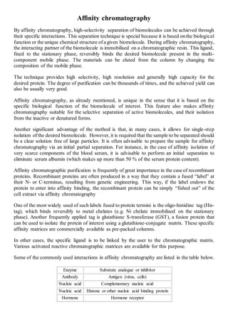
- 1. Affinity chromatography By affinity chromatography, high-selectivity separation of biomolecules can be achieved through their specific interactions. This separation technique is special because it is based on the biological function or the unique chemical structure of a given biomolecule. During affinity chromatography, the interacting partner of the biomolecule is immobilised on a chromatographic resin. This ligand, fixed to the stationary phase, reversibly binds the desired biomolecule present in the multi- component mobile phase. The materials can be eluted from the column by changing the composition of the mobile phase. The technique provides high selectivity, high resolution and generally high capacity for the desired protein. The degree of purification can be thousands of times, and the achieved yield can also be usually very good. Affinity chromatography, as already mentioned, is unique in the sense that it is based on the specific biological function of the biomolecule of interest. This feature also makes affinity chromatography suitable for the selective separation of active biomolecules, and their isolation from the inactive or denatured forms. Another significant advantage of the method is that, in many cases, it allows for single-step isolation of the desired biomolecule. However, it is required that the sample to be separated should be a clear solution free of large particles. It is often advisable to prepare the sample for affinity chromatography via an initial partial separation. For instance, in the case of affinity isolation of very scarce components of the blood serum, it is advisable to perform an initial separation to eliminate serum albumin (which makes up more than 50 % of the serum protein content). Affinity chromatographic purification is frequently of great importance in the case of recombinant proteins. Recombinant proteins are often produced in a way that they contain a fused “label” at their N- or C-terminus, resulting from genetic engineering. This way, if the label endows the protein to enter into affinity binding, the recombinant protein can be simply “fished out” of the cell extract via affinity chromatography One of the most widely used of such labels fused to protein termini is the oligo-histidine tag (His- tag), which binds reversibly to metal chelates (e.g. Ni chelate immobilised on the stationary phase). Another frequently applied tag is glutathione S-transferase (GST), a fusion protein that can be used to isolate the protein of interest using a glutathione-conjugate matrix. These specific affinity matrices are commercially available as pre-packed columns. In other cases, the specific ligand is to be linked by the user to the chromatographic matrix. Various activated reactive chromatographic matrices are available for this purpose. Some of the commonly used interactions in affinity chromatography are listed in the table below. Enzyme Substrate analogue or inhibitor Antibody Antigen (virus, cells) Nucleic acid Complementary nucleic acid Nucleic acid Histone or other nucleic acid binding protein Hormone Hormone receptor
- 2. Glutathione Glutathione S-transferase (GST) fusion protein Metal chelate His-tag fusion protein The phases of affinity chromatography are shown in Figure a. Figure a. The phases of affinity chromatography. In the upper left side of the figure, an illustrative representation of affinity chromatography is shown. Of the molecules present in the sample, only the ones having a “matching” binding site can bind to the matrix-conjugated specific ligand molecules. Other molecules can be readily washed off. By changing the composition of the mobile phase, the molecules of interest can be isolated in a pure form. The lower part of the figure shows a typical affinity chromatographic elution profile. 1. Sample preparation The sample must be a clear solution free from solid particles. This can be achieved by centrifugation or filtration. Protein solutions should be centrifuged at atleast 10000 g. Cell lysates should be centrifuged at 40-50000 g. A 0.45-µm pore size filter can be used for filtration. (Such preparation of samples is also necessary in FPLC and HPLC methods.) One must also consider how the solubility and stability of the sample or the desired protein can be influenced by the pH, the salt concentration, or the presence of any organic solvent. The factors affecting the interactions between the desired target protein and the matrix-bound ligand (pH, salt concentration, temperature) should also be determined. The composition of the initial binding buffer must be adjusted accordingly. Sample components interfering with the target protein and/or the ligand (e.g. metabolites in cell lysates) should be removed before loading onto the column. 2. Equilibration with a buffer facilitating the specific interaction The chromatographic column is washed with 3-4 column volumes of the starting (binding) buffer. The sample must also be equilibrated with this starting binding buffer (if necessary, via solvent exchange or dialysis).
- 3. 3. Binding of the molecule of interest and wash-out of the unbound material During sample loading, consider the strength of the interaction. In case of high-affinity samples, a high flow rate may be applied. In case of a weak interaction and/or a slow equilibration process, reduce the rate of sample loading. After sample application, the column should be further washed with binding buffer until all unbound components are removed. 4. Elution of the molecules of interest by changing the composition of the mobile phase Elution via pH and/or ionic strength changes: One possible and simple means of elution is achieved through decreasing the interaction strength between the ligand and the target protein. Changes in the pH will change the ionisation state of charged groups of the ligand and/or the target protein, thereby changing the strength of the interaction. Similarly, increasing the ionic strength (usually by raising the NaCl concentration) will generally reduce the interaction strength. In either case, the solubility and stability of the target protein should be considered. Competitive elution: For competitive elution, materials are applied that react with the target protein or the ligand, competing for the pre-existing interaction. For instance, His-Tag fusion proteins can be readily displaced from the metal chelate matrix by imidazole buffer (Figure b). GST-tagged target proteins will detach from their column-conjugated glutathione ligand upon mixing excess glutathione into the elution buffer. Figure b. Isolation of a His-Tag fusion protein. The cell extract containing the His-Tag fusion protein is purified using a Ni2+ chelate column. The upper panel shows the structure of the immobilised Ni2+chelate ligand. The sample is loaded onto the column in a neutral buffer. The fusion protein containing the His-Tag binds the Ni2+ chelate ligand. After washing off other proteins, competitive elution with imidazole buffer can be applied to isolate the pure fusion protein (lower right panel). Lower left panel shows the purity of fractions assessed by SDS gel electrophoresis. In all cases, the flow rate of the buffer should be reduced during elution, thereby avoiding excessive dilution of the target protein.
- 4. In cases when the target-ligand interaction is very strong, the above elution methods may turn out insufficient for eluting the protein of interest. In these cases, chaotropic agents (urea, guanidine) can be used to wash off the target protein from the column. This naturally will involve the denaturation of the protein, which can then be renatured in some (lucky) cases under suitable conditions—in the case of urea or guanidine there is a good chance for this. 5. Regeneration After successful completion of the elution, the column can be washed with several column volumes of binding buffer, and it can then be reused. For long-term storage, one must ensure that the column is not exposed to bacterial or fungal infection. The toxic compound sodium aside can be used to prevent such infection.