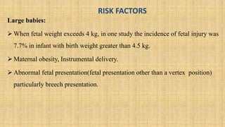
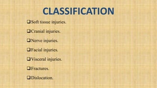
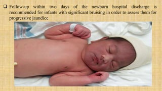
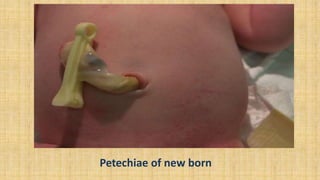
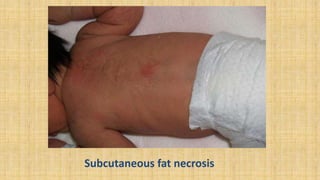
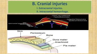
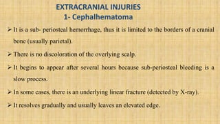
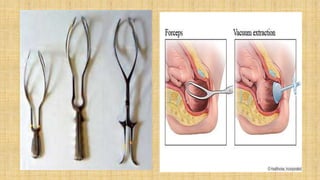
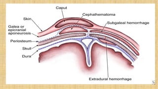
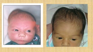
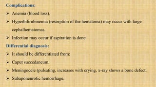
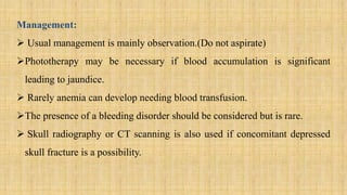
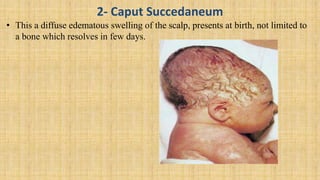
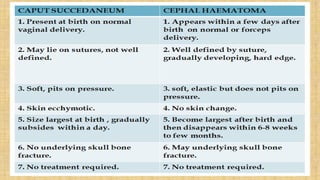
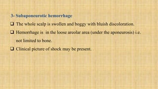
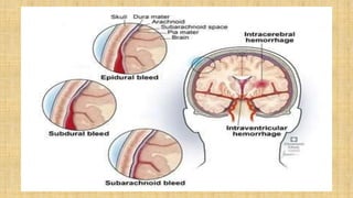
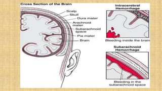
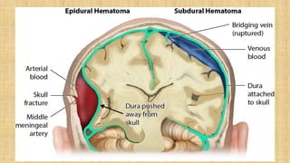
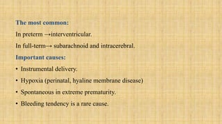
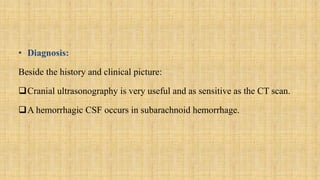
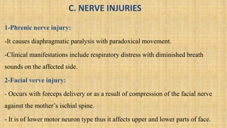
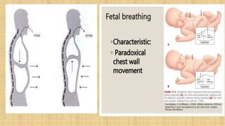
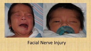
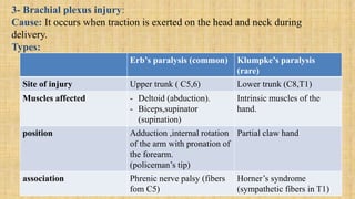
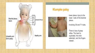
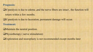
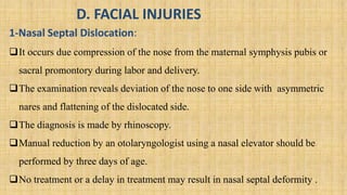
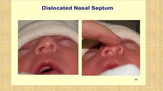
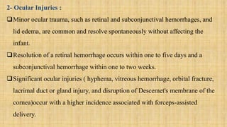
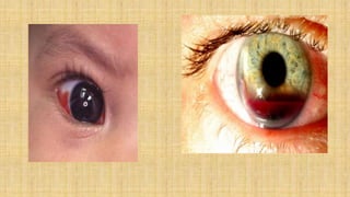
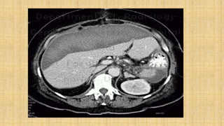
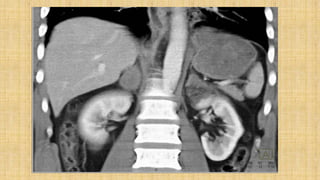
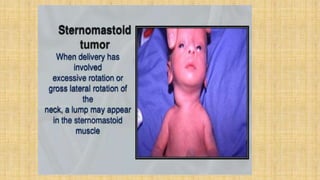
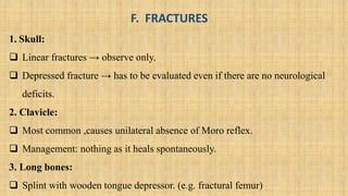
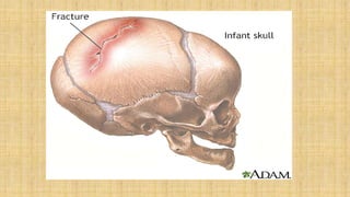
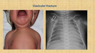
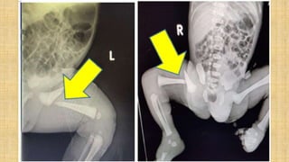
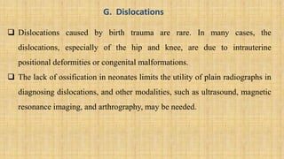
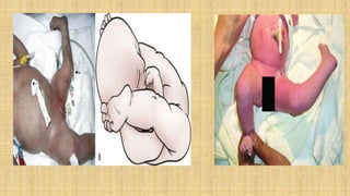
BIRTH INJURIES
Prepared by:
Ms. Lamnunnem Haokip
Senior Tutor
OBG Nursing Dept.
SSNSR, SU
• DEFINITION:
These are the injuries that occur during delivery.
It is defined as an impairment of the neonate's body function or structure due to
an adverse event that occurred at birth.
• INCIDENCE:
The overall incidence of birth injuries has declined with improvement in
obstetrical and prenatal diagnosis.
The reported incidence of birth injuries is :
 2% in vaginal deliveries (cephalic position).
1.1% in cesarean deliveries.
RISK FACTORS
Large babies:
 When fetal weight exceeds 4 kg, in one study the incidence of fetal injury was
7.7% in infant with birth weight greater than 4.5 kg.
 Maternal obesity, Instrumental delivery.
 Abnormal fetal presentation(fetal presentation other than a vertex position)
particularly breech presentation.
Other factors:
 Very small premature babies.
 Small maternal stature.
 CPD.
 Prolonged labor.
 Primiparous.
CLASSIFICATION
Soft tissue injuries.
Cranial injuries.
Nerve injuries.
Facial injuries.
Visceral injuries.
Fractures.
Dislocation.
SOFT TISSUE INJURIES
1. Bruising and petechiae
 Petechiae of the head and face are often seen in infants delivered from the
vertex position, especially with a face presentation.
 Most often, petechiae are present at birth, do not progress, and are not
associated with other bleeding.
 Significant bruising has been recognized as a major risk factor for the
development of severe hyperbilirubinemia.
 Follow-up within two days of the newborn hospital discharge is
recommended for infants with significant bruising in order to assess them for
progressive jaundice.
Petechiae of new born
2.Subcutaneous fat necrosis
 Subcutaneous fat necrosis (SFN) is uncommon
Usually occurs in the first few weeks of life as a result of ischemia to the
adipose tissue following a traumatic delivery.
SFN is characterized by firm, indurated nodules and plaques on the back,
buttocks, thighs, forearms, and cheeks.
Typically, this condition is self-limiting.
3. Lacerations
Fetal laceration has been reported as the most common birth injury
associated with cesarean delivery .
The lacerations occurred most often on the presenting part of the fetus,
typically the scalp and face.
The majority of fetal lacerations were mild, requiring repair with sterile
strips only.
Subcutaneous fat necrosis
B. Cranial injuries
I. Extracranial injuries.
II. Intracranial hemorrhage.
EXTRACRANIAL INJURIES
1- Cephalhematoma
 It is a sub- periosteal hemorrhage, thus it is limited to the borders of a cranial
bone (usually parietal).
 There is no discoloration of the overlying scalp.
 It begins to appear after several hours because sub-periosteal bleeding is a
slow process.
 In some cases, there is an underlying linear fracture (detected by X-ray).
 It resolves gradually and usually leaves an elevated edge.
Complications:
 Anemia (blood loss).
 Hyperbilirubinemia (resorption of the hematoma) may occur with large
cephalhematomas.
 Infection may occur if aspiration is done
Differential diagnosis:
 It should be differentiated from:
 Caput succedaneum.
 Meningocele (pulsating, increases with crying, x-ray shows a bone defect.
 Subaponeurotic hemorrhage.
Management:
 Usual management is mainly observation.(Do not aspirate)
Phototherapy may be necessary if blood accumulation is significant
leading to jaundice.
 Rarely anemia can develop needing blood transfusion.
The presence of a bleeding disorder should be considered but is rare.
 Skull radiography or CT scanning is also used if concomitant depressed
skull fracture is a possibility.
2- Caput Succedaneum
• This a diffuse edematous swelling of the scalp, presents at birth, not limited to
a bone which resolves in few days.
3- Subaponeurotic hemorrhage
 The whole scalp is swollen and boggy with bluish discoloration.
 Hemorrhage is in the loose areolar area (under the aponeurosis) i.e.
not limited to bone.
 Clinical picture of shock may be present.
II. Intracranial hemorrhage
• Definition: Hemorrhage inside the cranial cavity.
• Types
 Outside the brain (epidural, subdural, subarachnoid).
 In the brain ventricles (interventricular).
 In the brain parenchyma (e.g. intracerebral)
The most common:
In preterm →interventricular.
In full-term→ subarachnoid and intracerebral.
Important causes:
• Instrumental delivery.
• Hypoxia (perinatal, hyaline membrane disease)
• Spontaneous in extreme prematurity.
• Bleeding tendency is a rare cause.
Clinical manifestations:
 Pallor , cyanosis with irregular breathing, later on jaundice (resorption of
concealed blood).
 Lethargy , poor Moro reflex ,weak suckling, High pitched cry.
 Convulsions, usually tonic.
 Tense bulging anterior fontanel.
 Localizing neurological deficits may occur like occular palsies, unequal
pupils.
• N.B .Triad of pallor, high pitched cry, tense bulging ant. fontanel you
should suspect intracranial hemorrhage.
• Diagnosis:
Beside the history and clinical picture:
Cranial ultrasonography is very useful and as sensitive as the CT scan.
A hemorrhagic CSF occurs in subarachnoid hemorrhage.
Complications:
• Death from respiratory failure.
• Obstructive hydrocephalus.
• Cerebral palsy.
Treatment:
• Vitamin K to help coagulation.
• Ventilatory support (if there is respiratory difficulty).
• Phenobarbitone (if there is convulsions).
• Blood transfusion (if there is anemia).
• Treatment of complications like hydrocephalus.
C. NERVE INJURIES
1-Phrenic nerve injury:
-It causes diaphragmatic paralysis with paradoxical movement.
-Clinical manifestations include respiratory distress with diminished breath
sounds on the affected side.
2-Facial verve injury:
- Occurs with forceps delivery or as a result of compression of the facial nerve
against the mother’s ischial spine.
- It is of lower motor neuron type thus it affects upper and lower parts of face.
. Facial Nerve Injury
3- Brachial plexus injury:
Cause: It occurs when traction is exerted on the head and neck during
delivery.
Types:
Erb’s paralysis (common) Klumpke’s paralysis
(rare)
Site of injury Upper trunk ( C5,6) Lower trunk (C8,T1)
Muscles affected - Deltoid (abduction).
- Biceps,supinator
(supination)
Intrinsic muscles of the
hand.
position Adduction ,internal rotation
of the arm with pronation of
the forearm.
(policeman’s tip)
Partial claw hand
association Phrenic nerve palsy (fibers
fom C5)
Horner’s syndrome
(sympathetic fibers in T1)
Prognosis:
If paralysis is due to edema ,and the nerve fibers are intact , the function will
return within a few months.
If paralysis is due to laceration, permanent damage will occur.
Treatment:
Maintain the neutral position.
Physiotherapy ( nerve stimulation)
Exploration and neuroplasty is not recommended except months later
D. FACIAL INJURIES
1-Nasal Septal Dislocation:
It occurs due compression of the nose from the maternal symphysis pubis or
sacral promontory during labor and delivery.
The examination reveals deviation of the nose to one side with asymmetric
nares and flattening of the dislocated side.
The diagnosis is made by rhinoscopy.
Manual reduction by an otolaryngologist using a nasal elevator should be
performed by three days of age.
No treatment or a delay in treatment may result in nasal septal deformity .
2- Ocular Injuries :
Minor ocular trauma, such as retinal and subconjunctival hemorrhages, and
lid edema, are common and resolve spontaneously without affecting the
infant.
Resolution of a retinal hemorrhage occurs within one to five days and a
subconjunctival hemorrhage within one to two weeks.
Significant ocular injuries ( hyphema, vitreous hemorrhage, orbital fracture,
lacrimal duct or gland injury, and disruption of Descemet's membrane of the
cornea)occur with a higher incidence associated with forceps-assisted
delivery.
E. VISCERAL AND MUSCLE INJURIES
The important visceral and muscle injuries include:
 Subcapsular hematoma of the liver: The accumulation of blood between
Glisson’s Capsules and the liver parenchyma.
 Suprarenal hemorrhage: Suprarenal gland hemorrhage has been described in
several conditions (sepsis, local traumas, surgical procedures, anticoagulant
therapies, hypoxemia). The condition is rare in congenital bleeding disorders.
 Sternomastoid tumor: The sternomastoid "tumor" of infancy is a firm, fibrous
mass, appearing at two to three weeks of age. It may or may not be associated with
torticollis.
F. FRACTURES
1. Skull:
 Linear fractures → observe only.
 Depressed fracture → has to be evaluated even if there are no neurological
deficits.
2. Clavicle:
 Most common ,causes unilateral absence of Moro reflex.
 Management: nothing as it heals spontaneously.
3. Long bones:
 Splint with wooden tongue depressor. (e.g. fractural femur)
Clavicular fracture
G. Dislocations
 Dislocations caused by birth trauma are rare. In many cases, the
dislocations, especially of the hip and knee, are due to intrauterine
positional deformities or congenital malformations.
 The lack of ossification in neonates limits the utility of plain radiographs in
diagnosing dislocations, and other modalities, such as ultrasound, magnetic
resonance imaging, and arthrography, may be needed.
Management
If the baby is younger than 6 months of age and diagnosed with CHD, it’s
likely they’ll be fitted for a Pavlik harness. This harness presses their hip
joints into the sockets. The harness abducts the hip by securing their legs in
a froglike position. The baby may wear the harness for 6 to 12 weeks,
depending on their age and the severity of the condition
THANK YOU

Birth Injuries

  • 1.
    BIRTH INJURIES Prepared by: Ms.Lamnunnem Haokip Senior Tutor OBG Nursing Dept. SSNSR, SU
  • 2.
    • DEFINITION: These arethe injuries that occur during delivery. It is defined as an impairment of the neonate's body function or structure due to an adverse event that occurred at birth. • INCIDENCE: The overall incidence of birth injuries has declined with improvement in obstetrical and prenatal diagnosis. The reported incidence of birth injuries is :  2% in vaginal deliveries (cephalic position). 1.1% in cesarean deliveries.
  • 3.
    RISK FACTORS Large babies: When fetal weight exceeds 4 kg, in one study the incidence of fetal injury was 7.7% in infant with birth weight greater than 4.5 kg.  Maternal obesity, Instrumental delivery.  Abnormal fetal presentation(fetal presentation other than a vertex position) particularly breech presentation.
  • 4.
    Other factors:  Verysmall premature babies.  Small maternal stature.  CPD.  Prolonged labor.  Primiparous.
  • 5.
    CLASSIFICATION Soft tissue injuries. Cranialinjuries. Nerve injuries. Facial injuries. Visceral injuries. Fractures. Dislocation.
  • 6.
    SOFT TISSUE INJURIES 1.Bruising and petechiae  Petechiae of the head and face are often seen in infants delivered from the vertex position, especially with a face presentation.  Most often, petechiae are present at birth, do not progress, and are not associated with other bleeding.  Significant bruising has been recognized as a major risk factor for the development of severe hyperbilirubinemia.
  • 7.
     Follow-up withintwo days of the newborn hospital discharge is recommended for infants with significant bruising in order to assess them for progressive jaundice.
  • 8.
  • 9.
    2.Subcutaneous fat necrosis Subcutaneous fat necrosis (SFN) is uncommon Usually occurs in the first few weeks of life as a result of ischemia to the adipose tissue following a traumatic delivery. SFN is characterized by firm, indurated nodules and plaques on the back, buttocks, thighs, forearms, and cheeks. Typically, this condition is self-limiting.
  • 10.
    3. Lacerations Fetal lacerationhas been reported as the most common birth injury associated with cesarean delivery . The lacerations occurred most often on the presenting part of the fetus, typically the scalp and face. The majority of fetal lacerations were mild, requiring repair with sterile strips only.
  • 11.
  • 12.
    B. Cranial injuries I.Extracranial injuries. II. Intracranial hemorrhage.
  • 13.
    EXTRACRANIAL INJURIES 1- Cephalhematoma It is a sub- periosteal hemorrhage, thus it is limited to the borders of a cranial bone (usually parietal).  There is no discoloration of the overlying scalp.  It begins to appear after several hours because sub-periosteal bleeding is a slow process.  In some cases, there is an underlying linear fracture (detected by X-ray).  It resolves gradually and usually leaves an elevated edge.
  • 17.
    Complications:  Anemia (bloodloss).  Hyperbilirubinemia (resorption of the hematoma) may occur with large cephalhematomas.  Infection may occur if aspiration is done Differential diagnosis:  It should be differentiated from:  Caput succedaneum.  Meningocele (pulsating, increases with crying, x-ray shows a bone defect.  Subaponeurotic hemorrhage.
  • 18.
    Management:  Usual managementis mainly observation.(Do not aspirate) Phototherapy may be necessary if blood accumulation is significant leading to jaundice.  Rarely anemia can develop needing blood transfusion. The presence of a bleeding disorder should be considered but is rare.  Skull radiography or CT scanning is also used if concomitant depressed skull fracture is a possibility.
  • 19.
    2- Caput Succedaneum •This a diffuse edematous swelling of the scalp, presents at birth, not limited to a bone which resolves in few days.
  • 22.
    3- Subaponeurotic hemorrhage The whole scalp is swollen and boggy with bluish discoloration.  Hemorrhage is in the loose areolar area (under the aponeurosis) i.e. not limited to bone.  Clinical picture of shock may be present.
  • 23.
    II. Intracranial hemorrhage •Definition: Hemorrhage inside the cranial cavity. • Types  Outside the brain (epidural, subdural, subarachnoid).  In the brain ventricles (interventricular).  In the brain parenchyma (e.g. intracerebral)
  • 27.
    The most common: Inpreterm →interventricular. In full-term→ subarachnoid and intracerebral. Important causes: • Instrumental delivery. • Hypoxia (perinatal, hyaline membrane disease) • Spontaneous in extreme prematurity. • Bleeding tendency is a rare cause.
  • 28.
    Clinical manifestations:  Pallor, cyanosis with irregular breathing, later on jaundice (resorption of concealed blood).  Lethargy , poor Moro reflex ,weak suckling, High pitched cry.  Convulsions, usually tonic.  Tense bulging anterior fontanel.  Localizing neurological deficits may occur like occular palsies, unequal pupils. • N.B .Triad of pallor, high pitched cry, tense bulging ant. fontanel you should suspect intracranial hemorrhage.
  • 29.
    • Diagnosis: Beside thehistory and clinical picture: Cranial ultrasonography is very useful and as sensitive as the CT scan. A hemorrhagic CSF occurs in subarachnoid hemorrhage.
  • 30.
    Complications: • Death fromrespiratory failure. • Obstructive hydrocephalus. • Cerebral palsy. Treatment: • Vitamin K to help coagulation. • Ventilatory support (if there is respiratory difficulty). • Phenobarbitone (if there is convulsions). • Blood transfusion (if there is anemia). • Treatment of complications like hydrocephalus.
  • 31.
    C. NERVE INJURIES 1-Phrenicnerve injury: -It causes diaphragmatic paralysis with paradoxical movement. -Clinical manifestations include respiratory distress with diminished breath sounds on the affected side. 2-Facial verve injury: - Occurs with forceps delivery or as a result of compression of the facial nerve against the mother’s ischial spine. - It is of lower motor neuron type thus it affects upper and lower parts of face.
  • 33.
  • 34.
    3- Brachial plexusinjury: Cause: It occurs when traction is exerted on the head and neck during delivery. Types: Erb’s paralysis (common) Klumpke’s paralysis (rare) Site of injury Upper trunk ( C5,6) Lower trunk (C8,T1) Muscles affected - Deltoid (abduction). - Biceps,supinator (supination) Intrinsic muscles of the hand. position Adduction ,internal rotation of the arm with pronation of the forearm. (policeman’s tip) Partial claw hand association Phrenic nerve palsy (fibers fom C5) Horner’s syndrome (sympathetic fibers in T1)
  • 36.
    Prognosis: If paralysis isdue to edema ,and the nerve fibers are intact , the function will return within a few months. If paralysis is due to laceration, permanent damage will occur. Treatment: Maintain the neutral position. Physiotherapy ( nerve stimulation) Exploration and neuroplasty is not recommended except months later
  • 37.
    D. FACIAL INJURIES 1-NasalSeptal Dislocation: It occurs due compression of the nose from the maternal symphysis pubis or sacral promontory during labor and delivery. The examination reveals deviation of the nose to one side with asymmetric nares and flattening of the dislocated side. The diagnosis is made by rhinoscopy. Manual reduction by an otolaryngologist using a nasal elevator should be performed by three days of age. No treatment or a delay in treatment may result in nasal septal deformity .
  • 39.
    2- Ocular Injuries: Minor ocular trauma, such as retinal and subconjunctival hemorrhages, and lid edema, are common and resolve spontaneously without affecting the infant. Resolution of a retinal hemorrhage occurs within one to five days and a subconjunctival hemorrhage within one to two weeks. Significant ocular injuries ( hyphema, vitreous hemorrhage, orbital fracture, lacrimal duct or gland injury, and disruption of Descemet's membrane of the cornea)occur with a higher incidence associated with forceps-assisted delivery.
  • 41.
    E. VISCERAL ANDMUSCLE INJURIES The important visceral and muscle injuries include:  Subcapsular hematoma of the liver: The accumulation of blood between Glisson’s Capsules and the liver parenchyma.  Suprarenal hemorrhage: Suprarenal gland hemorrhage has been described in several conditions (sepsis, local traumas, surgical procedures, anticoagulant therapies, hypoxemia). The condition is rare in congenital bleeding disorders.  Sternomastoid tumor: The sternomastoid "tumor" of infancy is a firm, fibrous mass, appearing at two to three weeks of age. It may or may not be associated with torticollis.
  • 45.
    F. FRACTURES 1. Skull: Linear fractures → observe only.  Depressed fracture → has to be evaluated even if there are no neurological deficits. 2. Clavicle:  Most common ,causes unilateral absence of Moro reflex.  Management: nothing as it heals spontaneously. 3. Long bones:  Splint with wooden tongue depressor. (e.g. fractural femur)
  • 47.
  • 49.
    G. Dislocations  Dislocationscaused by birth trauma are rare. In many cases, the dislocations, especially of the hip and knee, are due to intrauterine positional deformities or congenital malformations.  The lack of ossification in neonates limits the utility of plain radiographs in diagnosing dislocations, and other modalities, such as ultrasound, magnetic resonance imaging, and arthrography, may be needed.
  • 51.
    Management If the babyis younger than 6 months of age and diagnosed with CHD, it’s likely they’ll be fitted for a Pavlik harness. This harness presses their hip joints into the sockets. The harness abducts the hip by securing their legs in a froglike position. The baby may wear the harness for 6 to 12 weeks, depending on their age and the severity of the condition
  • 53.