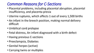
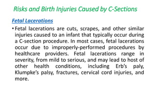
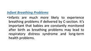
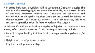
BIRTH INJURIES
Mrs. Gayathri R
First year MSc Nursing
Upasana College Of
Nursing
DEFINITION
An impairment of the infants body function or
structure due to adverse influences that occurred at
birth.
National Vital Statistics Report
Birth injuries is defined as those sustained during
labour and delivery. It may be severe enough to
cause neonatal deaths, still birth or number of
morbidities.
D C Dutta
CLASSIFICATION OF BIRTH INJURIES
• Soft tissue injuries
• Head and neck injuries
• Facial injuries
• Cranial nerve injuries
• Spinal cord injuries
• Peripheral nerve injuries
• Fractures
• Intra abdominal injury
HIGH RISK FACTORS
• Prolonged or obstructed labour
• Fetal macrosomia
• Cephalopelvic disproportion
• Abnormal presentation(breech)
• Instrumental delivery(forceps or ventouse)
• Difficult labour
• Shoulder dystocia
• Inadequate maternal pelvis
CAUSES OF BIRTH INJURIES
Prolonged or obstructed labour
Fetal macrosomia
Cephalopelvic disproportion
Abnormal presentation
Instrumental delivery
Difficult labour
Shoulder dystocia
Precipitate labour
Manipulative delivery
INTRACRANIAL HEMORRHAGE (ICH)
•Bleeding in the brain, also known as Intracranial
Hemorrhage has been known to affect
newborns, although it is much more prevalent
among premature infants. Intracranial
hemorrhage (ICH) may be-(a) External to the
brain (epidural, subdural or subarachnoid space)
(b) in the parenchyma of brain (cerebrum or
cerebellum) (c) into the ventricles from sub
ependymal germinal matrix or choroid plexus.
Traumatic
• Extradural hemorrhage: Usually associated with
fracture skull bone.
• Subdural hemorrhage: This condition occurs when
there is bleeding between the outer and inner layers
of the brain covering. Subdural hemorrhage is not as
common as it used to be, as there have been medical
advancements made in the childbirth process.
Anoxic
• Subarachnoid hemorrhage: This term is used to
describe bleeding that occurs below the innermost
area of the two membranes that cover the brain. It is
the most common type of bleeding in the skull.
• Intraventricular hemorrhage: This term describes
bleeding in the normal fluid-filled spaces, also known
as ventricles, in the brain. It affects the brain tissue.
Causes
• Preterm baby because of protection by their soft skull bones
and wide sutures.
• Trauma: Compression and stretching in moulding.
• Excessive compression of fetal head due to contracted pelvis,
occipito posterior position, and large baby.
• Rapid compression on fetal head, breech delivery, precipitate
labour.
• Upward compression as in breech delivery, face
presentation.
• Instrumental delivery.
Clinical features
• Baby cannot establish respiration himself.
• In severe cases, at birth, the infant is shocked, the eyes roll
upward.
• Trunk and limbs may be rigid, the fist clenched, limpness is
also common.
• Difficult grunting expiration after most due to excess
mucosa.
• Sometimes shallow, rapid and irregular with attack of apnea
and cyanosis.
• Worried and anxious expression, eyes are widen open for
long period, starring with a knowing lock, sunken eyes, rigid
neck, and spongy fontanelle.
Prevention
• Prevent or detect intrauterine fetal asphyxia in earliest by intensive
fetal monitoring.
• Liberal episiotomy and use of forceps to deliver the premature baby
minimize the intracranial disturbances.
• Avoid traumatic vaginal delivery in preference to caesarian section.
• Difficult forceps should be avoided.
• In vaccum delivery, traction is made only after proper cephalic
application.
• Extend the use of caesarian section in breech more liberally. Utmost
gentleness is to be executed in vaginal breech delivery. Never be at
haste especially during delivery of head. Forceps delivery of the after
coming head is preferable.
• Avoid prolonged and difficult labour.
Treatment and Management
• The baby should be nursed in quiet, warm and well ventilated
surrounding.
• Maintain cleanliness of the passage, suction immediately after birth
to remove the secretion that occludes the pharynx.
• Incubator nursery is preferable to supply oxygen and to maintain the
temperature and humidity.
• If respiration is established wrap properly and keep the infant on one
side turns.
• Restrict handling the baby. Bathing, weighing and measuring should
be withheld because it may provoke convulsions.
• Feeding by nasogastric tube is advisable, fluid balance is too
maintained, if necessary by parenteral route.
Treatment continue…
• Administer vitamin K 1mg intramuscularly to prevent further bleeding
due to hyoprothrombinaemia.
• Prophylactic antibiotics is to be administered as needed.
• Anticonvulsant may need to prevent convulsion i.e.
• Phenobarbitone 5-10mg/kg/day in divided doses at 6 hourly interval
intramuscularly.
• Phenytoin 10-15mg/kg intravenously as loading dose at the rate of
0.5mg/kg/min for maintenance dose of 5mg/kg/day with cardiac
monitoring.
• Diazepam 0.1mg/kg IM thrice daily.
• Subdural haematoma can be aspirated through lateral angles of the
anterior fontanelle if excessive haematoma is formed. Surgical
removal of clot may needed.
Treatment continue…
The following equipment should be at hand.
• Suction machine
• Oxygen
• Laryngoscope
• Endotracheal tube
• Keep close observation on:
• Vital signs Q4H as needed.
• Skin color.
• Respiration; type & regularity.
• Apex beat; type & regularity.
• Convulsion: spasm of muscles, part, duration.
INJURIES TO HEAD
Cephalhematoma
Caput succedaneum
Scalp injuries
Skull fracture
CAPUT SUCCEDAENUM
•A caput succedaneum is an edema of the scalp
at the neonates presenting part of the head. It
often appears over the vertex of the newborns
head as a result of pressure against the mother’s
cervix during labour. The edema in caput
succedaneum crosses the suture lines. It may
involve wide areas of the head or it may just be a
size of a large egg.
Causes
•Mechanical trauma of the initial portion of
scalp pushing through a narrowed cervix.
•Prolonged or difficult delivery.
•Vacuum extraction.
Signs & Symptoms
• Scalp swelling that extends across the midline and
over suture lines.
• Soft and puffy swelling of part of a scalp in a
newborns head.
• May be associated with increased molding of the
head.
• The swelling may or may not have some degree of
discoloration or bruising.
• Tends to disappear within 24-36 hours and tends to
reduce to size.
Management
• Needs no treatment. The edema is gradually absorbed and
disappears about the third day of life.
• Advice not to applying pressure over caput.
• Mother is very anxious so we must explain about what it is,
its causes in simple language.
• Baby should be handled gently apply dressing on abrasions.
• An abraded chignon usually heals rapidly if the area is kept
clean, dry & is irritated.
• Advice mother about not applying pressure over caput.
• Advice the mother that caput need no treatment and
disappear within 36 hours of birth.
Complication
Jaundice results as the bruise breaks
down into bilirubin.
CEPHALHEMATOMA
Cephalhematoma is a collection of blood between the
periosteum of a skull bone and the bone itself. It
occurs in one or both sides of head. It occasionally
forms over the occipital bone. The swelling with
Cephalhematoma is not present at birth rather it
develops within the first 24 to 48 hours after birth.
Causes
•Rupture of a periosteal capillary due to pressure
of birth.
•Instrumental delivery
•Precipitate delivery
•Prolonged pressure on the head
•Cephalopelvic disproportion
Signs & Symptoms
• Swelling of the infants head 24-48 hours after birth
• Discoloration of the swollen site due to presence of
coagulated blood
• Has clear edges that end at the suture lines
Management
• Observation and support of the affected part
• Transfusion and phototherapy may be necessary if blood
accumulation is significant
Complication
• Jaundice
Difference between a caput succedaneum and
Cephalhematoma
INDICATORS CAPUT SUCCEDANEUM CEPHALHEMATOMA
Location Presenting part of head Periosteum of skull
bone
Extent
of involvement
Both hemisphere; crosses
the suture lines
Individual bone; does
not cross the suture
line
Period
of absorption
3 to 4 days Few weeks to month
Treatment None Support
SCALP INJURIES
Minor injuries of the scalp such as abrasions in
forceps delivery (tip of blades), incised wound
inflicted during caesarean section or episiotomy
may be met with on occasion, the increased
wound may cause brisk hemorrhage and require
stitches. The wound should be dressed with an
antiseptic solution. E.g. Betadine
SKULL FRACTURE
• Fracture of the vault of the skull 9 frontal or anterior
part of the parietal bone may be of fissure or
depressed type.
Causes
• Effect of difficult forceps delivery in disproportion or
due to wrong application of the forceps.
• Projected sacral promontory of the flat pelvis may
produce depressed fracture even though the delivery
is spontaneous.
Treatment and Management
Treatment is conservative in symptom less cases.
In presence of symptom, the depressed bone has
to be elevated or subdural hematoma may have
to be aspirated or excised surgically.
INJURY TO THE NERVES
Inuries
to
nerves
Facial
palsy
Brachial
palsy
Erbs
palsy
Klumpke
’s palsy
FACIAL PALSY
It is also known as Bell’s palsy. The facial nerve may
injured by direct pressure of the forceps blades or by
hemorrhage or edema around the nerve. It may occur
in normal delivery with much pressure on the ramus of
the mandible where the nerve crosses superficially.
Facial nerve remain unprotected after its exit through
the stylomastoid foramen. It is involved by direct
pressure of the forceps blades or by hemorrhage and
edema around nerve.
Causes
• Forceps delivery.
• It may occur in spontaneous delivery when grasping the head or due
pressure is applied on the mastoid process or over the ramus of lower
jaw where the facial nerve lies superficially.
Clinical features
• There is unilateral facial weakness with the eyelid of the affected side
remaining open and mouth drawn over to the normal side.
• The paralyzed side is smooth.
• On crying the mouth is drawn to the uninjured side of the face.
• If the baby cannot form an effective seal on the nipple or treat, there
may be some initial feeding difficulties.
Management
• There is no special treatment, improve the conduction on 1 to 2
weeks.
• Protect the eyes, which remain open even during sleep, with
antiseptic ointment.
• Feeding difficulties are usually overcome by the baby’s own
adaptation, although alternative feeding position can be adopted.
• Maintain oral hygiene.
• If instrumental delivery and the baby have any injury, clean and dress
with antiseptic lotion.
• The condition usually disappears within weeks unless complicated by
intracranial hemorrhage.
BRACHIAL PALSY
The damage occur in the brachial nerve roots in the
trunk of the brachial plexus due to stretching or
effusion or hemorrhage inside the nerve sheath or
tearing of the fibers. Sometimes tearing of the fiber is
rare. This causes the hyperextension of the neck during
attempted delivery of shoulder dystocia or even in
spontaneous vaginal delivery or during difficult breech
extraction. Unilateral involvement is common. The two
common clinical types are:
• This is the commonest type when the 5th and 6th cervical
nerve roots are involved. The resulting paralysis causes the
arm to lie on the side with extension of the elbow, pronation
of the forearm and the flexion of the wrist. The Moro reflex
and biceps jerks are absent on the affected side. The arm is
inwardly rotated and the half closed hand turned outwards.
• The cause of Erb’s palsy are twisted on neck in delivery of
after coming head, excessive lateral flexion of the neck when
delivering the shoulder in vertex presentation and forceps
delivery.
Erb’s palsy
Treatment
• Use of a splint so as to hold the arm abducted to a
right angle and externally rotated, the forearm is
flexed at right angle and supinated and the hand is
dorsiflexed.
• Massage and passive movement are useful.
• Full recovery takes weeks or even months.
• Severe injury may produce permanent disability.
Klumpke’s palsy
•It occurs due to damage of 7th or 8th cervical or
1st thoracic nerve roots. The features are
paralysis of the muscles of the forearm with
wrist drop and flaccid digits. The arm is flexed at
the elbow, the wrist extended with flaccid hands
and flexed fingers. Mitosis, ptosis and anhidrosis
may present due to damage of cervical
sympathetic chain of the first thoracic root.
Management
• Splinting of arm and placing of cotton ball in the
baby’s hand to avoid contractures.
• Massage and passive movement are useful.
• Prognosis is usually good, but the permanent
deformity may persist in severe laceration of nerve
and hemorrhage. The lesions of upper brachial plexus
have a better prognosis than those of lower or total
plexus. If the paralysis persist more than 3 months,
neuroplasty is indicated.
MUSCLE TRAUMA
Torticollis (twisted neck)
• The most commonly damaged muscle is the sternomastoid muscle
during the birth of the anterior shoulder when the fetus assumes a
vertex presentation of during rotation of the shoulder when the fetus
is being born by breech. This damage causes torticollis, which means
twisted neck.
• Torticollis presents as a small lump over the sternomastoid muscle on
the affected side of the neck. The lump consist of blood and fibrous
tissue and appears to the painless for the baby.
• Stretching of the muscle can be achieved by lying the baby to sleep
on the unaffected side and by using muscle stretching exercise under
the guidance of a physiotherapist. The swelling will resolve over
several weeks.
Sternomastoid Hematoma (tumor)
• It appears about 7-10 days after birth and is usually situated
at the junction of upper and middle third of the muscle. It is
caused by rupture of the muscle fibers and blood vessel,
followed by a hematoma and cicatrices contraction. It is
associated with difficult breech delivery or attempted
delivery following shoulder dystocia or excessive lateral
flexion of the neck even during normal delivery. Gentle
movements with stretching of the neck muscles carried out
after feeds are helpful.
Necrosis of the subcutaneous tissues
• It may occur while the superficial skin remain intact.
After a few days, a small hard subcutaneous nodule
appears. It is the resultant of the fat necrosis due
pressure, and takes many weeks to disappear. No
treatment is required and it has no clinical
importance.
FRACTURES
Skull Fractures ( see under injuries to head )
Spine Fractures
• Fracture of the odentiod process or fracture dislocation of the 5th –
6th cervical vertebrae may occur due to acute bending of the spine
while delivering the after coming head, the result is instantaneous
death of the baby due to compression on the medulla.
Long Bone Fractures
• Bones commonly involved in fractures are humerus, clavicle and
femur. These occur in breech delivery.
• Fractures are usually of greenstick type but may be complete.
• Rapid union occurs with callus information.
• Deformity is a rarity even where the bone ends are not in good
aligment.
Treatment
• In clavicle fracture: A pad of cotton or wool is placed
in the axilla and the upper arm is lightly bandaged to
the side of the chest.
• In fracture Femur: The whole length of the affected
limbs may be bandaged to the front of the abdomen
or may be flexed by a posterior cast or treated by
vertical extension by fastering the baby’s ankles to the
crossbar placed above the cot. Healing usually occurs
in about 3 weeks.
• Fracture of the humerus is treated by bandagining the
arm to the side of the chest.
DISLOCATION
•The common site of dislocations of joints
are shoulder, hip, jaw and 5-6th cervical
vertebrae. Conformation is done by
radiology and the help of an orthopedic
surgeon should be sought.
Trauma to skin and superficial tissues
• Damage to the skin is often iatrogenic resulting from forceps
blades, vacuum extractor cups, scalp electrodes and scalpels.
The scalp may be edematous and bruished, if allowed to
remain on the perineum for a long period. Buttocks in
breech presentation, an eyelids, lip or nose in face
presentation, similarly become edematous and congested.
• The healing is perfect without leaving behind any trace of
the injury. Abrasion and laceration should be kept clean and
dry. If there is any indication of infection, medical advice
should be sought and antibiotics may be required.
Injury to the internal Organs
• Liver, kidney, adrenal or lungs are commonly injured
mainly during breech delivery. The most common
result of the injury is hemorrhage, severe hemorrhage
is fatal. In minor hemorrhage, the baby presents
features of blood loss in addition to the disturbed
function of the organ involved.
Treatment is directed
• To correct hypovolemia and anemia.
• Specific management – surgical or otherwise, to tackle
the injured viscera.
Persistent Pulmonary Hypertension of the Newborn (PPHN)
• Persistent pulmonary hypertension of the newborn (PPHN) occurs
when fetal circulation does not transition to life outside the womb.
Throughout pregnancy, the placenta provides oxygen to the fetus.
After birth, however, the newborn must learn to breathe on his or her
own. If this transition is unsuccessful, then the newborn may be
suffering from PPHN.
• PPHN is often the result of a difficult birth, yet in many instances it
arises due to medical negligence. For example, prescription-based
medications such as Zoloft, Celexa, and Paxil have been linked to an
increase in blood pressure and during pregnancy this can place stress
on the infant. Other causes include failure to treat maternal
infections, failure to detect and prevent infant asphyxia, and
performing an unnecessary C-section.
PLACENTAL BIRTH INJURIES
The placenta, an organ attached to the mother’s
womb while an infant is in utero, has one of the
most important functions during pregnancy. It not
only supplies nutrients to the baby, but it
transfers both oxygen and blood. If there are
placental birth injuries during pregnancy, the
consequences can be life-threatening, especially if
not diagnosed and treated as early as possible.
Types of Placental Injuries
Placental Abruption
• Placental abruption occurs when the placenta becomes
separated from the inner wall of the uterus, typically after 20
weeks gestation. Placental abruption can happen from a
variety of reasons, including previous pregnancies that had
placental problems, maternal age and infections, smoking
during pregnancy, diabetes, high blood pressure, and more.
In some cases, the cause is unknown, but women with the
risk factors of placental abruption should be monitored
carefully. If left undiagnosed and untreated, it can lead to a
host of long-term and life-threatening medical conditions
including cerebral palsy (CP), cognitive disorders, premature
birth, and a heightened risk of the infant dying.
Placenta Previa
• Although it’s a rare condition affecting less than 10% of all
pregnancies, placenta previa is a dangerous condition that
can lead to asphyxia, low birth weight, heart abnormalities,
SP, seizures, stillbirth, and more. Placenta previa occurs
when the placenta moves towards the bottom of the womb,
covering the cervix either marginally, partially, or fully. Bed
rest and medications are advised for the women who
experience partial or marginal placenta previa, but it will
greatly depend upon on how severe the symptoms. For
instance, excessive vaginal bleeding is one the most common
symptoms, and if doctors cannot get the bleeding under
control, they may schedule a C-section immediately.
Other treatment options include:
• Vitamin K injections to help promote blood clotting,
which in turn can reduce severe bleeding
• Steroid injections to strengthen the infant’s lungs
• Blood transfusions
• Medications to help stop labor, if applicable
Placental Insufficiency
•Placental insufficiency, also known as utero-placental
insufficiency, is marked by problems with blood flow
to the placenta during pregnancy. Consequently, the
placenta is unable to delivery the needed nutrients
and oxygen to the infant. There are several causes and
risk factors that can contribute to developing placental
insufficiency, including high blood pressure,
gestational diabetes, improper maternal weight gain,
smoking, maternal blood disorders, maternal
infections, and more. Placenta previa is more severe if
it develops early in pregnancy.
Treatment
• Typically consists of bed rest, getting high blood
pressure under control, patient education, and in
some cases, working with a high-risk maternal fetal
specialist. Other forms of treatment may include:
• Low dose aspirin
• Fetal monitoring
• No use of narcotics and/or anesthesia during labor
Failure to Treat Placental Problems
•As mentioned earlier, treatment must start as soon as
possible. If a physician fails to detect and treat these
issues in time, life-altering health issues may follow. In
addition to the aforementioned risks to infants,
mothers are also at risk of infection, hemorrhaging,
shock, and death. It’s extremely important to keep
prenatal appointments throughout your pregnancy,
and if you begin bleeding at any point, make sure to
inform your doctor immediately.
EPIDURAL BIRTH INJURIES
• An epidural is a popular pain-management method used by
millions of women each year while going through the labor
and delivery. In fact, an epidural is the most common type of
pain relief used during labor and delivery.
• An epidural, also known as epidural anesthesia, is a regional
anesthesia administered intravenously to block pain in the
lower part of the body. A long needle is typically inserted
into your back, around the spinal cord area, usually after the
cervix has dilated to at least 4 centimeters. A thin catheter is
threaded through the needle, which ensures that the
medication hits the epidural area and the catheter stays in
place. Once the catheter is in place, medication will fluidly
disperse either intermittently or continuously.
Types of Medications Used in Epidurals
Medications used is epidurals are known as local anesthetics,
meaning medicines used to reduce pain sensation in the
targeted areas without you being fully unaware or unfeeling of
other local senses. The most common types of medications
used in epidurals include:
• Bupivacaine
• Lidocaine, or
• Chloroprocaine
In the majority of instances, these medications are used in
combinations with narcotics or opoids to ensure maximum
pain relief and to help reduce the dosage amount of the local
anesthetics.
Risks Associated with Epidurals
Maternal risks associated with epidurals include:
• Seizures and dizziness
• Infections
• Spinal membrane injuries
• Arachnoiditis
• Breathing problems
• Nerve damage
• Long-term back pain
• Increased risk of vacuum extraction or forceps use during delivery
• Lower blood pressure
Infant risks include:
• Difficulties with breastfeeding
• Lack of oxygen, leading to more serious health problems,
such as cognitive disorders and cerebral palsy
• Brain injuries
• Infant stroke
• Coma
• Low Apgar scores
• Poor muscle tone
Medical Mistakes and Epidural Injuries
• Administering too much medication
• Administering medication the mother is allergic to
• Injecting the needle into the wrong area
• Failure to monitor maternal and fetal distress
• Administering the epidural before proper cervix
dilation
• Administering an epidural to a mother who uses blood
thinners or has a low platelet counts
CESAREAN SECTION INJURIES
Cesarean sections (C-section) have increased in
popularity over the past decade. Although the numbers
have remained steady during the past few years, in
2010 alone, a little over 32% of all deliveries in the
United States were C-sections. There are a myriad of
reasons why C-sections are performed, but one thing
each C-section has is common in the risk of C-Section
injuries, to both mother and infant.
Common Reasons for C-Sections
• Placental problems, including placental abruption, placental
insufficiency, and placenta previa
• Uterine ruptures, which affects 1 out of every 1,500 births
• An infant in the breech position, making normal delivery
difficult
• Umbilical cord prolapse
• Fetal distress, An infant diagnosed with a birth defect
• Having previous C-sections
• Preeclampsia, Diabetes
• Genital herpes (active)
• Carrying twins or multiples
Risks and Birth Injuries Caused by C-Sections
Fetal Lacerations
• Fetal lacerations are cuts, scrapes, and other similar
injuries caused to an infant that typically occur during
a C-section procedure. In most cases, fetal lacerations
occur due to improperly-performed procedures by
healthcare providers. Fetal lacerations range in
severity, from mild to serious, and may lead to host of
other health conditions, including Erb’s paly,
Klumpke’s palsy, fractures, cervical cord injuries, and
more.
Infant Breathing Problems
•Infants are much more likely to experience
breathing problems if delivered by C-section. It’s
important that babies are constantly monitored
after birth as breathing problems may lead to
respiratory distress syndrome and long-term
health problems.
Delayed C-Section
• In some instances, physicians fail to schedule a C-section despite the
fact that the warning signs are there. For example, fetal distress is one
of the most common reasons that C-sections are scheduled and
carried out. A delayed C-section can also be caused by failure to
closely monitor the mother for distress, and in some cases, failure to
secure an operation room in time to perform the surgery.
A delayed C-section can lead to a myriad of injuries. In the most severe
cases, infant death may occur. Other consequences may include:
• Lack of oxygen, leading to infant brain damage, cerebral palsy, and/or
autism
• Heightened risk of physical injuries
• Physical developmental delays
Anesthesia Injuries
•Extremely low blood pressure
•Internal bleeding
•Blood clots
•Severe headaches
•Placenta previa
•Placental abruption
Maternal Surgical Injuries
•Maternal surgical injuries are extremely rare, but
if they occur, life-threatening health issues may
follow. Surgical injuries happen when a nearby
organ is cut or affected in some way during the
C-section, such as the bladder. Additional
surgery to repair the damaged organ may be
required.
Maternal Infections
•Streptococcus
•Endometritis
•Intra-amniotic infection
•Extremely high fever
Blood Clots
•Blood clots are a common risk after a C-section.
However, they can be prevented in many cases if
the mother is monitored and allowed to walk
within 24 hours after the surgery. If blood clots
become too severe, they can break apart and
travel to other parts of the body, including the
brain, heart, and lungs.
Hemorrhaging
•There is always a chance of increased
bleeding, but if it isn’t kept under control, a
mother may hemorrhage. Transfusions are
rare, but if the bleeding is uncontrollable
and severe, it may become necessary.
BIRTH INJURY TREATMENT
Surgery
The most common types of birth injuries that generally
require surgery include:
• Severe cases of brachial plexus injuries, when other
forms of treatment, such as physical therapy, didn’t
work
• Brain hemorrhaging
• A fractured skull
Medications
• The type of medication will depend the type and severity of the birth
injury. The most common types of medication include:
• Pain management and anti-inflammatory medications, such as aspirin
and corticosteroids
• Anti-spastic medication, such as baclofen, tazidine, and dantrolene
• Seizure medication, such as gabapentin and topiramate
• Anticholinergic medication, including trihexyphenidyl hydrochloride
and benzotropine mesylate
• Botox, to weaken injured muscles in an attempt the “catch up” the
injured muscles to the other muscles
• Stool softeners
Physical Therapy
• Physical therapy is one of the most common treatment options for
children who have brachial plexus injuries, cerebral palsy (CP),
shoulder dystocia, and any injury that resulted in weakened muscles,
coordination problems, lack of voluntary muscle control, and more.
• Strength and balance
• Coordination
• Flexibility
• Reducing physical limitations
• Increasing fitness, gait, and posture
Hyperbaric Oxygen Therapy
• In recent years, studies and research have suggested that hyperbaric
oxygen therapy (HBOT) can help reduce the symptoms associated
with brain damage in infants who experienced oxygen deprivation
during childbirth.
• HBOT consists of placing an infant in a hyperbaric chamber that’s
filled with 100% pure oxygen. The air pressure is generally raised up
to at least three times normal air pressure, allowing the the baby to
breathe in pure oxygen three times higher than normal.
• Although more research is needed to understand how effective HBOT
is for infants, there is indication that it may play an important role in
treating symptoms associated with CP and autism.
Neonatal Therapeutic Hypothermia
• Neonatal therapeutic hypothermia is a clinical treatment
that reduces an infant’s body temperature in attempt to slow
down injuries and diseases. It’s most often used for newborn
babies who are at a heightened risk of developing severe
brain damage.
• Neonatal therapeutic hypothermia works by placing the
infant in a cooling blanket. The temperature of the blanket is
lowered significantly so that the infant’s entire body
temperature is lowered. The therapy usually takes place in a
neonatal intensive care unit.
Occupational Therapy
• Developing fine motor skills
• Learning basic skills tasks such as brushing teeth and
hair
• Developing positive behavior
• Reducing outbursts and impulsiveness
• Improving focus skills and social skills
• Developing and improving hand-eye coordination
• Assisting with learning disabilities
PREVENTION OF INJURY IN
NEWBORN BABY
Antenatal periods
•Screen out the risk babies.
•Employ liberal use of C/S, malpresentation
should be included and manage accordingly.
Intranatal Period
During normal delivery
• Continuous fetal monitoring to detect fetal distress, extract
baby before he become compromised. This can prevent
traumatic cerebral anoxia.
• Episiotomy is to be done carefully after placing two fingers in
between the head the stretched perineum to prevent injury
to the scalp.
• The neck should not be unduly stretched while delivering the
shoulders to minimize injuries to the brachial plexus or
sternomastoid.
Special care in preterm delivery
• Prevent anoxia.
• Avoid strong sedation.
• Liberal episiotomy and use of forceps to minimize
intracranial compression.
• Administer vitamin K 1mg intramuscularly to prevent
or minimize haemorrhage from the traumatized area.
Forceps delivery
• Difficult forceps are to be withheld in preference to
the safer caesarean section.
• Never apply traction unless the application is a correct
one.
Ventouse delivery
• It is relatively less traumatic, but it should be avoided
in preterm babies.
Vaginal breech delivery
To prevent intracranial injury
• The crucial period in breech delivery is during delivery
of the after – coming head.
• Never be in haste during delivery of the head which
find little time to mould.
• Episiotomy should be done as a routine to minimize
head compression.
• Controlled delivery of the head by forceps is
preferable.
To prevent spinal injury
•Acute bending at the neck is to be
prevented while forceps are being applied
to the after coming head or delivery of the
bead by Burn’s Marshall Techniques.
To prevent injury to the brachial plexus and sternomastoid
muscle
• The trunk should not be pulled to one side as to cause too
much stretching to the neck.
To prevent fracture – dislocation and visceral injuries
• The limbs are delivered in a manner described in breech
delivery.
• Rotating the trunk by grasping the thoracic cage not only
prevents fracture of the head by jaw flexion is preferably and
shoulder traction, the flexion is preferably achieved by
placing the fingers over the molar prominences.
CONCLUSION
The incidence of birth injuries has dramatically decreased in
the last 2 decades. Erb palsy is the most common brachial
plexus injury, and management should include close follow-up
evaluation and physical therapy until 3 to 4 months of age.
Shoulder dystocia is a major risk factor for brachial plexus
injury. The birth of a baby is a complex medical event that
carries with it great risk and the possibility of infant injury.
Unfortunately, too often birth injuries are caused by medical
mistakes.
BIBLIOGRAPHY
• Nima Bhaskar, Text book of midwifery and obstetrics,
EMMESS medical publishers, 1st edition.
• D.C Dutta, Text book of obstetrics and gynecology, New
central agency, 6th edition.
• Wongs;Merilyn,Essentials of Pediatric Nursing,8th
edition,Elsievier Publication.
• Rimple Sharma, Essentials of Pediatric Nursing,2th
edition,Jaypee Brothers Medical Publishers.
• Manoj Yadav,A Text Book Of ChildhealthNursing,2011
edition,Choice books & printers (P) ltd.
THANK YOU….

Birth injuries

  • 1.
    BIRTH INJURIES Mrs. GayathriR First year MSc Nursing Upasana College Of Nursing
  • 2.
    DEFINITION An impairment ofthe infants body function or structure due to adverse influences that occurred at birth. National Vital Statistics Report Birth injuries is defined as those sustained during labour and delivery. It may be severe enough to cause neonatal deaths, still birth or number of morbidities. D C Dutta
  • 3.
    CLASSIFICATION OF BIRTHINJURIES • Soft tissue injuries • Head and neck injuries • Facial injuries • Cranial nerve injuries • Spinal cord injuries • Peripheral nerve injuries • Fractures • Intra abdominal injury
  • 4.
    HIGH RISK FACTORS •Prolonged or obstructed labour • Fetal macrosomia • Cephalopelvic disproportion • Abnormal presentation(breech) • Instrumental delivery(forceps or ventouse) • Difficult labour • Shoulder dystocia • Inadequate maternal pelvis
  • 5.
    CAUSES OF BIRTHINJURIES Prolonged or obstructed labour Fetal macrosomia Cephalopelvic disproportion Abnormal presentation Instrumental delivery Difficult labour Shoulder dystocia Precipitate labour Manipulative delivery
  • 6.
    INTRACRANIAL HEMORRHAGE (ICH) •Bleedingin the brain, also known as Intracranial Hemorrhage has been known to affect newborns, although it is much more prevalent among premature infants. Intracranial hemorrhage (ICH) may be-(a) External to the brain (epidural, subdural or subarachnoid space) (b) in the parenchyma of brain (cerebrum or cerebellum) (c) into the ventricles from sub ependymal germinal matrix or choroid plexus.
  • 7.
    Traumatic • Extradural hemorrhage:Usually associated with fracture skull bone. • Subdural hemorrhage: This condition occurs when there is bleeding between the outer and inner layers of the brain covering. Subdural hemorrhage is not as common as it used to be, as there have been medical advancements made in the childbirth process.
  • 8.
    Anoxic • Subarachnoid hemorrhage:This term is used to describe bleeding that occurs below the innermost area of the two membranes that cover the brain. It is the most common type of bleeding in the skull. • Intraventricular hemorrhage: This term describes bleeding in the normal fluid-filled spaces, also known as ventricles, in the brain. It affects the brain tissue.
  • 9.
    Causes • Preterm babybecause of protection by their soft skull bones and wide sutures. • Trauma: Compression and stretching in moulding. • Excessive compression of fetal head due to contracted pelvis, occipito posterior position, and large baby. • Rapid compression on fetal head, breech delivery, precipitate labour. • Upward compression as in breech delivery, face presentation. • Instrumental delivery.
  • 10.
    Clinical features • Babycannot establish respiration himself. • In severe cases, at birth, the infant is shocked, the eyes roll upward. • Trunk and limbs may be rigid, the fist clenched, limpness is also common. • Difficult grunting expiration after most due to excess mucosa. • Sometimes shallow, rapid and irregular with attack of apnea and cyanosis. • Worried and anxious expression, eyes are widen open for long period, starring with a knowing lock, sunken eyes, rigid neck, and spongy fontanelle.
  • 11.
    Prevention • Prevent ordetect intrauterine fetal asphyxia in earliest by intensive fetal monitoring. • Liberal episiotomy and use of forceps to deliver the premature baby minimize the intracranial disturbances. • Avoid traumatic vaginal delivery in preference to caesarian section. • Difficult forceps should be avoided. • In vaccum delivery, traction is made only after proper cephalic application. • Extend the use of caesarian section in breech more liberally. Utmost gentleness is to be executed in vaginal breech delivery. Never be at haste especially during delivery of head. Forceps delivery of the after coming head is preferable. • Avoid prolonged and difficult labour.
  • 12.
    Treatment and Management •The baby should be nursed in quiet, warm and well ventilated surrounding. • Maintain cleanliness of the passage, suction immediately after birth to remove the secretion that occludes the pharynx. • Incubator nursery is preferable to supply oxygen and to maintain the temperature and humidity. • If respiration is established wrap properly and keep the infant on one side turns. • Restrict handling the baby. Bathing, weighing and measuring should be withheld because it may provoke convulsions. • Feeding by nasogastric tube is advisable, fluid balance is too maintained, if necessary by parenteral route.
  • 13.
    Treatment continue… • Administervitamin K 1mg intramuscularly to prevent further bleeding due to hyoprothrombinaemia. • Prophylactic antibiotics is to be administered as needed. • Anticonvulsant may need to prevent convulsion i.e. • Phenobarbitone 5-10mg/kg/day in divided doses at 6 hourly interval intramuscularly. • Phenytoin 10-15mg/kg intravenously as loading dose at the rate of 0.5mg/kg/min for maintenance dose of 5mg/kg/day with cardiac monitoring. • Diazepam 0.1mg/kg IM thrice daily. • Subdural haematoma can be aspirated through lateral angles of the anterior fontanelle if excessive haematoma is formed. Surgical removal of clot may needed.
  • 14.
    Treatment continue… The followingequipment should be at hand. • Suction machine • Oxygen • Laryngoscope • Endotracheal tube • Keep close observation on: • Vital signs Q4H as needed. • Skin color. • Respiration; type & regularity. • Apex beat; type & regularity. • Convulsion: spasm of muscles, part, duration.
  • 15.
    INJURIES TO HEAD Cephalhematoma Caputsuccedaneum Scalp injuries Skull fracture
  • 16.
    CAPUT SUCCEDAENUM •A caputsuccedaneum is an edema of the scalp at the neonates presenting part of the head. It often appears over the vertex of the newborns head as a result of pressure against the mother’s cervix during labour. The edema in caput succedaneum crosses the suture lines. It may involve wide areas of the head or it may just be a size of a large egg.
  • 17.
    Causes •Mechanical trauma ofthe initial portion of scalp pushing through a narrowed cervix. •Prolonged or difficult delivery. •Vacuum extraction.
  • 18.
    Signs & Symptoms •Scalp swelling that extends across the midline and over suture lines. • Soft and puffy swelling of part of a scalp in a newborns head. • May be associated with increased molding of the head. • The swelling may or may not have some degree of discoloration or bruising. • Tends to disappear within 24-36 hours and tends to reduce to size.
  • 19.
    Management • Needs notreatment. The edema is gradually absorbed and disappears about the third day of life. • Advice not to applying pressure over caput. • Mother is very anxious so we must explain about what it is, its causes in simple language. • Baby should be handled gently apply dressing on abrasions. • An abraded chignon usually heals rapidly if the area is kept clean, dry & is irritated. • Advice mother about not applying pressure over caput. • Advice the mother that caput need no treatment and disappear within 36 hours of birth.
  • 20.
    Complication Jaundice results asthe bruise breaks down into bilirubin.
  • 21.
    CEPHALHEMATOMA Cephalhematoma is acollection of blood between the periosteum of a skull bone and the bone itself. It occurs in one or both sides of head. It occasionally forms over the occipital bone. The swelling with Cephalhematoma is not present at birth rather it develops within the first 24 to 48 hours after birth.
  • 22.
    Causes •Rupture of aperiosteal capillary due to pressure of birth. •Instrumental delivery •Precipitate delivery •Prolonged pressure on the head •Cephalopelvic disproportion
  • 23.
    Signs & Symptoms •Swelling of the infants head 24-48 hours after birth • Discoloration of the swollen site due to presence of coagulated blood • Has clear edges that end at the suture lines Management • Observation and support of the affected part • Transfusion and phototherapy may be necessary if blood accumulation is significant Complication • Jaundice
  • 24.
    Difference between acaput succedaneum and Cephalhematoma INDICATORS CAPUT SUCCEDANEUM CEPHALHEMATOMA Location Presenting part of head Periosteum of skull bone Extent of involvement Both hemisphere; crosses the suture lines Individual bone; does not cross the suture line Period of absorption 3 to 4 days Few weeks to month Treatment None Support
  • 25.
    SCALP INJURIES Minor injuriesof the scalp such as abrasions in forceps delivery (tip of blades), incised wound inflicted during caesarean section or episiotomy may be met with on occasion, the increased wound may cause brisk hemorrhage and require stitches. The wound should be dressed with an antiseptic solution. E.g. Betadine
  • 26.
    SKULL FRACTURE • Fractureof the vault of the skull 9 frontal or anterior part of the parietal bone may be of fissure or depressed type. Causes • Effect of difficult forceps delivery in disproportion or due to wrong application of the forceps. • Projected sacral promontory of the flat pelvis may produce depressed fracture even though the delivery is spontaneous.
  • 27.
    Treatment and Management Treatmentis conservative in symptom less cases. In presence of symptom, the depressed bone has to be elevated or subdural hematoma may have to be aspirated or excised surgically.
  • 28.
    INJURY TO THENERVES Inuries to nerves Facial palsy Brachial palsy Erbs palsy Klumpke ’s palsy
  • 29.
    FACIAL PALSY It isalso known as Bell’s palsy. The facial nerve may injured by direct pressure of the forceps blades or by hemorrhage or edema around the nerve. It may occur in normal delivery with much pressure on the ramus of the mandible where the nerve crosses superficially. Facial nerve remain unprotected after its exit through the stylomastoid foramen. It is involved by direct pressure of the forceps blades or by hemorrhage and edema around nerve.
  • 30.
    Causes • Forceps delivery. •It may occur in spontaneous delivery when grasping the head or due pressure is applied on the mastoid process or over the ramus of lower jaw where the facial nerve lies superficially. Clinical features • There is unilateral facial weakness with the eyelid of the affected side remaining open and mouth drawn over to the normal side. • The paralyzed side is smooth. • On crying the mouth is drawn to the uninjured side of the face. • If the baby cannot form an effective seal on the nipple or treat, there may be some initial feeding difficulties.
  • 31.
    Management • There isno special treatment, improve the conduction on 1 to 2 weeks. • Protect the eyes, which remain open even during sleep, with antiseptic ointment. • Feeding difficulties are usually overcome by the baby’s own adaptation, although alternative feeding position can be adopted. • Maintain oral hygiene. • If instrumental delivery and the baby have any injury, clean and dress with antiseptic lotion. • The condition usually disappears within weeks unless complicated by intracranial hemorrhage.
  • 32.
    BRACHIAL PALSY The damageoccur in the brachial nerve roots in the trunk of the brachial plexus due to stretching or effusion or hemorrhage inside the nerve sheath or tearing of the fibers. Sometimes tearing of the fiber is rare. This causes the hyperextension of the neck during attempted delivery of shoulder dystocia or even in spontaneous vaginal delivery or during difficult breech extraction. Unilateral involvement is common. The two common clinical types are:
  • 33.
    • This isthe commonest type when the 5th and 6th cervical nerve roots are involved. The resulting paralysis causes the arm to lie on the side with extension of the elbow, pronation of the forearm and the flexion of the wrist. The Moro reflex and biceps jerks are absent on the affected side. The arm is inwardly rotated and the half closed hand turned outwards. • The cause of Erb’s palsy are twisted on neck in delivery of after coming head, excessive lateral flexion of the neck when delivering the shoulder in vertex presentation and forceps delivery. Erb’s palsy
  • 34.
    Treatment • Use ofa splint so as to hold the arm abducted to a right angle and externally rotated, the forearm is flexed at right angle and supinated and the hand is dorsiflexed. • Massage and passive movement are useful. • Full recovery takes weeks or even months. • Severe injury may produce permanent disability.
  • 35.
    Klumpke’s palsy •It occursdue to damage of 7th or 8th cervical or 1st thoracic nerve roots. The features are paralysis of the muscles of the forearm with wrist drop and flaccid digits. The arm is flexed at the elbow, the wrist extended with flaccid hands and flexed fingers. Mitosis, ptosis and anhidrosis may present due to damage of cervical sympathetic chain of the first thoracic root.
  • 36.
    Management • Splinting ofarm and placing of cotton ball in the baby’s hand to avoid contractures. • Massage and passive movement are useful. • Prognosis is usually good, but the permanent deformity may persist in severe laceration of nerve and hemorrhage. The lesions of upper brachial plexus have a better prognosis than those of lower or total plexus. If the paralysis persist more than 3 months, neuroplasty is indicated.
  • 37.
    MUSCLE TRAUMA Torticollis (twistedneck) • The most commonly damaged muscle is the sternomastoid muscle during the birth of the anterior shoulder when the fetus assumes a vertex presentation of during rotation of the shoulder when the fetus is being born by breech. This damage causes torticollis, which means twisted neck. • Torticollis presents as a small lump over the sternomastoid muscle on the affected side of the neck. The lump consist of blood and fibrous tissue and appears to the painless for the baby. • Stretching of the muscle can be achieved by lying the baby to sleep on the unaffected side and by using muscle stretching exercise under the guidance of a physiotherapist. The swelling will resolve over several weeks.
  • 38.
    Sternomastoid Hematoma (tumor) •It appears about 7-10 days after birth and is usually situated at the junction of upper and middle third of the muscle. It is caused by rupture of the muscle fibers and blood vessel, followed by a hematoma and cicatrices contraction. It is associated with difficult breech delivery or attempted delivery following shoulder dystocia or excessive lateral flexion of the neck even during normal delivery. Gentle movements with stretching of the neck muscles carried out after feeds are helpful.
  • 39.
    Necrosis of thesubcutaneous tissues • It may occur while the superficial skin remain intact. After a few days, a small hard subcutaneous nodule appears. It is the resultant of the fat necrosis due pressure, and takes many weeks to disappear. No treatment is required and it has no clinical importance.
  • 40.
    FRACTURES Skull Fractures (see under injuries to head ) Spine Fractures • Fracture of the odentiod process or fracture dislocation of the 5th – 6th cervical vertebrae may occur due to acute bending of the spine while delivering the after coming head, the result is instantaneous death of the baby due to compression on the medulla. Long Bone Fractures • Bones commonly involved in fractures are humerus, clavicle and femur. These occur in breech delivery. • Fractures are usually of greenstick type but may be complete. • Rapid union occurs with callus information. • Deformity is a rarity even where the bone ends are not in good aligment.
  • 41.
    Treatment • In claviclefracture: A pad of cotton or wool is placed in the axilla and the upper arm is lightly bandaged to the side of the chest. • In fracture Femur: The whole length of the affected limbs may be bandaged to the front of the abdomen or may be flexed by a posterior cast or treated by vertical extension by fastering the baby’s ankles to the crossbar placed above the cot. Healing usually occurs in about 3 weeks. • Fracture of the humerus is treated by bandagining the arm to the side of the chest.
  • 42.
    DISLOCATION •The common siteof dislocations of joints are shoulder, hip, jaw and 5-6th cervical vertebrae. Conformation is done by radiology and the help of an orthopedic surgeon should be sought.
  • 43.
    Trauma to skinand superficial tissues • Damage to the skin is often iatrogenic resulting from forceps blades, vacuum extractor cups, scalp electrodes and scalpels. The scalp may be edematous and bruished, if allowed to remain on the perineum for a long period. Buttocks in breech presentation, an eyelids, lip or nose in face presentation, similarly become edematous and congested. • The healing is perfect without leaving behind any trace of the injury. Abrasion and laceration should be kept clean and dry. If there is any indication of infection, medical advice should be sought and antibiotics may be required.
  • 44.
    Injury to theinternal Organs • Liver, kidney, adrenal or lungs are commonly injured mainly during breech delivery. The most common result of the injury is hemorrhage, severe hemorrhage is fatal. In minor hemorrhage, the baby presents features of blood loss in addition to the disturbed function of the organ involved. Treatment is directed • To correct hypovolemia and anemia. • Specific management – surgical or otherwise, to tackle the injured viscera.
  • 45.
    Persistent Pulmonary Hypertensionof the Newborn (PPHN) • Persistent pulmonary hypertension of the newborn (PPHN) occurs when fetal circulation does not transition to life outside the womb. Throughout pregnancy, the placenta provides oxygen to the fetus. After birth, however, the newborn must learn to breathe on his or her own. If this transition is unsuccessful, then the newborn may be suffering from PPHN. • PPHN is often the result of a difficult birth, yet in many instances it arises due to medical negligence. For example, prescription-based medications such as Zoloft, Celexa, and Paxil have been linked to an increase in blood pressure and during pregnancy this can place stress on the infant. Other causes include failure to treat maternal infections, failure to detect and prevent infant asphyxia, and performing an unnecessary C-section.
  • 46.
    PLACENTAL BIRTH INJURIES Theplacenta, an organ attached to the mother’s womb while an infant is in utero, has one of the most important functions during pregnancy. It not only supplies nutrients to the baby, but it transfers both oxygen and blood. If there are placental birth injuries during pregnancy, the consequences can be life-threatening, especially if not diagnosed and treated as early as possible.
  • 47.
    Types of PlacentalInjuries Placental Abruption • Placental abruption occurs when the placenta becomes separated from the inner wall of the uterus, typically after 20 weeks gestation. Placental abruption can happen from a variety of reasons, including previous pregnancies that had placental problems, maternal age and infections, smoking during pregnancy, diabetes, high blood pressure, and more. In some cases, the cause is unknown, but women with the risk factors of placental abruption should be monitored carefully. If left undiagnosed and untreated, it can lead to a host of long-term and life-threatening medical conditions including cerebral palsy (CP), cognitive disorders, premature birth, and a heightened risk of the infant dying.
  • 48.
    Placenta Previa • Althoughit’s a rare condition affecting less than 10% of all pregnancies, placenta previa is a dangerous condition that can lead to asphyxia, low birth weight, heart abnormalities, SP, seizures, stillbirth, and more. Placenta previa occurs when the placenta moves towards the bottom of the womb, covering the cervix either marginally, partially, or fully. Bed rest and medications are advised for the women who experience partial or marginal placenta previa, but it will greatly depend upon on how severe the symptoms. For instance, excessive vaginal bleeding is one the most common symptoms, and if doctors cannot get the bleeding under control, they may schedule a C-section immediately.
  • 49.
    Other treatment optionsinclude: • Vitamin K injections to help promote blood clotting, which in turn can reduce severe bleeding • Steroid injections to strengthen the infant’s lungs • Blood transfusions • Medications to help stop labor, if applicable
  • 50.
    Placental Insufficiency •Placental insufficiency,also known as utero-placental insufficiency, is marked by problems with blood flow to the placenta during pregnancy. Consequently, the placenta is unable to delivery the needed nutrients and oxygen to the infant. There are several causes and risk factors that can contribute to developing placental insufficiency, including high blood pressure, gestational diabetes, improper maternal weight gain, smoking, maternal blood disorders, maternal infections, and more. Placenta previa is more severe if it develops early in pregnancy.
  • 51.
    Treatment • Typically consistsof bed rest, getting high blood pressure under control, patient education, and in some cases, working with a high-risk maternal fetal specialist. Other forms of treatment may include: • Low dose aspirin • Fetal monitoring • No use of narcotics and/or anesthesia during labor
  • 52.
    Failure to TreatPlacental Problems •As mentioned earlier, treatment must start as soon as possible. If a physician fails to detect and treat these issues in time, life-altering health issues may follow. In addition to the aforementioned risks to infants, mothers are also at risk of infection, hemorrhaging, shock, and death. It’s extremely important to keep prenatal appointments throughout your pregnancy, and if you begin bleeding at any point, make sure to inform your doctor immediately.
  • 53.
    EPIDURAL BIRTH INJURIES •An epidural is a popular pain-management method used by millions of women each year while going through the labor and delivery. In fact, an epidural is the most common type of pain relief used during labor and delivery. • An epidural, also known as epidural anesthesia, is a regional anesthesia administered intravenously to block pain in the lower part of the body. A long needle is typically inserted into your back, around the spinal cord area, usually after the cervix has dilated to at least 4 centimeters. A thin catheter is threaded through the needle, which ensures that the medication hits the epidural area and the catheter stays in place. Once the catheter is in place, medication will fluidly disperse either intermittently or continuously.
  • 54.
    Types of MedicationsUsed in Epidurals Medications used is epidurals are known as local anesthetics, meaning medicines used to reduce pain sensation in the targeted areas without you being fully unaware or unfeeling of other local senses. The most common types of medications used in epidurals include: • Bupivacaine • Lidocaine, or • Chloroprocaine In the majority of instances, these medications are used in combinations with narcotics or opoids to ensure maximum pain relief and to help reduce the dosage amount of the local anesthetics.
  • 55.
    Risks Associated withEpidurals Maternal risks associated with epidurals include: • Seizures and dizziness • Infections • Spinal membrane injuries • Arachnoiditis • Breathing problems • Nerve damage • Long-term back pain • Increased risk of vacuum extraction or forceps use during delivery • Lower blood pressure
  • 56.
    Infant risks include: •Difficulties with breastfeeding • Lack of oxygen, leading to more serious health problems, such as cognitive disorders and cerebral palsy • Brain injuries • Infant stroke • Coma • Low Apgar scores • Poor muscle tone
  • 57.
    Medical Mistakes andEpidural Injuries • Administering too much medication • Administering medication the mother is allergic to • Injecting the needle into the wrong area • Failure to monitor maternal and fetal distress • Administering the epidural before proper cervix dilation • Administering an epidural to a mother who uses blood thinners or has a low platelet counts
  • 58.
    CESAREAN SECTION INJURIES Cesareansections (C-section) have increased in popularity over the past decade. Although the numbers have remained steady during the past few years, in 2010 alone, a little over 32% of all deliveries in the United States were C-sections. There are a myriad of reasons why C-sections are performed, but one thing each C-section has is common in the risk of C-Section injuries, to both mother and infant.
  • 59.
    Common Reasons forC-Sections • Placental problems, including placental abruption, placental insufficiency, and placenta previa • Uterine ruptures, which affects 1 out of every 1,500 births • An infant in the breech position, making normal delivery difficult • Umbilical cord prolapse • Fetal distress, An infant diagnosed with a birth defect • Having previous C-sections • Preeclampsia, Diabetes • Genital herpes (active) • Carrying twins or multiples
  • 60.
    Risks and BirthInjuries Caused by C-Sections Fetal Lacerations • Fetal lacerations are cuts, scrapes, and other similar injuries caused to an infant that typically occur during a C-section procedure. In most cases, fetal lacerations occur due to improperly-performed procedures by healthcare providers. Fetal lacerations range in severity, from mild to serious, and may lead to host of other health conditions, including Erb’s paly, Klumpke’s palsy, fractures, cervical cord injuries, and more.
  • 61.
    Infant Breathing Problems •Infantsare much more likely to experience breathing problems if delivered by C-section. It’s important that babies are constantly monitored after birth as breathing problems may lead to respiratory distress syndrome and long-term health problems.
  • 62.
    Delayed C-Section • Insome instances, physicians fail to schedule a C-section despite the fact that the warning signs are there. For example, fetal distress is one of the most common reasons that C-sections are scheduled and carried out. A delayed C-section can also be caused by failure to closely monitor the mother for distress, and in some cases, failure to secure an operation room in time to perform the surgery. A delayed C-section can lead to a myriad of injuries. In the most severe cases, infant death may occur. Other consequences may include: • Lack of oxygen, leading to infant brain damage, cerebral palsy, and/or autism • Heightened risk of physical injuries • Physical developmental delays
  • 63.
    Anesthesia Injuries •Extremely lowblood pressure •Internal bleeding •Blood clots •Severe headaches •Placenta previa •Placental abruption
  • 64.
    Maternal Surgical Injuries •Maternalsurgical injuries are extremely rare, but if they occur, life-threatening health issues may follow. Surgical injuries happen when a nearby organ is cut or affected in some way during the C-section, such as the bladder. Additional surgery to repair the damaged organ may be required.
  • 65.
  • 66.
    Blood Clots •Blood clotsare a common risk after a C-section. However, they can be prevented in many cases if the mother is monitored and allowed to walk within 24 hours after the surgery. If blood clots become too severe, they can break apart and travel to other parts of the body, including the brain, heart, and lungs.
  • 67.
    Hemorrhaging •There is alwaysa chance of increased bleeding, but if it isn’t kept under control, a mother may hemorrhage. Transfusions are rare, but if the bleeding is uncontrollable and severe, it may become necessary.
  • 68.
    BIRTH INJURY TREATMENT Surgery Themost common types of birth injuries that generally require surgery include: • Severe cases of brachial plexus injuries, when other forms of treatment, such as physical therapy, didn’t work • Brain hemorrhaging • A fractured skull
  • 69.
    Medications • The typeof medication will depend the type and severity of the birth injury. The most common types of medication include: • Pain management and anti-inflammatory medications, such as aspirin and corticosteroids • Anti-spastic medication, such as baclofen, tazidine, and dantrolene • Seizure medication, such as gabapentin and topiramate • Anticholinergic medication, including trihexyphenidyl hydrochloride and benzotropine mesylate • Botox, to weaken injured muscles in an attempt the “catch up” the injured muscles to the other muscles • Stool softeners
  • 70.
    Physical Therapy • Physicaltherapy is one of the most common treatment options for children who have brachial plexus injuries, cerebral palsy (CP), shoulder dystocia, and any injury that resulted in weakened muscles, coordination problems, lack of voluntary muscle control, and more. • Strength and balance • Coordination • Flexibility • Reducing physical limitations • Increasing fitness, gait, and posture
  • 71.
    Hyperbaric Oxygen Therapy •In recent years, studies and research have suggested that hyperbaric oxygen therapy (HBOT) can help reduce the symptoms associated with brain damage in infants who experienced oxygen deprivation during childbirth. • HBOT consists of placing an infant in a hyperbaric chamber that’s filled with 100% pure oxygen. The air pressure is generally raised up to at least three times normal air pressure, allowing the the baby to breathe in pure oxygen three times higher than normal. • Although more research is needed to understand how effective HBOT is for infants, there is indication that it may play an important role in treating symptoms associated with CP and autism.
  • 72.
    Neonatal Therapeutic Hypothermia •Neonatal therapeutic hypothermia is a clinical treatment that reduces an infant’s body temperature in attempt to slow down injuries and diseases. It’s most often used for newborn babies who are at a heightened risk of developing severe brain damage. • Neonatal therapeutic hypothermia works by placing the infant in a cooling blanket. The temperature of the blanket is lowered significantly so that the infant’s entire body temperature is lowered. The therapy usually takes place in a neonatal intensive care unit.
  • 73.
    Occupational Therapy • Developingfine motor skills • Learning basic skills tasks such as brushing teeth and hair • Developing positive behavior • Reducing outbursts and impulsiveness • Improving focus skills and social skills • Developing and improving hand-eye coordination • Assisting with learning disabilities
  • 74.
    PREVENTION OF INJURYIN NEWBORN BABY Antenatal periods •Screen out the risk babies. •Employ liberal use of C/S, malpresentation should be included and manage accordingly.
  • 75.
    Intranatal Period During normaldelivery • Continuous fetal monitoring to detect fetal distress, extract baby before he become compromised. This can prevent traumatic cerebral anoxia. • Episiotomy is to be done carefully after placing two fingers in between the head the stretched perineum to prevent injury to the scalp. • The neck should not be unduly stretched while delivering the shoulders to minimize injuries to the brachial plexus or sternomastoid.
  • 76.
    Special care inpreterm delivery • Prevent anoxia. • Avoid strong sedation. • Liberal episiotomy and use of forceps to minimize intracranial compression. • Administer vitamin K 1mg intramuscularly to prevent or minimize haemorrhage from the traumatized area.
  • 77.
    Forceps delivery • Difficultforceps are to be withheld in preference to the safer caesarean section. • Never apply traction unless the application is a correct one. Ventouse delivery • It is relatively less traumatic, but it should be avoided in preterm babies.
  • 78.
    Vaginal breech delivery Toprevent intracranial injury • The crucial period in breech delivery is during delivery of the after – coming head. • Never be in haste during delivery of the head which find little time to mould. • Episiotomy should be done as a routine to minimize head compression. • Controlled delivery of the head by forceps is preferable.
  • 79.
    To prevent spinalinjury •Acute bending at the neck is to be prevented while forceps are being applied to the after coming head or delivery of the bead by Burn’s Marshall Techniques.
  • 80.
    To prevent injuryto the brachial plexus and sternomastoid muscle • The trunk should not be pulled to one side as to cause too much stretching to the neck. To prevent fracture – dislocation and visceral injuries • The limbs are delivered in a manner described in breech delivery. • Rotating the trunk by grasping the thoracic cage not only prevents fracture of the head by jaw flexion is preferably and shoulder traction, the flexion is preferably achieved by placing the fingers over the molar prominences.
  • 81.
    CONCLUSION The incidence ofbirth injuries has dramatically decreased in the last 2 decades. Erb palsy is the most common brachial plexus injury, and management should include close follow-up evaluation and physical therapy until 3 to 4 months of age. Shoulder dystocia is a major risk factor for brachial plexus injury. The birth of a baby is a complex medical event that carries with it great risk and the possibility of infant injury. Unfortunately, too often birth injuries are caused by medical mistakes.
  • 82.
    BIBLIOGRAPHY • Nima Bhaskar,Text book of midwifery and obstetrics, EMMESS medical publishers, 1st edition. • D.C Dutta, Text book of obstetrics and gynecology, New central agency, 6th edition. • Wongs;Merilyn,Essentials of Pediatric Nursing,8th edition,Elsievier Publication. • Rimple Sharma, Essentials of Pediatric Nursing,2th edition,Jaypee Brothers Medical Publishers. • Manoj Yadav,A Text Book Of ChildhealthNursing,2011 edition,Choice books & printers (P) ltd.
  • 83.